

Lewy body co-pathology in Alzheimer's disease and primary age-related tauopathy contributes to differential neuropathological, cognitive, and brain atrophy patterns
- PMID: 39711133
- PMCID: PMC11772724
- DOI: 10.1002/alz.14191
Lewy body co-pathology in Alzheimer's disease and primary age-related tauopathy contributes to differential neuropathological, cognitive, and brain atrophy patterns
Abstract
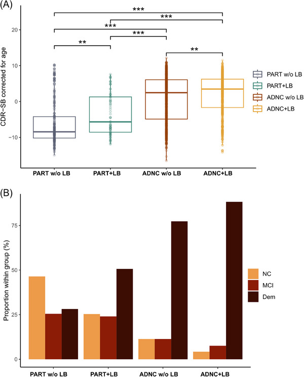
Introduction: Alzheimer's disease (AD) co-pathology with Lewy bodies (LB) is frequent and influences clinical manifestations and outcomes. Its significance in primary age-related tauopathy (PART) is unknown. We investigated the influence of LB on cognition and brain atrophy in AD and PART.
Methods: We performed a retrospective cohort study in a large sample of autopsied participants with AD neuropathological change (ADNC) with and without LB and PART with and without LB, with corresponding ante mortem magnetic resonance imaging (MRI) data from the National Alzheimer's Coordinating Center dataset.
Results: LB co-pathology worsened cognitive impairment in both PART and ADNC. On longitudinal follow-up, LB impacted cognitive decline in multiple domains. Additionally, LB influenced brain atrophy on MRI across groups and LB regional staging was different in PART and ADNC, accompanying tauopathy progression.
Discussion: These results suggest that LB co-pathology is associated with divergent patterns of cognitive impairment, brain atrophy, and regional pathological distribution in PART and AD.
Highlights: Lewy body (LB) co-pathology is frequent in Alzheimer's disease (AD) with important clinical implications. LB co-pathology is also present in primary age-related tauopathy (PART), but its significance is still understudied. In PART and AD, LB leads to higher cognitive impairment and brain regional atrophy. In PART and AD, LB tends to accompany neurofibrillary tangle progression, suggesting amyloid pathology might be a trigger for regional pathology progression.
Keywords: Alzheimer's disease; Lewy bodies; MRI; co‐pathology; primary age‐related tauopathy.
© 2024 The Author(s). Alzheimer's & Dementia published by Wiley Periodicals LLC on behalf of Alzheimer's Association.
Conflict of interest statement
T.G.O. is a scientific advisor and shareholder of Ceracuity Inc., has been a consultant for Sonae and Guidepoint, and has received fees as a speaker from Eisai and conference fees covered from Roche. The remaining authors have no disclosures to report. Author disclosures are available in the Supporting Information.
Figures


References
MeSH terms
Grants and funding
- P20 AG068053/AG/NIA NIH HHS/United States
- P30 AG066515/AG/NIA NIH HHS/United States
- P30 AG062421/AG/NIA NIH HHS/United States
- P30 AG066508/AG/NIA NIH HHS/United States
- P30 AG066519/AG/NIA NIH HHS/United States
- P30 AG072973/AG/NIA NIH HHS/United States
- P30 AG066462/AG/NIA NIH HHS/United States
- R01 NS095252/NS/NINDS NIH HHS/United States
- P30 AG066509/AG/NIA NIH HHS/United States
- P20 AG068077/AG/NIA NIH HHS/United States
- P30 AG072972/AG/NIA NIH HHS/United States
- P30 AG072979/AG/NIA NIH HHS/United States
- R01 AG054008/AG/NIA NIH HHS/United States
- P20 AG068082/AG/NIA NIH HHS/United States
- P30 AG072975/AG/NIA NIH HHS/United States
- P30 AG066444/AG/NIA NIH HHS/United States
- P30 AG066507/AG/NIA NIH HHS/United States
- P30 AG072946/AG/NIA NIH HHS/United States
- P30 AG066518/AG/NIA NIH HHS/United States
- P30 AG066511/AG/NIA NIH HHS/United States
- U24 AG072122/AG/NIA NIH HHS/United States
- P30 AG066512/AG/NIA NIH HHS/United States
- P30 AG072978/AG/NIA NIH HHS/United States
- P30 AG062429/AG/NIA NIH HHS/United States
- Tau Consortium
- P30 AG062422/AG/NIA NIH HHS/United States
- FAM/2022/Fundação Amélia de Mello
- R01 AG079280/AG/NIA NIH HHS/United States
- R01 NS086736/NS/NINDS NIH HHS/United States
- P30 AG066530/AG/NIA NIH HHS/United States
- P30 AG066546/AG/NIA NIH HHS/United States
- UIDP/50026/2020/Foundation for Science and Technology
- UIDB/50026/2020/Foundation for Science and Technology
- P30 AG072977/AG/NIA NIH HHS/United States
- P30 AG062677/AG/NIA NIH HHS/United States
- RF1 AG060961/AG/NIA NIH HHS/United States
- P20 AG068024/AG/NIA NIH HHS/United States
- P30 AG072958/AG/NIA NIH HHS/United States
- P30 AG062715/AG/NIA NIH HHS/United States
- P30 AG066506/AG/NIA NIH HHS/United States
- P30 AG066468/AG/NIA NIH HHS/United States
- P30 AG072976/AG/NIA NIH HHS/United States
- P30 AG072947/AG/NIA NIH HHS/United States
- P30 AG072931/AG/NIA NIH HHS/United States
- P30 AG066514/AG/NIA NIH HHS/United States
- P30 AG072959/AG/NIA NIH HHS/United States
LinkOut - more resources
Full Text Sources
Medical
