Review
. 2025 Jan;23(1):65-86.
doi: 10.1111/ddg.15541. Epub 2024 Dec 23.

Update vulval dermatology - diagnostics and therapy

Affiliations
Review

Update vulval dermatology - diagnostics and therapy

Christine Brägelmann et al. J Dtsch Dermatol Ges. 2025 Jan.

Abstract

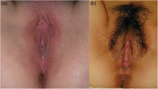
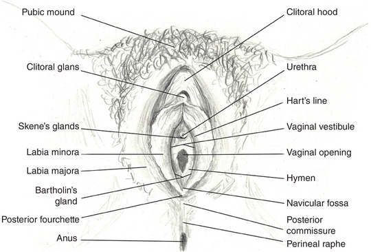
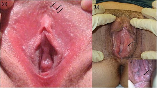
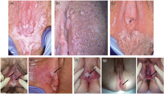
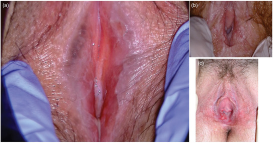
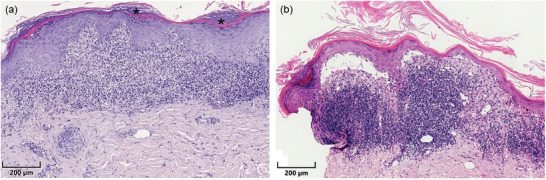
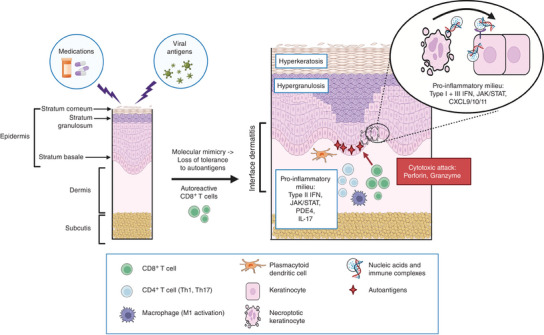
The vulva is a periorificial skin area and as such represents a transitional zone with unique functional and physiological characteristics. Knowledge of its anatomy is limited among both the general population and healthcare professionals, and unrealistic expectations of normal proportions are common. Ignorance of anatomical variations can cause unnecessary anxiety. In Germany, specialists in gynecology and obstetrics most commonly treat neoplastic vulvar dermatoses, while chronic inflammatory dermatoses commonly affecting the female genitalia (such as psoriasis, atopic dermatitis, hidradenitis suppurativa, and vitiligo) are typically treated by dermatologists. Both specialties treat infectious vulvar dermatoses and sexually transmitted infections. Certain dermatoses, such as lichen sclerosus, lichen planus, and lichen simplex chronicus, tend to affect the vulva preferentially; however, terminology can be confusing. Therefore, this article provides basic information on vulvar anatomy and physiology and summarizes recommendations for the diagnosis and management of the most common vulvar dermatoses, with a special focus on chronic inflammatory dermatoses, to provide a useful guide for all involved specialists in daily practice. Interdisciplinary collaboration and the establishment of dedicated consultation hours may help to improve the clinical care of vulvar dermatoses.

Keywords: Vulva; eczema; lichen planus; lichen sclerosus; lichen simplex chronicus; vagina.


Conflict of interest statement

The authors have received honoraria as speakers, advisory board members, travel reimbursements, or research support, or are involved in conducting clinical trials for the following companies: C.B.: L'Oréal, Novartis, Pfizer, UCB Pharma. L.W.: MSD, Roche, Eisai, Pfizer, Seagen, Novartis, GSK, AstraZeneca. D.N.: AbbVie, Almirall, AstraZeneca, Biogen, BMS, Boehringer‐Ingelheim, Eli Lilly, GlaxoSmithKline, Incyte, Kyowa Kirin, LaRoche/Cerave, L'Oréal, MSD, Novartis, Pfizer, Regeneron, Sanofi, UCB Pharma. The remaining authors declare no relevant conflicts of interest.

Figures

FIGURE 1
FIGURE 1
Normal vulvar anatomy. Vulvar anatomy varies greatly between individuals. Hormonal changes throughout life influence the pubic hair, the size of the mons pubis and labia majora, as well as the condition of the mucosa. In individuals with fair skin, a faint erythema is physiological, whereas mild hyperpigmentation is normal in darker skin types. (a) Normal adult premenopausal vulva (Caucasian female) (adapted from: Remas6, Public Domain, via Wikimedia Commons). (b) Normal adult premenopausal vulva (Asian female) (adapted from: Alexkhandria2K, Public Domain, via Wikimedia Commons).
FIGURE 2
FIGURE 2
Schematic illustration of vulvar anatomy. Depiction of the vulva in the lithotomy position, including anatomical terms (reproduced with the kind permission of Dr. med. Ingeborg Voß‐Heine).
FIGURE 3
FIGURE 3
Normal variants. (a) Fordyce spots: ectopic sebaceous glands appearing clinically as white‐yellow papules of variable size on mucosal tissue (License; Montana69, via Wikimedia Commons). (b) Micropapillomatosis vulvae: defined by multiple hyperkeratotic epithelial protrusions up to 1 mm in size, located distal to the hymenal ring. (c) Micropapillomatosis vulvae, detail from another patient: In sporadic cases, distinguishing it from condylomata acuminata can be very difficult, giving rise to the term “pseudocondyloma.” Micropapillomatosis vulvae is often symmetrically located at the vaginal vestibule, and human papillomavirus is not detectable.
FIGURE 4
FIGURE 4
Epithelial neoplasms. (a) Condylomata acuminata: confluent, broadly based, skin‐colored papules with a hyperkeratotic surface in an asymmetric array. (b) Condylomata acuminata: multiple skin‐colored, uneven papules on both hair‐bearing and hair‐free skin. (c) Condylomata acuminata: confluent patches of flat hyperkeratotic lesions located at the posterior commissure and perineum. (d) High‐grade squamous intraepithelial lesion (HSIL): metaplastic, sharply defined hyperkeratotic white plaques at the vaginal introitus. (e) High‐grade squamous intraepithelial lesion (HSIL): sharply defined atrophic plaque with erosion. (f) Keratinizing cutaneous squamous cell carcinoma: elevated whitish tumor associated with long‐standing lichen sclerosus. (g) Cutaneous squamous cell carcinoma: polypoid nodule associated with lichen sclerosus. (h) Bowen's carcinoma (confirmed association with human papillomavirus 16).
FIGURE 5
FIGURE 5
Melanocytic neoplasms. (a) Benign vulvar melanosis associated with topical estrogen therapy for lichen sclerosus. (b) Benign vulvar melanosis associated with lichen sclerosus. (c) Melanoma in situ: ill‐defined, uneven hyperpigmentation of the vaginal vestibule and clitoral glans. (d) Local recurrence of vulvar melanoma. (e) Nodular vulvar melanoma of the labium minus with satellite metastasis. (f) Dermoscopic findings of mucosal melanoma: structureless pattern, unevenly distributed globules, gray hue, and atypical vessels.
FIGURE 6
FIGURE 6
Infectious vulvar dermatoses. (a) Herpes genitalis (overview): grouped, painful blisters on an erythematous background. (b) Herpes genitalis (detail): the blisters tend to rupture due to friction, leaving subtle erosions. (c) Lipschütz ulcer (acute genital ulceration): a very painful, well‐circumscribed ulcer with fibrin crusts. (d) Lipschütz ulcer (acute genital ulceration): another patient. (e) Candidal vulvovaginitis: florid erosive dermatitis with vaginal discharge. (f) Tinea vulvae: depending on the causal fungus (anthropophilic vs. zoophilic), there may be slight scaling with a predilection for follicles or a suppurative pustular reaction with potential scarring. In this case, Tinea mentagrophytes acquired from a guinea pig was the causative agent. (g) Tinea corporis (another patient): septated hyphae in the stratum corneum (Periodic acid‐Schiff [PAS] stain, original magnification ×  400).
FIGURE 7
FIGURE 7
Clinical findings in hidradenitis suppurativa and vitiligo. (a) Hidradenitis suppurativa: multiple follicular atrophic scars on the mons pubis and inner thighs (bikini zone). The area within the labia majora is typically spared. (b) Non‐segmental vitiligo: sharply circumscribed depigmentations with a predilection for periorificial skin sites, including the vulva. Note the depigmented pubic hair.
FIGURE 8
FIGURE 8
Acute vulvar dermatitis. (a) Acute vulvar dermatitis: erosive, pruritic erythema with peripheral scatter (allergic contact dermatitis due to the topical application of estradiol cream). (b) Acute vulvar dermatitis: sharply circumscribed erythema with slight erosions (irritative contact dermatitis).
FIGURE 9
FIGURE 9
Chronic vulvar dermatitis. (a) Chronic vulvar dermatitis (lichen simplex chronicus): thickening and lichenification of the labia majora, only slight erythema. (b) Chronic vulvar dermatitis (detail): Note the alteration of the skin surface and pronounced folds in absence of Wickham striae or whitish shine.
FIGURE 10
FIGURE 10
Histology of vulvar dermatitis. (a) Acute dermatitis with spongiosis and acanthosis of the epithelium of the labia minora. Superficial edema and numerous dilated vessels. (b) Lichen simplex chronicus with compact orthohyperkeratosis, hypergranulosis and acanthosis of the labia minora. In the dermis fibrosis, interstitial cellularity and numerous dilated vessels. ((a, b) hematoxylin‐eosin stain, scale bar: 200 µm).
FIGURE 11
FIGURE 11
Clinical findings in lichen sclerosus. (a) Lichen sclerosus: acute inflammatory reaction with painful erosions along the labia minora and chronic areas laterally, characterized by porcelain‐like skin thickening. (b) Lichen sclerosus: the characteristic figure‐eight configuration involves the vulva, perineum, and perianal area. Chronic lichen sclerosus frequently leads to fissures.
FIGURE 12
FIGURE 12
Histology of lichen sclerosus. (a) Early‐stage lichen sclerosus with a slightly atrophic epidermis, superficial lichenoid inflammatory infiltrate, and dilated vessels. (b) Rarefied elastic fibers within the inflammatory infiltrate. (c) Late‐stage lichen sclerosus: atrophic epidermis, subepidermal hyalinization of connective tissue, and sparse inflammatory infiltrate. (d) Absence of elastic fibers within the hyalinized zone. ((a, c) Hematoxylin‐eosin stain, scale bar: 200 µm; (b, d) Elastica stain, scale bar: 200 µm).
FIGURE 13
FIGURE 13
Pathophysiology of lichen sclerosus. A certain genetic predisposition, combined with risk factors, initiates the inflammatory early stage of lichen sclerosus, characterized by “interface dermatitis.” This reaction involves a Th1‐dominated cytokine milieu and a dense infiltrate of primarily CD4+ and CD8+ T cells in the upper dermis. In the late stage of the disease, pro‐sclerotic factors (e.g., miRNA155, TGF‐β, BMP2) predominate, leading to epidermal atrophy, dermal sclerosis, and reduction and dilation of dermal vessels, which explains the common occurrence of hemorrhage. Inflammation and vascular sclerosis result in oxidative stress and downregulation of tumor suppressor genes, potentially contributing to carcinogenesis. (This figure was created with BioRender.com and adapted from, , .)
FIGURE 14
FIGURE 14
Clinical findings in lichen planus. (a) Mucosal lichen planus: erosive mucosal alteration with Wickham striae. Note involvement of vaginal epithelium. (b) Mucosal lichen planus: hyperkeratotic mucosal alteration with Wickham striae. (c) Mucosal lichen planus: long‐standing inflammatory reaction may lead to strictures.
FIGURE 15
FIGURE 15
Histology of lichen planus. (a) Lichen planus mucosae showing orthohyperkeratosis, hypergranulosis, and irregular sawtooth acanthosis of the epidermis, with singular basal dyskeratoses. Dense lichenoid lymphocytic infiltrate in the dermis. (b) Bullous lichen planus: the severity of the inflammatory reaction may cause disruption of epidermal adhesion at the basal membrane, accompanied by pigmentary incontinence. ((a, b) Hematoxylin‐eosin stain, scale bar: 200 µm).
FIGURE 16
FIGURE 16
Pathophysiology of lichen planus. A genetic predisposition, combined with triggers such as viral antigens, provokes a loss of tolerance to autoantigens through molecular mimicry, leading to a cytotoxic T‐cell‐mediated anti‐epithelial inflammatory reaction (interface dermatitis) and subsequent keratinocyte destruction. Released immunostimulatory immune complexes and nucleic acids perpetuate the interface dermatitis. Clinically, this chronic process can result in hyperkeratosis and erosions. (This figure was created with BioRender.com and adapted from, .)

Similar articles

References

    1. Paarlberg KM, Weijenborg PTM. Request for operative reduction of the labia minora; a proposal for a practical guideline for gynecologists. J Psychosom Obstet Gynaecol. 2008;29:230‐234. - PubMed
    1. Lykkebo AW, Drue HC, Lam JUH, Guldberg R. The Size of Labia Minora and Perception of Genital Appearance: A Cross‐Sectional Study. J Low Genit Tract Dis. 2017;21:198‐203. - PubMed
    1. Pauls RN. Anatomy of the clitoris and the female sexual response. Clin Anat. 2015;28:376‐384. - PubMed
    1. Baskin L, Shen J, Sinclair A, et al. Development of the human penis and clitoris. Differentiation. 2018;103:74‐85. - PMC - PubMed
    1. Emmanuel M, Bokor BR. Tanner Stages. [Updated 2022 Dec 11]. In: StatPearls [Internet]. Treasure Island (FL): StatPearls Publishing; 2024. Jan. Available from: https://www.ncbi.nlm.nih.gov/books/NBK470280/ [Last accessed June 1, 2024].