[Preprint]. 2024 Dec 11:rs.3.rs-5538058.
doi: 10.21203/rs.3.rs-5538058/v1.

Design of allosteric modulators that change GPCR G protein subtype selectivity

Affiliations

Design of allosteric modulators that change GPCR G protein subtype selectivity

Madelyn N Moore et al. Res Sq. .

Abstract

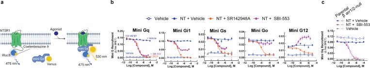
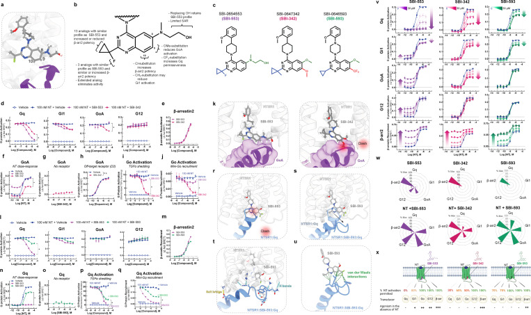
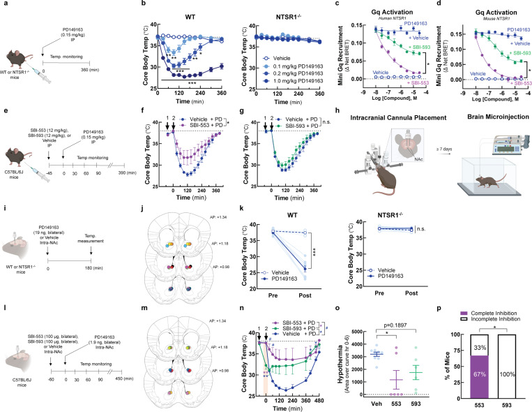
G protein-coupled receptors (GPCRs), the largest family of drug targets, can signal through 16 subtypes of Gα proteins. Biased compounds that selectively activate therapy-relevant pathways promise to be safer, more effective medications. The determinants of bias are poorly understood, however, and rationally-designed, G protein-subtype-selective compounds are lacking. Here, using the prototypical class A GPCR neurotensin receptor 1 (NTSR1), we find that small molecules binding the intracellular GPCR-transducer interface change G protein coupling by subtype-specific and predictable mechanisms, enabling rational drug design. We demonstrate that the compound SBI-553 switches NTSR1 G protein preference by acting both as a molecular bumper and a molecular glue. Structurally, SBI-553 occludes G protein binding determinants on NTSR1, promoting association with select G protein subtypes for which an alternative, shallow-binding conformation is energetically favorable. Minor modifications to the SBI-553 scaffold produce allosteric modulators with distinct G protein subtype selectivity profiles. Selectivity profiles are probe-independent, conserved across species, and translate to differences in in vivo activity. These studies demonstrate that G protein selectivity can be tailored with small changes to a single chemical scaffold targeting the receptor-transducer interface and, as this pocket is broadly conserved, present a strategy for pathway-selective drug discovery applicable to the diverse GPCR superfamily.

Keywords: G protein selectivity; G protein-coupled receptor; allosteric modulation; biased signaling; neurotensin receptor 1; β-arrestin.


Figures

Figure 1.
Figure 1.. The NTSR1 allosteric modulator SBI-553 exhibits transducer-specific efficacy.
Ligand-directed NTSR1 signaling was assessed in HEK293T cells transiently expressing NTSR1 and G protein or β-arrestin activation sensors. (A-E) NTSR1 ligand-induced G protein activation by TRUPATH. (A) Illustration of BRET2-based TRUPATH assay of G protein activation. (B) Depiction of TRUPATH data transformation. G protein activation results in reduced BRET as a BRET donor tagged-Gα and a BRET acceptor tagged-Gγ subunit dissociate. Curves were inverted such that G protein activation resulted in upward sloping curves. (C) G protein activation was assessed following treatment with the endogenous agonist NT, the NT peptide analog PD149163, the β-arrestin-biased ligand SBI-553, and the orthosteric antagonist SR142948A. (D) Maximal ligand-induced G protein activation. Colored asterisks over each bar indicate the treatments from which that compound significantly differed. Treatment vs NT (*), SR142948A (*), PD149163 (*), SBI-553(*). (E) Maximal SBI-553-induced G protein activation in cells transiently expressing NTSR1 or an empty control vector. (F-G) NTSR1 ligand-induced β-arrestin recruitment by BRET. (F) Illustration of BRET1-based assay for β-arrestin recruitment. (G) β-arrestin1/2 recruitment was assessed following treatment with NT, PD149163, SBI-553, or SR142948A. (H) Summary of compound efficacy and potency. Radar plots depict the extent of transducer activation relative to NT (i.e., fold change NT Emax for each transducer). Heat maps depict ligand potency. (I-K) NTSR1 ligand-induced G protein activation by TGFα shedding. (I) Illustration of TGFα shedding assay for G protein activation. (J) Ligand-induced G protein activation. (K) Maximal G protein activation following treatment with NT or SBI-553. Asterisks (*) indicate a change from the GΔC negative control, unless otherwise indicated. For curve parameters, sample size, and statistical comparisons, see Table S1.
Figure 2.
Figure 2.. Unlike competitive antagonist SR142948A, SBI-553 biases NT-NTSR1 signaling away from Gq/11 and toward both β-arrestin recruitment and alternative G protein activation.
(A-E) SR142948A uniformly competitively antagonizes NT-induced NTSR1 transducer activation. (A) Illustration of NT and SR142948 competing to occupy the same binding site on NTSR1. (B) Characteristic agonist dose-response curve (DRC) shifts in the presence of increasing concentrations of a competitive antagonist. (C) NT-induced β-arrestin1/2 recruitment to the NTSR1 was assessed by BRET in the absence and presence of SR142948A. (D) NT-induced G protein activation was assessed by TRUPATH in the absence and presence of SR142948A. (E) NT DRC EC50 values for the 12 G proteins evaluated in D in the presence of increasing concentrations of SR142948A. (F-L) SBI-553 exerts transducer-specific effects on NT-induced NTSR1 transducer activation. (F) Illustration of NT and SBI-553 binding distinct sites on the NTSR1. (G) (Top) Characteristic agonist DRC shifts in the presence of a noncompetitive antagonist. (Bottom) Characteristic agonist DRC shifts in the presence of an allosteric agonist. (H) NT-induced human β-arrestin1/2 recruitment to the NTSR1 in the absence and presence of SBI-553. (I) NT-induced G protein activation was assessed by TRUPATH in the absence and presence of SBI-553. (J) NT DRC EC50 values for the 12 G proteins evaluated in H in the presence of increasing concentrations of SBI-553. (K) NT DRC EC50 values for each G protein at the maximal SBI-553 concentration for which a sigmoidal curve could be fit. Data are represented as fold-change from vehicle. (L) Radar plots depict the extent of activation of each transducer in the presence NT or SBI-553 alone and both NT and SBI-553 in combination. Fold-change values are standardized to NT Emax for each transducer. (M) Effect of SR142948A and SBI-553 on NT-induced G protein activation, second assay validation. NT-induced G protein activation was assessed by TGFα shedding. NT-induced activation of Gq, Gi1/2, Go, and G12 sensors in the presence of SR142948A, SBI-553, or vehicle. (N) Illustration of NTSR1 G protein activation following application of NT alone and in the presence of SR149163 or SBI-553. (O) Rank order of NT-NTSR1 G protein preference in the absence and presence of SBI-553. For curve parameters, N, and statistical comparisons, see Table S2.
Figure 3.
Figure 3.. SBI-553 blocks NTSR1 coupling with a subset of G proteins by a β-arrestin-independent mechanism.
(A) Illustration of BRET1-based assay for mini-G recruitment. (B) NT (100nM)-induced Mini-Gq, Gi1, Gs, Go, or G12 recruitment to the NTSR1 was assessed in the presence of Vehicle (HBSS with 0.5%HP-β-cyclodextrin), SBI-553 or SR142948A. Controls received Vehicle alone. (C) NT induced Mini-Gq recruitment in the presence of SBI-553 was assessed in β-arrestin1/2-null HEK293 cells and their parental control line. Cells were pretreated with either SBI-553 or its vehicle prior to application of 100 nM NT or its vehicle. For curve parameters, N, and statistical comparisons, see Table S3. For supporting data, see Figure S1.
Figure 4.
Figure 4.. The sensitivity of a Gα protein to SBI-553 antagonism is determined by the ability of its C-terminus to adopt an alternative shallow-binding conformation.
(A-E) Sensitivity of G proteins to SBI-553 antagonism can be reversed by exchanging their C-termini. (A) Location of the GoA/Gq 5 C-terminal amino acid residue swap. (B) Swapping GoA’s 5 C-terminal amino acids for those of Gq confers sensitivity to SBI-553 antagonism. Inset, effect of SBI-553 on NT-induced activation of WT GoA for reference. (C) Swapping Gq’s 5 C-terminal amino acids for those of GoA reduces the antagonist efficacy of SBI-553. Inset, effect of SBI-553 on NT-induced activation of WT Gq for reference. (D) Location of Gq/GoA 13 C-terminal amino acid residue swap, GqΔ234–246. (E) Swapping Gq’s 13 C-terminal amino acids for those of GoA reduces the antagonist potency and efficacy of SBI-553. (F) Alignment of G protein C-termini. (G-I) Single amino acid substitutions on Gq’s C-terminus are insufficient to render Gq permissive of SBI-553. (G) Location of Gq point mutants on the NTSR1-Gq structure. (H) NT-induced activation of WT and mutant Gq constructs. (I) Effect of Gq mutagenesis on SBI-553’s ability to antagonize Gq activation by 100 nM NT. (J-L) Identification of an SBI-553-induced shallow-binding ‘open’ GoA conformation. (A) Cryo-EM structures showing NTSR1 bound by the NT active fragment and G protein in the absence and presence of SBI-553. (Left) NTSR1, NT, mini-GoA (PDB 8FN1). (Middle) NTSR1, NT, SBI-553, mini-GoA (PDB 8FN0). (Right) NTSR1, NT, mini-Gq (PDB 8FMZ). (K) Docking of SBI-553 in the 8FN1 and 8FMZ structures suggests that both GoA and Gq should clash with SBI-553. (L) Registration of the 8FN1 and 8FN0 structures illustrates that, in the presence of SBI-553, GoA adopts an alternative ‘open’ conformation to accommodate SBI-553 binding. (M-P) Homology modeling indicates that some NTSR1-SBI-553-’open’ position G protein complexes are more energetically favorable than others. In silico homology models were created based on the ‘open’ NTSR1-SBI-553-bound GoA conformation by changing the 13 C-terminal amino acids. Free energy of dissociation changes (ΔGdiss) were calculated, and models are positioned from most stable (right) to least stable (left). (N) NTSR1-SBI-553-G protein ‘open’ conformation ΔGdiss presented for individual G proteins, categorized by extent of sensitivity to SBI-553 antagonism. (O) Correlation of NTSR1-SBI-553-G protein ‘open’ conformation ΔGdiss with experimental SBI-553-induced fold change in NT G protein activation potency, as presented in Fig 2K. (P) Correlation of NTSR1-SBI-553-G protein ‘open’ conformation ΔGdiss with experimental max SBI-553-induced G protein activation, as presented in Fig 2D. (Q) Model of SBI-555 G protein subtype selectivity. For curve parameters, sample size, and statistical comparisons, see Table S4. For supporting data, see Figure S2, S3.
Figure 5.
Figure 5.. Discovery of SBI-553 analogs with distinct G protein selectivity profiles.
(A) Position of SBI-553 in the NTSR1 intracellular core. Numbers mark quinazoline C8, C9, and C10. GoA shown in purple. (B) Summary of structure-activity relationship (SAR) study findings. (C) Structures of SBI-553 and analogs SBI-342 and SBI-593. (D-K) Analog SBI-342 exhibits Go antagonism, not agonism. (D) Screen evaluating SBI-342 antagonism of NT-induced Gq, Gi1, GoA, and G12 activation by TRUPATH. (E) Screen evaluating SBI-342 β-arrestin2 agonism by BRET. (F) Effect of 30 μM SBI-553 vs SBI-342 across the NT GoA DRC. (G) Assessment of SBI-342 on GoA TRUPATH activation sensor activity in HEK293T cells not expressing NTSR1. (H) Assessment of SBI-342 (30 μM) on GoA activation stimulated by the Gi/o-coupled dopamine receptor D2. (I) Comparison of SBI-553 and SBI-342 antagonism of NT-induced Go activation in the AP-TGFα shedding assay. (J) Comparison of SBI-553 and SBI-342 antagonism of NT-induced miniGoA recruitment to the NTSR1 by BRET. (K) SBI-553 can co-occupy NTSR1 with GoA’s C-terminus in its ‘open’ position, while the 9-methoxy of SBI-342 clashes with GoA. (L-U) Analog SBI-593 exhibits partial rather than full Gq antagonism. (L) Screen evaluating SBI-593 antagonism of NT-induced Gq, Gi1, GoA, and G12 activation by TRUPATH. (M) Screen evaluating SBI-593 β-arrestin2 agonism by BRET. (N) Effect of 30 μM SBI-553 vs SBI-593 across the NT Gq DRC. (O) Assessment of SBI-593 on Gq TRUPATH activation sensor activity in HEK293T cells not expressing NTSR1. (P) Comparison of SBI-553 and SBI-593 antagonism of NT-induced Gq activation in the AP-TGFα shedding assay. (Q) Comparison of SBI-553 and SBI-593 antagonism of NT-induced miniGoA recruitment to the NTSR1 by BRET. (R) Docking SBI-593 into the SBI-553 binding site in the NTSR1:Gq structure (PDB 8FMZ) indicates a clash between Gq and SBI-593, as for SBI-553. (S) Molecular dynamics simulations indicate a repositioning of the Gq C-terminus within the NTSR1 core. (T) Interactions between NTSR1 and Gq stabilizing this new position are shown. (U) Attractive van der Waals contacts between SBI-593 and the Gq C-terminus its new position are illustrated as dotted green lines. (V-X) SBI-553 analogs have distinct G protein selectivity profiles. (V) NT-induced G protein activation was assessed in the absence and presence of SBI-553, −342, and −593. (W) Radar plots depict extent of transducer activation induced by SBI-553, −342, and −593 alone (top) and in the presence of NT (bottom). Values reflect maximal % Emax relative to NT (G proteins) or SBI-553 (β-arrestin2). (X) Summary of NTSR1 G protein activation following application of NT in the presence of SBI-553, −342, and −593. For curve parameters, N, and statistical comparisons, see Table S5. For supporting data, see Figures S3–9.
Figure 6.
Figure 6.. SBI-593 does not produce a complete block of NTSR1 agonist-induced, Gq-mediated hypothermia in mice.
(A-B) Systemic PD142948A induces an NTSR1-dependent hypothermia in mice. (A) Experimental timeline. (B) Effect of PD149163 (0.1–1.0 mg/kg, i.p.) on core body temperature in WT and NTSR1−/− mice. (C-D) SBI-593 partially antagonizes PD149163-induced Gq activation by the NTSR1. (C) Effect of SBI-553 and SBI-593 on PD149163-induced Gq activation by the (C) human and (D) mouse NTSR1. In HEK293T cells transiently expressing NTSR1, the NT-induced association between NTSR1 and miniGq was assessed by BRET. (E-G) Systemic SBI-593 administration does not attenuate PD149163-induced hypothermia. (E) Experimental timeline. (F) Effect of SBI-553 (12 mg/kg, i.p.) vs. Vehicle (5% HP-β-cyclodextrin) pretreatment on PD149163 (15 mg/kg,i.p.)-induced hypothermia. (G) Effect of SBI-593 (12 mg/kg, i.p.) vs. Vehicle (20% DMSO, 5% Tween 80) pretreatment on PD149163 (0.15 mg/kg, ,i.p.)-induced hypothermia. (H) Overview of bilateral intracranial cannula placement in mice. Cannulas targeted the nucleus accumbens (NAc). (I-K) Intra-NAc PD142948A induces an NTSR1-dependent hypothermia in mice. (I) Experimental timeline. (J) Ink-verified cannula placements for mice in panel K. Numbers denote mm in front of bregma. AP, anterior-posterior. (K) Effect of microinjection of PD149163 (93 ng, bilateral) into the NAc on core body temperature in WT and NTSR1−/− mice. (L-P) Intra-NAc SBI-593 administration partially but does not fully attenuate PD149163-induced hypothermia. (L) Experimental timeline. (M) Ink-verified cannula placements for mice in panel N. Numbers denote mm in front of bregma. AP, anterior-posterior. (N) Effect of intra-NAc SBI-553 (100 μg, bilateral) and SBI-593 (100 μg, bilateral) on PD149163 (9.3 ng, bilateral, intra-NAc)-induced hypothermia. (O) Area of the curve for data in panel N. Baseline set at 37°C. (P) Proportion of mice exhibiting complete vs. incomplete blockade of PD149163-induce hypothermia by treatment group. For information on N, statistical comparisons, and curve parameters, see Table S6.

References

    1. Sriram K. & Insel P. A. G Protein-Coupled Receptors as Targets for Approved Drugs: How Many Targets and How Many Drugs? Molecular Pharmacology 93, 251–258 (2018). 10.1124/mol.117.111062 - DOI - PMC - PubMed
    1. Smith J. S., Lefkowitz R. J. & Rajagopal S. Biased signalling: from simple switches to allosteric microprocessors. Nat Rev Drug Discov 17, 243–260 (2018). 10.1038/nrd.2017.229 - DOI - PMC - PubMed
    1. Kolb P. et al. Community guidelines for GPCR ligand bias: IUPHAR review 32. Br J Pharmacol 179, 3651–3674 (2022). 10.1111/bph.15811 - DOI - PMC - PubMed
    1. Slosky L. M., Caron M. G. & Barak L. S. Biased Allosteric Modulators: New Frontiers in GPCR Drug Discovery. Trends Pharmacol Sci 42, 283–299 (2021). 10.1016/j.tips.2020.12.005 - DOI - PMC - PubMed
    1. Wingler L. M. et al. Angiotensin and biased analogs induce structurally distinct active conformations within a GPCR. Science 367, 888–892 (2020). 10.1126/science.aay9813 - DOI - PMC - PubMed

Publication types

LinkOut - more resources