[Preprint]. 2024 Dec 13:rs.3.rs-5494070.
doi: 10.21203/rs.3.rs-5494070/v1.

Covalent-fragment screening identifies selective inhibitors of multiple Staphylococcus aureus serine hydrolases important for growth and biofilm formation

Affiliations

Covalent-fragment screening identifies selective inhibitors of multiple Staphylococcus aureus serine hydrolases important for growth and biofilm formation

Matthew Bogyo et al. Res Sq. .

Update in

Abstract

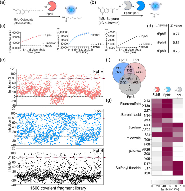
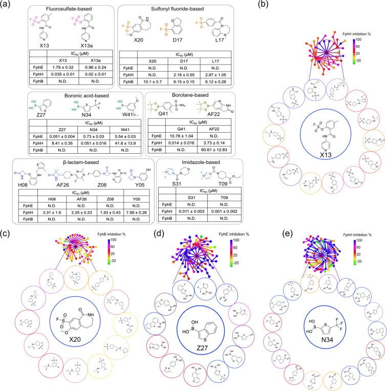
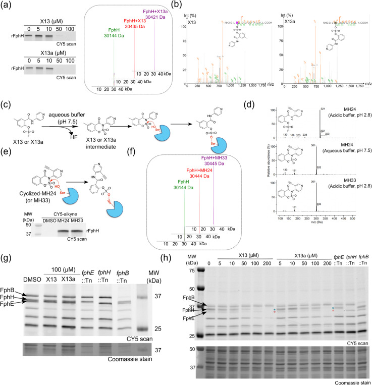
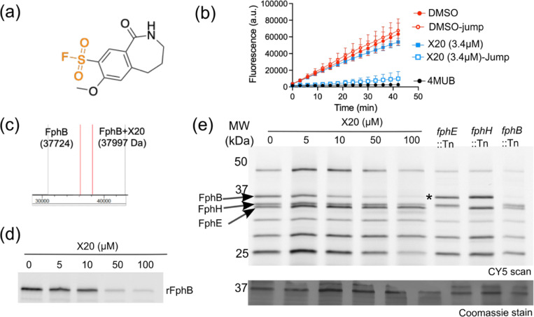
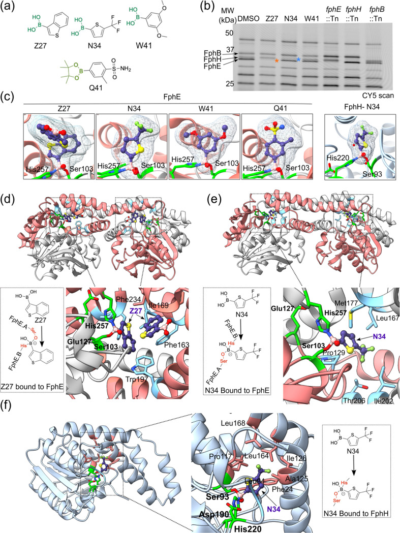
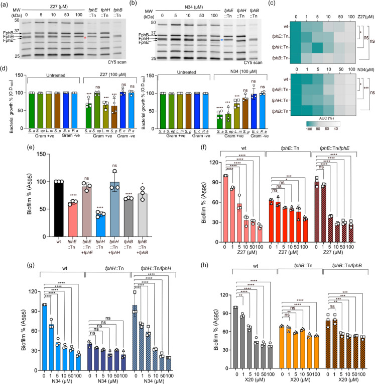
Staphylococcus aureus is a leading cause of bacteria-associated mortality worldwide. This is largely because infection sites are often difficult to localize and the bacteria forms biofilms which are not effectively cleared using classical antibiotics. Therefore, there is a need for new tools to both image and treat S. aureus infections. We previously identified a group of S. aureus serine hydrolases known as fluorophosphonate-binding hydrolases (Fphs), which regulate aspects of virulence and lipid metabolism. However, because their structures are similar and their functions overlap, it remains challenging to distinguish the specific roles of individual members of this family. In this study, we applied a high-throughput screening approach using a library of covalent electrophiles to identify inhibitors for FphB, FphE, and FphH. We identified inhibitors that irreversibly bind to the active-site serine residue of each enzyme with high potency and selectivity without requiring extensive medicinal chemistry optimization. Structural and biochemical analysis identified novel binding modes for several of the inhibitors. Selective inhibitors of FphH impaired both bacterial growth and biofilm formation while Inhibitors of FphB and FphE had no impact on cell growth and only limited impact on biofilm formation. These results suggest that all three hydrolases likely play functional, but non-equivalent roles in biofilm formation and FphH is a potential target for development of therapeutics that have both antibiotic and anti-biofilm activity. Overall, we demonstrate that focused covalent fragment screening can be used to rapidly identify highly potent and selective electrophiles targeting bacterial serine hydrolases. This approach could be applied to other classes of lipid hydrolases in diverse pathogens or higher eukaryotes.


Figures

Figure 1
Figure 1
High-throughput screening against S. aureus serine hydrolases. (a-b) Schematic representation of the enzyme assay used in the high-throughput screening of purified rFphE, rFphH and rFphB using the substrates 4-methylumbelliferyl octanoate for FphE and 4-methylumbelliferyl butyrate for FphB and FphH. (c) Progress curves for each serine hydrolase (0.5 nM rFphE, 5 nM rFphH and 100 nM rFphB) using 20 μM of the substrate for each assay. (d) Calculated Z’ values (n = 8, mean ± SD) for each enzyme. (e) Plot of the percent inhibition values (at 50 μM) for each compound in the primary screening fragment library for FphE (red), FphH (blue) and FphB (black). An FP-based inhibitor was used as a positive control. (f) Venn diagram showing the distribution of the top hits with ≥80% inhibition among the three targets. (g) Heat maps for the top hits arranged according to their electrophile type and percent inhibition for each target enzyme.
Figure 2
Figure 2
Identification of multiple electrophile-classes for S. aureus serine hydrolases (a) Structures of hit compounds, selected based on their selectivity against each Fph enzyme, organized by electrophile-class. The measured IC50 values for each target are shown for each set of hits. N.D. indicates that the IC50 value could not be determined due to incomplete inhibition at the highest concentration tested. (b-e) Generation of structure activity relationship data using the neighborhood tree generated from the SALI plot (shown in Figure S1b–d). Plots show closely related structures based on the OrgFunction descriptor for each electrophile and their corresponding activities for each target Fph enzyme.
Figure 3
Figure 3
Biochemical characterization of fluorosulfate-based fragments. (a) Fluorescent SDS-PAGE gel images showing concentration-dependent inhibition of the labeling of rFphH (500 nM) by FP-Cy5 probe (300 nM) for X13 and X13a. Deconvoluted intact protein mass spectra of 1μM rFphH alone or after 1hr incubation with 10 μM X13 or X13a. (b) MS/MS spectra confirming covalent modification of the active site Ser113 of rFphH by X13 and X13a. (c) Proposed mechanism of action of compounds X13 and X13a involving initial cyclization to eliminate HF, followed by ring opening by the active site serine. (d) LC/MS profiles for X13d and X13e in LC/MS buffer or aqueous buffer. (e) Proposed reaction mechanism of X13d or X13e binding to rFphH, Fluorescence SDS-PAGE image of rFphH for direct visualization of labelling by clickable probes X13d and X13e with Cy5-azide. (f) Native mass spectrometry (MS) characterization of 1μM rFphH alone or after 1hr incubation with 10 μM X13d or X13e. (g) Competition of FP-Cy5 labeling of live S. aureus cells (USA300 LAC) or the individual transposon mutants for fphE, fphH and fphB after preincubated with 100 μM X13 and X13a for 120 min, incubated with clickable 1μM FP, lysis and labeling with Cy5-azide and analysis by SDS-PAGE and fluorescence scanning. (h) Competition of FP-Cy5 labeling of S. aureus lysates. S. aureus USA300 WT cells were lysed prior to preincubation with the indicated doses of X13 and X13a for 120 min, labelled with clickable FP-Cy5, and analyzed by SDS-PAGE and fluorescence scan. The indicated Fph transposon mutants (::Tn) lysates are included to highlight the location of the three enzymes in the gel. All data shown are representative of three independent experiments.
Figure 4
Figure 4
A sulfonyl fluoride-based irreversible electrophile is selective for FphB. (a) The structure of FphB-selective hit compound, X20. (b) Jump dilution assay of rFphB inhibited by X20. rFphB was incubated for 60 min with 101 μM (10X IC50) of X20, followed by a 30-fold dilution prior to performing enzyme activity assay. Activity from this jump-dilution was compared with a DMSO control and 3.75 μM of X20. (c) Deconvoluted mass spectra of 1μM rFphB alone or after 1hr incubation with 10 μM X20. (d) Fluorescent SDS-PAGE gel image showing the competition of FP-Cy5 labelling of rFphB preincubated with the indicated doses of X20 and subsequently labelled with the FP-Cy5 probe (500 nM). (e) Competition of FP-Cy5 labeling of live S. aureus cells. USA300 WT cells were preincubated with the indicated concentrations of X20 for 120 min. The individual transposon mutants of fphB, fphH and fphE are included to highlight the location of the three enzymes in the gel. The cells were incubated with 1μM FP-Alkyne, lysed, labelled with Cy5-Azide, analyzed by SDS-PAGE and analyzed by fluorescence scan. All data shown are representative of three independent experiments.
Figure 5
Figure 5
Boron based inhibitors of FphE and FphH. (a) The structure of the three top boronic acid hits, as well as one of the promiscuous borolane hits. (b) Fluorescent SDS-PAGE gel image showing competition of FP labeling in intact S. aureus cells by 100 μM Z27, N34 and W41. The individual transposon mutants of fphB, fphH and fphE are included to highlight the location of the three enzymes in the gel. (c) Close-up view of FphE covalently inhibited by Z27 (also includes an unreacted non covalently bound molecule; PDB ID 8UGM, 1.65 Å), N34 (8TFW, 1.93 Å), W41 (8UIX, 2.39 Å) and Q41 (8UWM, 1.97 Å). Boron binds to the active site FphE-serine and histidine from different homodimer FphE monomers. FphH binds N34 using the active site serine and histidine residues from the same monomer (8TAV, 1.39 Å). Ligand bound to FphE-Ser103 and FphE-His257 or FphH-Ser93-His220 are shown as a blue mesh. (d) Structure of FphE bound by Z27 showing the dual attack mechanism using serine and histidine. The structure also shows a second unreacted bound Z27 molecule in the active site that does not engage the catalytic residues. (e) Structure of FphE bound by N34 showing the dual attack mechanism using serine and histidine. (f) Structure of FphH bound by N34 through a di-covalent interaction with the active site serine and histidine.
Figure 6
Figure 6
Boronic acid-based inhibitors show antibacterial and antibiofilm activity. Fluorescence SDS-PAGE gel images of live S. aureusUSA300 WT cell preincubated for 120 min with the indicated concentrations of Z27 (a) and N34 (b) and then labeled with the broad-spectrum serine hydrolase FP probe. The individual transposon mutants of fphB, fphH and fphE are included to indicate the location of the three enzymes in the gel. All the cells were labeled with the clickable FP-alkyne, lysed and labelled with Cy5-azide and analyzed by SDS-PAGE followed by fluorescence scanning. (c) Heat maps derived from growth curves to compare the area under the curve (AUC) for S. aureus wild type and S. aureus fph transposon mutants after treatment with the indicated concentrations of Z27 or N34 (Generated from Figure S5d & e). AUCs are normalized such that 100% represents the AUC for untreated wild-type S. aureus. The graphs show mean ± standard deviation of data from n = 2 biological replicates (each as technical duplicates). (d) Effects of Z27 and N34 on the indicated bacteria. Gram-positive bacteria tested: S. a: S. aureus, S. ep: S. epidermidis, L. m: Listeria monocytogenes, S. p: Streptococcus pyogenes. Gram-negative bacteria tested E. c: Escherichia coli (E. coli), P. a: Pseudomonas aeruginosa. The data plotted are growth percent relative to the untreated control at the 10hr time point for each strain. The graphs show mean ± standard deviation of data from n = 2 biological replicates (each as technical duplicates). (e) Percentage biofilm formation measured by crystal violet staining for S. aureus wild type, fph mutants, and complement strains. Results are normalized such that 100% represents the signal from WT S. aureus (f) Percentage of biofilm inhibition by Z27 measured for wild-type S. aureus, fphE mutant, and the corresponding fphE complement. Results are normalized such that 100% represents levels for untreated WT S. aureus (g) Percentage of biofilm inhibition by N34 measured for wild-type S. aureus, fphH mutant, and fphH complement. (h) Percentage biofilm inhibition by X20 measured for wild-type S. aureus, fphB mutant, and FphB complement. Results are normalized such that 100% represents untreated WT S. aureus. Bars represent means ± standard deviation of data from n = 3 biological replicates. P values were determined using ordinary one-way ANOVA multiple comparisons. *P < 0.05, **P < 0.01, ***P < 0.001 and ****P < 0.0001.

Similar articles

References

    1. Lee A. S. et al. Methicillin-resistant Staphylococcus aureus. Nat. Rev. Dis. Primer 4, 1–23 (2018). - PubMed
    1. Howden B. P. et al. Staphylococcus aureus host interactions and adaptation. Nat. Rev. Microbiol. 21, 380–395 (2023). - PMC - PubMed
    1. Hamdan-Partida A., Sainz-Espuñes T. & Bustos-Martínez J. Characterization and persistence of Staphylococcus aureus strains isolated from the anterior nares and throats of healthy carriers in a Mexican community. J Clin Microbiol 48, 1701–1705 (2010). - PMC - PubMed
    1. Bhattacharya M., Wozniak D. J., Stoodley P. & Hall-Stoodley L. Prevention and treatment of Staphylococcus aureus biofilms. Expert Rev. Anti Infect. Ther. 13, 1499–1516 (2015). - PMC - PubMed
    1. Wu X. et al. Staphylococcus aureus biofilm: Formulation, regulatory, and emerging natural products-derived therapeutics. Biofilm 7, 100175 (2024). - PMC - PubMed

Publication types

LinkOut - more resources