[Preprint]. 2024 Dec 11:rs.3.rs-5529783.
doi: 10.21203/rs.3.rs-5529783/v1.

Adolescent-like Processing of Behaviorally Salient Cues in Sensory and Prefrontal Cortices of Adult Preterm-Born Mice

Affiliations

Adolescent-like Processing of Behaviorally Salient Cues in Sensory and Prefrontal Cortices of Adult Preterm-Born Mice

Adema Ribic et al. Res Sq. .

Abstract

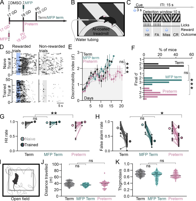
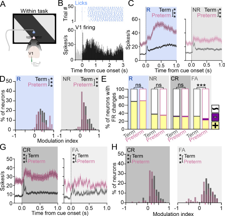
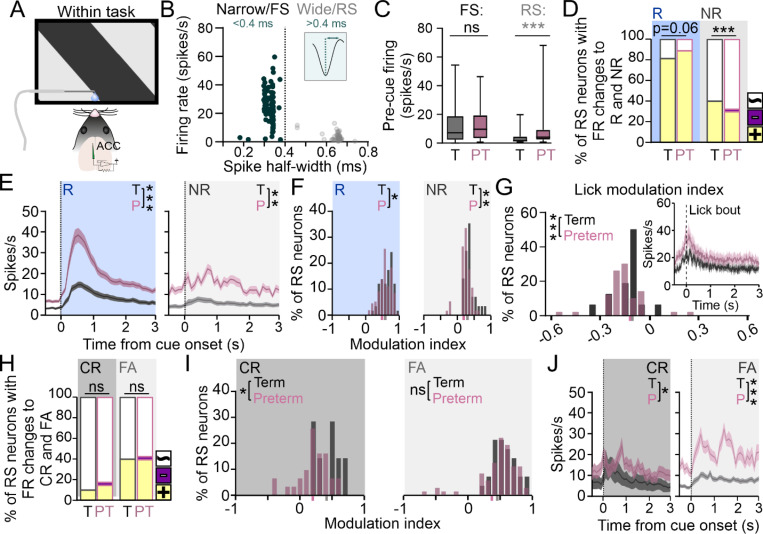
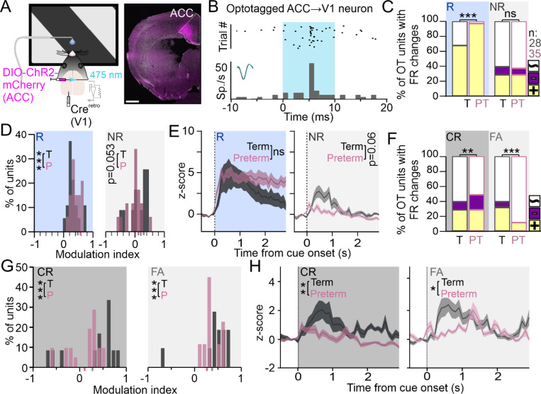
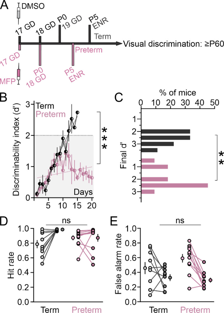
Preterm birth is a leading risk factor for atypicalities in cognitive and sensory processing, but it is unclear how prematurity impacts circuits that support these functions. To address this, we trained adult mice born a day early (preterm mice) on a visual discrimination task and found that they commit more errors and fail to achieve high levels of performance. Using in vivo electrophysiology, we found that the neurons in the primary visual cortex (V1) and the V1-projecting prefrontal anterior cingulate cortex (ACC) are hyper-responsive to the reward, reminiscent of cue processing in adolescence. Moreover, the non-rewarded cue fails to robustly activate the V1 and V1-projecting ACC neurons during error trials, in contrast to prefrontal fast-spiking (FS) interneurons which show elevated error-related activity, suggesting that preterm birth impairs the function of prefrontal circuits for error monitoring. Finally, environmental enrichment, a well-established paradigm that promotes sensory maturation, failed to improve the performance of preterm mice, suggesting limited capacity of early interventions for reducing the risk of cognitive deficits after preterm birth. Altogether, our study for the first time identifies potential circuit mechanisms of cognitive atypicalities in the preterm population and highlights the vulnerability of prefrontal circuits to advanced onset of extrauterine experience.

Keywords: prefrontal; preterm; processing; representation; task; visual.


Figures

Figure 1
Figure 1. Adult preterm mice have intact orientation selectivity.
A) Preterm mice were generated through subcutaneous injection of mifepristone (MFP) to timed pregnant dams at gestational day (GD) 17. Preterm mice were delivered at GD18, a day early. Control term mice were delivered at GD19 by dams injected with vehicle (DMSO) at GD17. B) Primary visual cortex (V1) of mice was recorded while mice were presented with 12 different orientations 30º apart. Preterm mice had no significant differences in C) orientation selectivity index (OSI: Term=0.74±0.02, Preterm=0.74±0.04, Welch’s t-test p=0.83, t=0.23, df=11.82;), D) fraction of neurons with OSI<0.3 (% selective neurons: Term=77.59±3.81, Preterm=72.04±5.22, t-test p=0.4, t=0.87, df=19), E) tuning width of selective neurons (Term=17.96±1.1, Preterm=21.57±3.3, Welch’s t-test p=0.32, t=1.04, df=10.99), or F) the distribution of preferred frequencies (p=0.19, X2 test). N=414 neurons from 11 term mice and 306 neurons from 10 preterm mice.
Figure 2
Figure 2. Preterm mice show impaired discriminability and commit more errors during visual discrimination.
A) Schematics of gestation length for experimental groups. Preterm mice were generated through subcutaneous injection of MFP to timed pregnant dams at GD17, and control term mice were delivered at GD19 by dams injected with vehicle (DMSO) at GD17 or with MFP at GD18. B) Head-fixed term and preterm mice were trained to discriminate two orientations while locomoting on a treadmill positioned in front of a screen displaying the task cues. The reward (water) was delivered through a spout positioned near the mouth. C) Task structure: a single session consisted of 50 randomized, 3 s long presentations of rewarded and non-rewarded cue each, with an intertrial interval of 15 s. The detection window was 10 s. Four possible outcomes are Hit, False Alarm (FA), Miss and Correct Rejection (CR), as indicated. D) Representative lick raster of naïve (top) and trained (bottom) mice to the rewarded (left) and non-rewarded (right) cues, indicated with a blue and grey rectangle, respectively. Note the reduced reaction times after the presentation of the rewarded cue and withholding of licks during and after the non-rewarded cue in trained mice. E) Behavioral performance was measured as d’, which showed linear improvements in both groups of term mice (linear regression: R2 Term=0.73, MFP Term=0.93; p=0.32, F=0.97, DFn=1, DFd=354). Learning trajectory of preterm mice, however, had a significantly fatter slope, indicating impaired learning (R2 Preterm=0.48; p<0.0001, F=16.92, DFn=2, DFd=43; N=22 Term/19 MFP Term and 29 Preterm mice). F) A significant fraction of preterm mice (17.24%) failed to reach the criterion, in contrast to both groups of term mice (p<0.0001, Χ2 test). G) Hit rates of naïve preterm mice are significantly lower at the onset of training (2-way ANOVA birth x training p=0.004, F(1, 68)=9.16; birth p=0.003, F=9.25; training p<0.0001, F=36.88; Sidak post hoc: Term/MFP Term vs Preterm naïve p<0.0001, trained p=0.9). H) False Alarm rates were significantly higher in trained preterm mice, indicating impaired response inhibition (2-way ANOVA birth x training p=0.002, F(1, 68)=10.30; birth p=0.44, F=0.6; training p<0.0001, F=203.8; Sidak post hoc: Term/MFP Term vs Preterm naïve p=0.19, trained p=0.01). I) Schematics of the open field test. Mice were released into an open arena and their locomotor activity was recorded and quantified. J) Term and preterm mice crossed a similar distance while in the open field, confirming similar levels of locomotor activity (Welch’s t-test Term/MFP Term vs Preterm p=0.86, t=0.18, df=52.07; N=30 Term, 25 MFP Term and 35 Preterm mice). K) Term and preterm mice had comparable preference for the walls of the open field arena, indicating comparable exploratory drive (t-test p=0.44, t=0.78, df=88).
Figure 3
Figure 3. Processing and representation of task cues are impaired in the V1 of preterm mice.
A) V1 activity of trained term and preterm mice was recorded while the mice were engaged in the task. B) Top: lick raster, and Bottom: peristimulus time histogram (PSTH) of a representative V1 neuron showing the transient and sustained components of the response during the cue presentation. C) PSTHs of V1 neurons signifcantly modulated by task cues revealed profound hyper-responsiveness of V1 neurons to the rewarded cue (left) and elevated firing rates during the presentation of the non-rewarded cue (right) in preterm mice (area under the curve (AUC): TermR=22±1.4, N=296 units from 12 mice and PretermR=50.23±3.32, N=187 units from 10 mice, Welch’s t-test p<0.0001, t=7.81, df=251.8; TermNR=18.82±1.24, N=189 units and PretermNR=34.52±2.55, N=121 units, p<0.0001, t=5.5, df=177). Data are represented as mean±SEM. D) Histogram of modulation indices of neurons in (C) revealed a significant shift to the right during in response to the rewarded cue (left) and a shift to the left in response to the non-rewarded cue (right), indicating stronger modulation by the rewarded cue and weaker by the non-rewarded cue. p<0.0001, Χ2 test. Mean modulation indices are indicated in grey (term) and pink (preterm) on X axis. E) Fractions of neurons significantly modulated by task cues are not significantly different between term and preterm mice, but the representation of the non-rewarded cue is significantly reduced in preterm mice during error trials (FA). Colors indicate positive (yellow), negative (violet) and while (unmodulated) neurons. p<0.0001, Χ2 test. G) PSTHs of neurons significantly modulated by the non-rewarded cue during associated behavioral outcomes revealed elevated firing rates during Correct Rejections and an almost complete absence of evoked activity during the False Alarms (AUC: TermCR=14.08±1.78, N=149 units from 12 mice and PretermCR=36±2, N=119 units from 10 mice, Welch’s t-test p<0.0001, t=9.44, df=195.2; TermFA=13.05±1.6, N=162 units and PretermFA=25.08±2.14, N=110 units, Welch’s t-test p<0.0001, t=4.51, df=218.8). Data are represented as mean±SEM. H) Modulation of neurons by the non-rewarded cue was shifted to smaller values for both behavioral outcomes. p<0.0001, Χ2 test. Mean modulation indices are indicated in grey (term) and pink (preterm) on X axis.
Figure 4
Figure 4. ACC of preterm mice is hyperactive and strongly reward-driven.
A) ACC activity of trained term and preterm mice was recorded while the mice were engaged in the task. B) Isolated neurons were sorted into putative fast-spiking (FS, narrow waveform) and regular-spiking (RS, wide waveform) based on waveform width, with most narrow waveform units (<0.4 ms) had high firing rates. C) Mean firing rates of isolated FS and RS spiking units revealed increased firing rates of RS neurons in the ACC of preterm mice (FS Term=12.18±0.98, Preterm=12.15±0.96 spikes/s; N=131 and 134 units from 12 term and 10 preterm mice; RS Term=2.8±0.5, Preterm=7.4±0.8 spikes/s, N=63 and 154 units; Mann-Whitney t-test p<0.0001, U=3797). D) Representation of the rewarded cue is marginally increased, and that of the non-rewarded cue is significantly decreased in RS ACC neurons of preterm mice (p=0.06 and <0.0001, X2 test). Colors indicate positive (yellow), negative (violet) and while (unmodulated) neurons. E) PSTHs revealed robust activation of neurons in response to the rewarded cue (left) and highly irregular responses (right) to the non-rewarded cue in preterm mice (AUC: TermR =26±3.61, N=57 units from 12 mice; PretermR=61.29±10.10, N=151 units from 10 mice; Welch’s t-test p=0.0013, t=3.21, df=182.8; TermNR=18±2.36, N=28 units from 12 mice and PretermNR=48.81±6.11, N=56 units from 12 mice; Welch’s t-test, p<0.0001, t=4.57, df=69.54). Data are represented as mean±SEM. F) Modulation by both cues is weaker in preterm mice (p=0.02 and 0.003, Χ2 test), with values of their modulation indices shifted to the left. Mean modulation indices are indicated in grey (term) and pink (preterm) on X axis. G) Modulation of RS unit firing by licks revealed that most isolated neurons ramp-up their firing rates prior to lick onset, resulting in negative modulation indices, which were significantly shifted to the left in preterm mice (p=0.0001, Χ2 test). Inset: PSTH of units modulated by licks, with the onset of lick bout at 0 s. Data are represented as mean±SEM. H) Representation of the non-rewarded cue during both behavioral outcomes is similar in term and preterm mice, with higher activation of neurons during the False Alarm trials (p=0.25 and 0.95, X2 test). Colors indicate positive (yellow), negative (violet) and while (unmodulated) neurons. I) Modulation of RS neurons by the non-rewarded cue is weaker during the Correct Rejection trials in preterm mice, with modulation index values significantly shifted to the left (p=0.025 and 0.44, X2 test). Mean modulation indices are indicated in grey (term) and pink (preterm) on X axis. J) PSTHs revealed highly irregular firing to the non-rewarded cue during both behavioral outcomes in preterm mice, with significantly elevated firing rates AUC: TermCR=21.73±3.39, N=7 units from 12 mice and PretermCR=37.28±5.94, N=31 units from 10 mice; p=0.03, t=2.28, df=34.40; TermFA=21.58±2.12, N=28 units from 12 mice; PretermFA=57.15±8.9, N=72 units from 10 mice; p=0.0002, t=3.85, df=78.46). Data are represented as mean±SEM.
Figure 5
Figure 5. V1-projecting ACC neurons of preterm mice have a deficit in the representation of the non-rewarded cue during error trials.
A) ACC→V1 neurons were labeled using AAV-Creretro and DIO-ChR2-mCherry and their activity recorded during task performance (left). Representative image of labeled neurons (magenta) with ACC indicated. Nuclei are labeled with DAPI (white). Scale bar: 500 μm. B) Representative raster and PSTH of an optotagged ACC→V1 neuron, with robust, regular and short latency firing in response to a 10 ms pulse of blue (473 nm) laser (indicated with blue rectangle). C) Rewarded cue activates almost all ACC→V1 neurons in preterm mice, whereas the non-rewarded cue activates an equal fraction of neurons in term and preterm mice (p<0.0001 and p=0.78, X2 test). Colors indicate positive (yellow), negative (violet) and while (unmodulated) neurons, and the numbers the total number of neurons isolated from both groups of mice. D) Modulation indices of ACC→V1 neurons by the rewarded cue in preterm mice are shifted to the right, indicating stronger modulation (p<0.0001, X2 test). Modulation indices by the non-rewarded cue were marginally lower in preterm mice (p=0.053, X2 test). Mean modulation indices are indicated in grey (term) and pink (preterm) on X axis. E) z-scored PSTHs revealed a faster ramp-up and slower ramp-down of ACC→V1 neurons after the presentation of the rewarded cue in preterm mice, with no significant difference in the firing rates between term and preterm mice (left; AUC: TermR=8.82±1.72, N=17 units from 7 mice, PretermR=12.51±1.41, N=34 units from 6 mice, Welch’s t-test p=0.11, t=1.66, df=36.84). PSTHs of responses evoked by the non-rewarded cue (right) revealed an almost absent peak in activity in preterm mice, with marginal differences in the overall firing rate (AUC: TermNR=2.67±0.58, N=12 units, PretermNR=1.28±0.39, N=18 units, Welch’s t-test p=0.06, t=1.99, df=20.35). Data are represented as mean±SEM. F) Representation of the non-rewarded cue during the associated behavioral outcomes is severely impaired in ACC→V1 neurons of preterm mice, with more suppressed neurons during correct responses and very few neurons activated by the non-rewarded cue during errors (p=0.008 and <0.0001, Χ2 test). Colors indicate positive (yellow), negative (violet) and while (unmodulated) neurons. G) Modulation indices of ACC→V1 neurons in preterm mice are shifted to the left, indicating weakened modulation by the non-rewarded cue during both behavioral outcomes (p<0.0001 for both, Χ2 test). Mean modulation indices are indicated in grey (term) and pink (preterm) on X axis. H) z-scored PSTHs to the non-rewarded cue revealed almost absent peaks in evoked activity in ACC→V1 neurons during both behavioral outcomes (AUC: TermCR=2.56±0.54, N=12 units and PretermCR=0.65±0.3, N=20 units, Welch’s t-test p=0.006, t=3.12, df=17.78; TermFA=3.12±0.48, N=11 units and PretermFA=1.86±0.29, N=9 units, Welch’s t-test p=0.039, t=2.25, df=15.88). Data are represented as mean±SEM.
Figure 6
Figure 6. Optogenetically identified prefrontal FS neurons show impaired representation of task cues in preterm mice.
A) FS neurons were labeled using S5E2-ChR2-mCherry and their activity recorded during task performance. B) Representative image of labeled neurons (magenta) with ACC indicated. Sections were counterstained with anti-Parvalbumin (PV) antibody (yellow). Yellow arrows indicate neurons with overlapping signals, white arrows indicate neurons with only mCherry fluorescence. Scale bars: 400 and 100 μm. C) Representative raster and PSTH of an optotagged S5E2+ FS neuron, with robust, rhythmic firing in response to a 10 ms pulse of blue (473 nm) laser (indicated with blue rectangle). D) Pre-cue firing of isolated neurons was not significantly different between term and preterm mice (Term=4.03±0.83 spikes/s, Preterm=4.48±1.2 spikes/s; Welch’s t-test p=0.76, t=0.3, df=89.14; N=50 units from 5 term and 52 units from 4 preterm mice). E) Rewarded cue activates a larger fraction of FS interneurons in preterm mice, with no differences in the representation of the non-rewarded cue (p<0.0001 and p=0.73, Χ2 test). Colors indicate positive (yellow), negative (violet) and while (unmodulated) neurons. F) z-scored PSTHs revealed a significant deficit in the activity of preterm FS interneurons evoked by the rewarded cue (AUC: TermR=13.36±1.36, N=13 units, PretermR=4.03±0.68, N=37 units; Welch’s t-test p<0.0001). Data are represented as mean±SEM. G) Modulation indices of FS neurons by both cues were shifted to the left (p<0.0001 and p=0.034, Χ2 test). Mean modulation indices are indicated in grey (term) and pink (preterm) on X axis. H) Representation of the non-rewarded cue is severely impaired in preterm mice during both behavioral outcomes, with very few neurons activated during correct responses and more neurons activated during errors compared to term mice (<0.0001 and p=0.0006, Χ2 test). Colors indicate positive (yellow), negative (violet) and while (unmodulated) neurons. I) Modulation indices of FS unit during behavioral outcomes associated with the non-rewarded cues revealed significantly impaired modulation by the non-rewarded cue during correct responses (Correct Rejections) and stronger modulation during errors (False Alarms) in preterm mice (p<0.0001 for both, Χ2 test). Mean modulation indices are indicated in grey (term) and pink (preterm) on X axis. J) PSTHs revealed almost absent cue evoked responses during Correct Rejections (AUC: TermCR=3.1±0.47, N=10 units, PretermCR=0.33±0.24, N=2 units; Welch’s t-test p<0.0001). Responses to the non-rewarded cue during False Alarms were irregular and non-selective, but not significantly different between term and preterm mice (AUC: TermFA=2.14±0.3, N=10 units, PretermFA=2.8±0.41, N=21 units; Welch’s t-test p=0.2). Data are represented as mean±SEM.
Figure 7
Figure 7. Lifelong environmental enrichment fails to improve the learning trajectory of preterm mice.
A) Experimental timeline. Term and preterm mice were housed in enriched environment from postnatal day 5 (P5) until the end of the training. B) The learning trajectory of term mice reared in enriched environment was steep (R2 TermENR=0.94, PretermENR=0.09; p<0.0001, F(1, 31)=63.11). Data are represented as mean±SEM of 9 term mice and 11 preterm mice. C) A significant proportion of preterm mice reared in enriched environment failed to reach the criterion, unlike term mice who reached very high values (p=0.004, Χ2 test). D) Hit rates were not significantly different between term and preterm mice reared in enriched environment (2-way ANOVA: training x birth interaction p=0.021, F(1, 18)=6.45; birth p=0.77, F=0.084, training p=0.02, F=6.339; no significant differences between term and preterm mice in the first or last session, Šídák’s multiple comparisons test). E) False Alarm rates were not significantly different between term and preterm mice reared in enriched environment (2-way ANOVA: training x birth interaction p=0.13, F(1, 18)=2.49; birth p=0.42, F=0.7, training p=0.0008, F=16.1; no significant differences between term and preterm mice in the first or last session, Šídák’s multiple comparisons test).

Similar articles

References

    1. Heuvelman H et al. (2018) Gestational age at birth and risk of intellectual disability without a common genetic cause. Eur J Epidemiol 33:667–678 - PMC - PubMed
    1. Perapoch J et al. (2021) Prematurity and ADHD in Childhood: An Observational Register-Based Study in Catalonia. J Atten Disord 25:933–941 - PubMed
    1. Crump C, Sundquist J, Sundquist K (2023) Preterm or early term birth and risk of attention-deficit/hyperactivity disorder: a national cohort and co-sibling study. Ann Epidemiol 86:119–125e4 - PMC - PubMed
    1. Allen L, Leon-Attia O, Shaham M, Shefer S, Gabis L (2020) V. Autism risk linked to prematurity is more accentuated in girls. PLoS ONE 15:e0236994. - PMC - PubMed
    1. Crump C, Sundquist J, Sundquist K (2021) Preterm or early term birth and risk of autism. Pediatrics 148 - PMC - PubMed

Publication types

LinkOut - more resources