

Treatment Effectiveness of Volar Plates in Distal Radius Dorsal Rim Fractures
- PMID: 39711624
- PMCID: PMC11663069
- DOI: 10.1055/s-0044-1790579
Treatment Effectiveness of Volar Plates in Distal Radius Dorsal Rim Fractures
Abstract
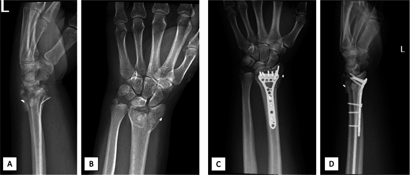
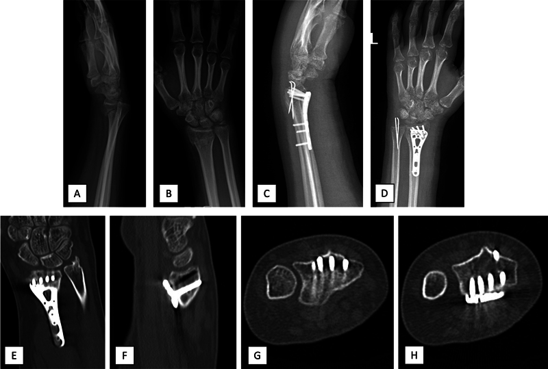
Objective To assess the efficacy of distal radius volar plates in cases involving dorsal fragments at the Ulnar Corner (UC) and Lister Tubercle (LT). Methods A retrospective study that included patients with distal radius fractures (DRFs) featuring UC and LT dorsal fragments treated with volar plates. The exclusion criteria comprised lunate facet fractures, UC fragment ratio below 25%, and patients treated with dorsal plates. Radiographic and tomographic measurements included radial length (RL), radial inclination (RI), ulnar variance (UV), palmar tilt (PT), fragment areas, UC fragment ratio, and displacement. The scores on the Gartland Werley (GW) classification and on the Disabilities of the Arm, Shoulder, and Hand (DASH) questionnaire, as well as grip strength, and range of motion (ROM), were assessed. Results The study involved 17 male and 5 female subjects (mean age: 39.7 ± 10.7 years). The UC and LT fragments displayed mean areas of 1.6 ± 0.7cm 2 and UC fragment ratio of 0.4 ± 0.1. The fixation rates for UC and LT fragments were of 18.2% and 31.8% respectively. Improved RI, UV, and PT were noted postoperatively. The mean GW and DASH scores were of 2.1 ± 2.0 and 4.3 ± 3.2 respectively. Grip strength on the operated side was of 89.5 ± 9.8% of the healthy side, and at least 90.9% of the patients achieved adequate ROM. Conclusion While volar plates are the standard treatment for intra-articular DRFs, displaced dorsal fragments can impact the outcomes. Mini dorsal incisions may aid in the fixation of UC fragments that are challenging to secure with volar plates, preserving joint health.
Keywords: bone plates; fracture fixation, internal; radius fractures; retrospective studies.
The Author(s). This is an open access article published by Thieme under the terms of the Creative Commons Attribution 4.0 International License, permitting copying and reproduction so long as the original work is given appropriate credit ( https://creativecommons.org/licenses/by/4.0/ ).
Conflict of interest statement
Conflito de Interesses Os autores não têm conflito de interesses a declarar.
Figures


Similar articles
-
A new distal radius fracture classification depending on the specific fragments through machine learning clustering method.BMC Musculoskelet Disord. 2024 Dec 30;25(1):1085. doi: 10.1186/s12891-024-08215-1. BMC Musculoskelet Disord. 2024. PMID: 39736589 Free PMC article.
-
Open Reduction and Internal Fixation by Volar Locking Plates and the "Poking Reduction" Technique in Distal Radius Fractures with Displaced Dorsal Ulnar Fragments: A Retrospective Study.Orthop Surg. 2022 Oct;14(10):2489-2498. doi: 10.1111/os.13468. Epub 2022 Aug 26. Orthop Surg. 2022. PMID: 36017756 Free PMC article.
-
The Effect of Displaced Dorsal Rim Fragment in a Distal Radius Fracture.J Wrist Surg. 2016 Mar;5(1):31-5. doi: 10.1055/s-0035-1571184. Epub 2016 Jan 6. J Wrist Surg. 2016. PMID: 26855833 Free PMC article.
-
Is it really necessary to restore radial anatomic parameters after distal radius fractures?Injury. 2014 Dec;45 Suppl 6:S21-6. doi: 10.1016/j.injury.2014.10.018. Epub 2014 Oct 27. Injury. 2014. PMID: 25457314 Review.
-
Combined Approach for Intra-articular Distal Radius Fracture: A Case Series and Literature Review.Clin Orthop Surg. 2021 Dec;13(4):529-538. doi: 10.4055/cios20291. Epub 2021 Nov 15. Clin Orthop Surg. 2021. PMID: 34868503 Free PMC article. Review.
References
-
- Chang H C, Poh S Y, Seah S C, Chua D T, Cha B K, Low C O. Fragment-specific fracture fixation and double-column plating of unstable distal radial fractures using AO mini-fragment implants and Kirschner wires. Injury. 2007;38(11):1259–1267. - PubMed
-
- Musgrave D S, Idler R S. Volar fixation of dorsally displaced distal radius fractures using the 2.4-mm locking compression plates. J Hand Surg Am. 2005;30(04):743–749. - PubMed
-
- Ng C Y, McQueen M M. What are the radiological predictors of functional outcome following fractures of the distal radius? J Bone Joint Surg Br. 2011;93(02):145–150. - PubMed
-
- Knirk J L, Jupiter J B. Intra-articular fractures of the distal end of the radius in young adults. J Bone Joint Surg Am. 1986;68(05):647–659. - PubMed
LinkOut - more resources
Full Text Sources