. 2024 Dec 18:18:6115-6132.
doi: 10.2147/DDDT.S480514. eCollection 2024.

Ciprofol Ameliorates Myocardial Ischemia/Reperfusion Injury by Inhibiting Ferroptosis Through Upregulating HIF-1α

Affiliations

Ciprofol Ameliorates Myocardial Ischemia/Reperfusion Injury by Inhibiting Ferroptosis Through Upregulating HIF-1α

Jun Ding et al. Drug Des Devel Ther. .

Abstract

Purpose: Ciprofol is a novel intravenous anesthetic that has been increasingly used in clinical anesthesia and sedation. Studies suggested that ciprofol reduced oxidative stress and inflammatory responses to alleviate cerebral ischemia/reperfusion (I/R) injury, but whether ciprofol protects the heart against I/R injury and the mechanisms are unknown. Herein, we assessed the effects of ciprofol on ferroptosis during myocardial I/R injury.

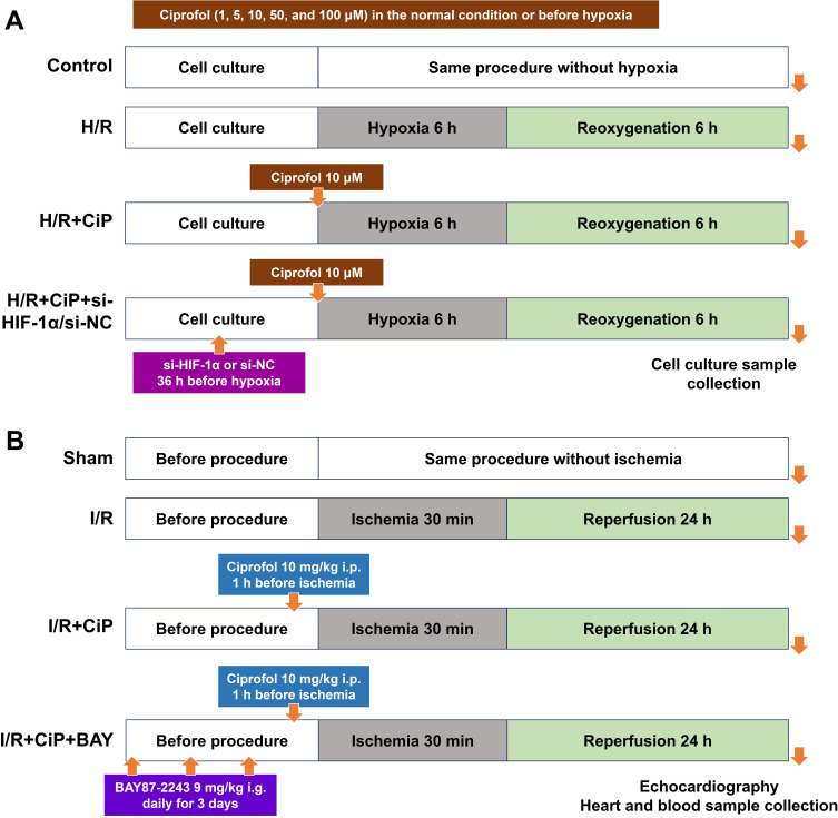
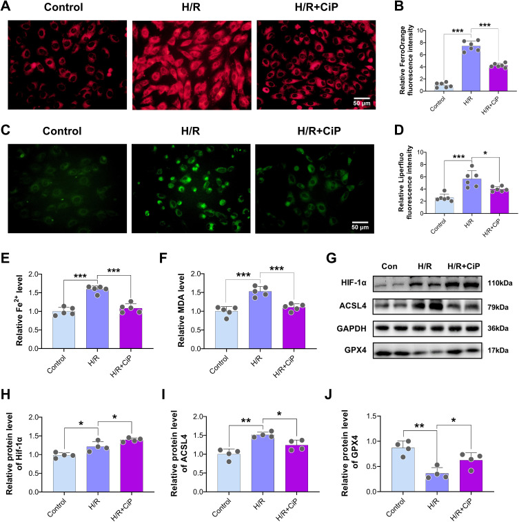
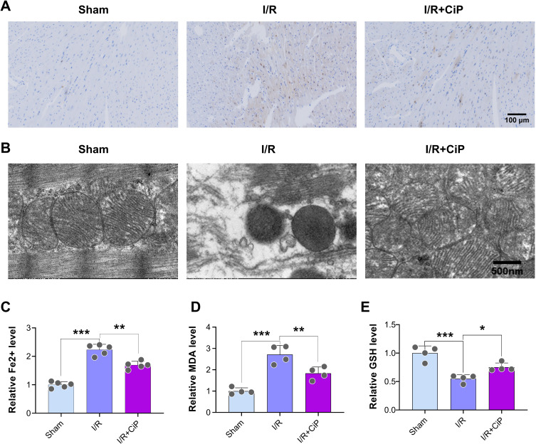
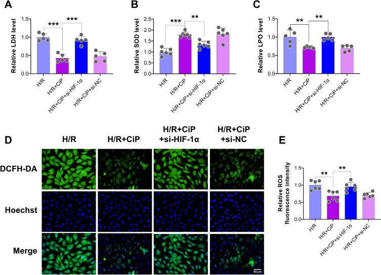
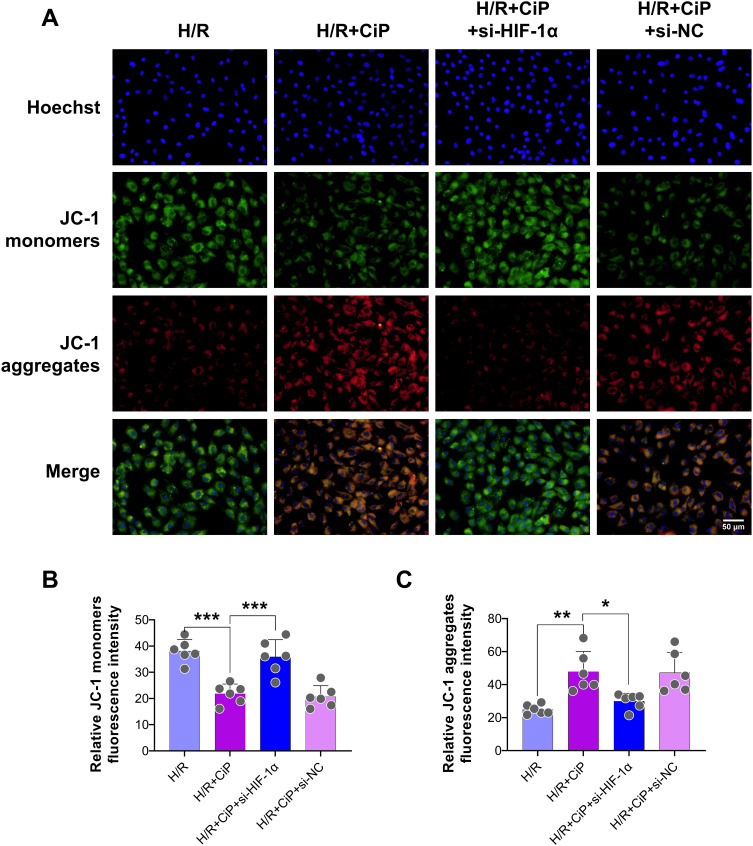
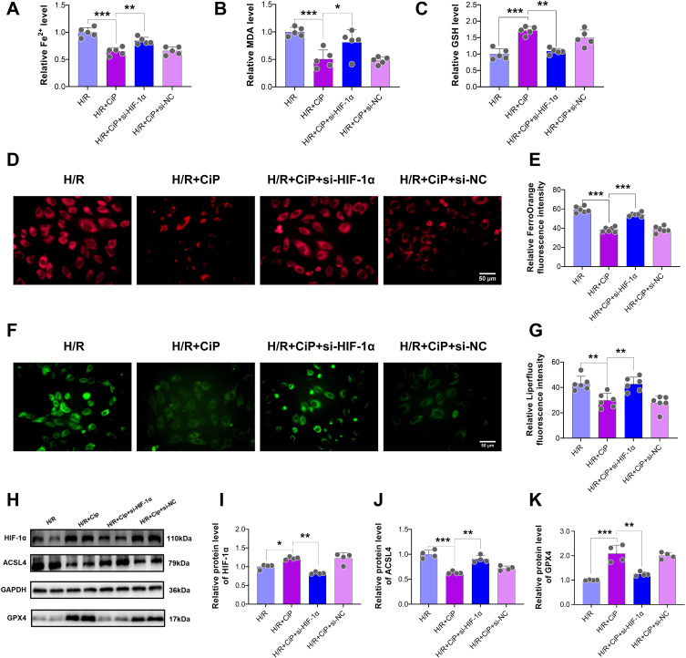
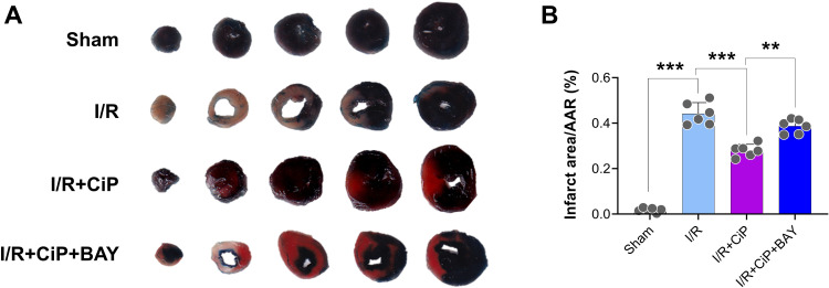
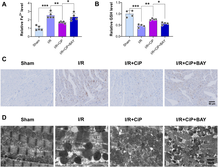
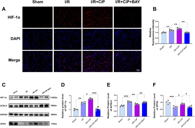
Methods: Experimental models of myocardial I/R injury in mice (ischemia for 30 min and reperfusion for 24 h) and hypoxia/reoxygenation (H/R) injury in H9c2 cardiomyocytes (hypoxia for 6 h followed by 6 h of reoxygenation) were established. Ciprofol was used prior to ischemia or hypoxia. Echocardiography, myocardial TTC staining, HE staining, DAB-enhanced Perl's staining, transmission electron microscopy, FerroOrange staining, Liperfluo staining, JC-1 staining, Rhodamine-123 staining, DCFH-DA staining, and Western blot were performed. Cell viability, serum cardiac enzymes, and oxidative- and ferroptosis-related biomarkers were measured. HIF-1α siRNA transfection and the specific inhibitor BAY87-2243 were utilized for mechanistic investigation.

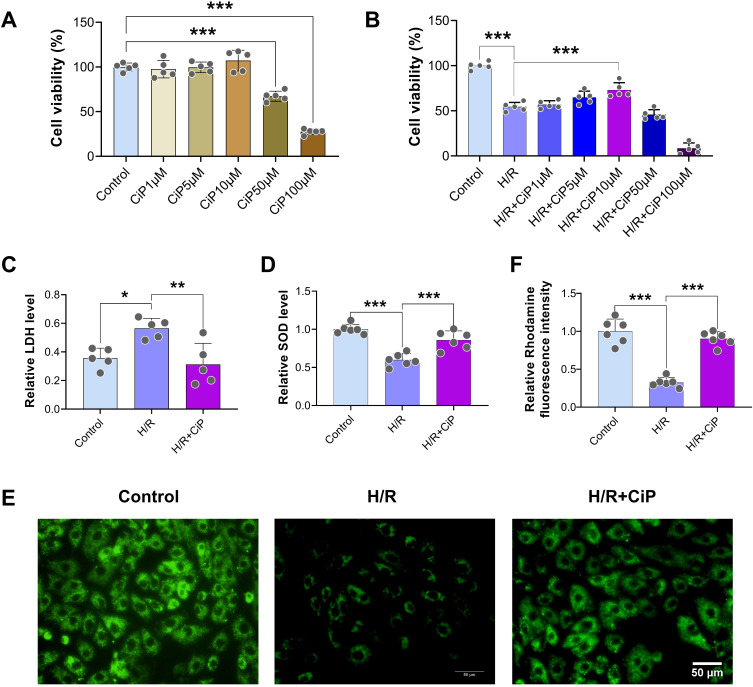
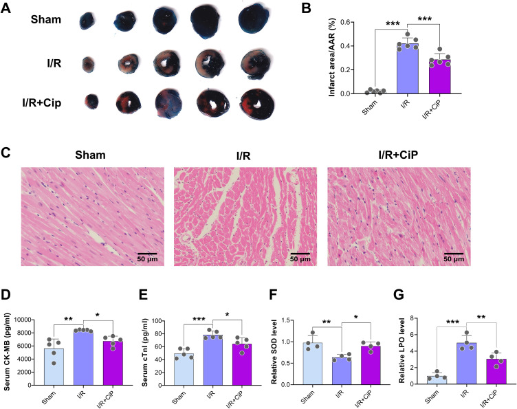
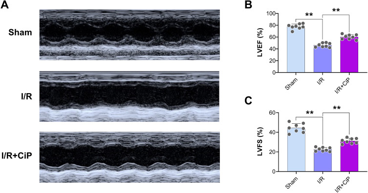
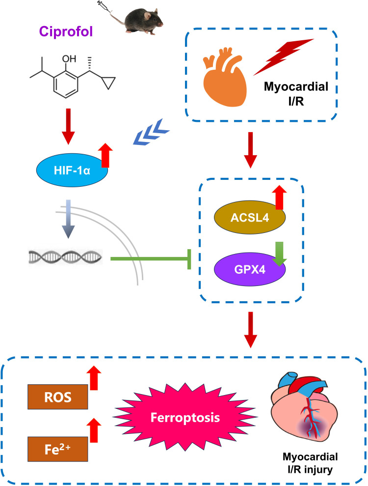
Results: Ciprofol treatment reduced myocardial infarct area and myocardium damage, alleviated oxidative stress and mitochondrial injury, suppressed Fe2+ accumulation and ferroptosis, and improved cardiac function in mice with myocardial I/R injury. Ciprofol also increased cell viability, attenuated mitochondrial damage, and reduced intracellular Fe2+ and lipid peroxidation in cardiomyocytes with H/R injury. Ciprofol enhanced the protein expression of HIF-1α and GPX4 and reduced the expression of ACSL4. Specifically, the protective effects of ciprofol against I/R or H/R injury were abolished by downregulating the expression of HIF-1α using siRNA transfection or the inhibitor BAY87-2243.

Conclusion: Ciprofol ameliorated myocardial I/R injury in mice and H/R injury in cardiomyocytes by inhibiting ferroptosis via the upregulation of HIF-1α expression.

Keywords: GPX4/ACSL4; HIF-1α; ciprofol; ferroptosis; myocardial ischemia/reperfusion injury; oxidative stress.


Conflict of interest statement

The authors declare that they have no competing interests within this work.

Figures

Figure 1
Figure 1
Schematic diagram. (A) H9c2 cardiomyocytes underwent H/R injury. First, cells were treated with ciprofol (1, 5, 10, 50, and 100 μM) in the normal condition or before hypoxia. Next, ciprofol (10 μM) was used, and si-HIF-1α or si-NC was transfected 36 h before ciprofol treatment. At the end of reoxygenation, cell culture samples were collected. (B) Mice underwent myocardial I/R injury. Ciprofol (10 mg/kg i.p.) was given 1 h before ischemia. BAY87-2243 (9 mg/kg i.g.) was administered daily for 3 days prior to ischemia. At the end of reperfusion, echocardiography was conducted, and heart and blood samples were collected.
Figure 2
Figure 2
Ciprofol attenuated H/R-induced injury in H9c2 cardiomyocytes. (A) Cell viability after ciprofol (1, 5, 10, 50, and 100 μM) in the normal condition. (B) Cell viability after ciprofol (1, 5, 10, 50, and 100 μM) during H/R. (C and D) Levels of LDH and SOD. Ciprofol (10 μM) was added 1 h before H/R. (E and F) Representative images and fluorescence intensity of Rhodamine 123 staining. Ciprofol (10 μM) was added 1 h before H/R. Scale bar = 50 µm. Data are shown as mean ± SD (n = 5‒6). *P < 0.05, **P < 0.01, ***P < 0.001.
Figure 3
Figure 3
Ciprofol alleviated I/R-induced myocardial injury in mice. Ciprofol (10 mg/kg i.p.) was given 1 h prior to ischemia. (A and B) Representative myocardial TTC staining images and quantification of infarction size. (C) Representative HE staining images of myocardium. Scale bar = 50 μm. (D and E) Serum levels of cardiac enzymes (CK-MB and cTnI). (F and G) Serum levels of SOD and LPO. Data are shown as mean ± SD (n = 4‒6). *P < 0.05, **P < 0.01, ***P < 0.001.
Figure 4
Figure 4
Ciprofol improved cardiac function in mice with myocardial I/R injury. Ciprofol (10 mg/kg i.p.) was given 1 h prior to ischemia. (A) Representative echocardiography images. (B and C) Values of LVEF and LVFS. Data are shown as mean ± SD (n = 8). **P < 0.01.
Figure 5
Figure 5
Ciprofol suppressed H/R-induced ferroptosis and increased HIF-1α expression in H9c2 cardiomyocytes. Ciprofol (10 μM) was added 1 h before H/R. (A and B) Representative images and fluorescence intensity of FerroOrange staining. Scale bar = 50 µm. (C and D) Representative images and fluorescence intensity of Liperfluo staining. Scale bar = 50 µm. (E and F) Levels of Fe2+ and MDA. (G–J) Western blot bands and quantification of HIF-1α, ACSL4, and GPX4 protein expression. Data are shown as mean ± SD (n = 4‒6). *P < 0.05, **P < 0.01, ***P < 0.001.
Figure 6
Figure 6
Ciprofol reduced Fe2+ accumulation and mitochondrial damage in mice with myocardial I/R injury. Ciprofol (10 mg/kg i.p.) was given 1 h prior to ischemia. (A) Representative Perl’s staining images of myocardium. Scale bar = 100 µm. (B) Representative mitochondrial ultrastructure images under transmission electron microscopy. Scale bar = 500 nm. (C‒E) Serum levels of Fe2+, MDA, and GSH. Data are shown as mean ± SD (n = 4‒5). *P < 0.05, **P < 0.01, ***P < 0.001.
Figure 7
Figure 7
Knockdown of HIF-1α abolished the protective effects of ciprofol against H/R-induced injury in H9c2 cardiomyocytes. si-HIF-1α or si-NC was transfected 36 h before ciprofol (10μM) treatment. (A–C) Levels of LDH, SOD, and LPO. (D and E) Representative DCFH-DA staining images and fluorescence intensity of intracellular ROS. Scale bar = 50 µm. Data are shown as mean ± SD (n = 5‒6). **P < 0.01, ***P < 0.001.
Figure 8
Figure 8
Knockdown of HIF-1α reversed the protective effects of ciprofol on mitochondrial function in H9c2 cardiomyocytes subjected to H/R injury. si-HIF-1α or si-NC was transfected 36 h before ciprofol (10μM) treatment. (A) Representative JC‐1 staining images showing mitochondrial membrane potential. Scale bar = 50 µm. (B and C) Fluorescence intensity of JC-1 monomers (green) and aggregates (red). Data are shown as mean ± SD (n = 6). *P < 0.05, **P < 0.01, ***P < 0.001.
Figure 9
Figure 9
Ciprofol inhibited H/R-induced ferroptosis in H9c2 cardiomyocytes by upregulating HIF-1α. si-HIF-1α or si-NC was transfected 36 h before ciprofol (10μM) treatment. (AC) Levels of Fe2+, MDA, and GSH. (D and E) Representative images and fluorescence intensity of FerroOrange staining. Scale bar = 50 µm. (F and G) Representative images and fluorescence intensity of Liperfluo staining. Scale bar = 50 µm. (H‒K) Western blot bands and quantification of HIF-1α, ACSL4, and GPX4 protein expression. Data are shown as mean ± SD (n = 4‒6). *P < 0.05, **P < 0.01, ***P < 0.001.
Figure 10
Figure 10
Inhibiting HIF-1α reversed the protective effect of ciprofol on I/R-induced myocardial infarction in mice. BAY87-2243 (9 mg/kg i.g.) was administered daily for 3 days prior to ischemia. (A) Representative myocardial TTC staining images. (B) Quantification of myocardial infarction size. Data are shown as mean ± SD (n = 6). **P < 0.01, ***P < 0.001.
Figure 11
Figure 11
Inhibiting HIF-1α reversed the protective effects of ciprofol on Fe2+ accumulation and mitochondrial damage in mice with myocardial I/R injury. BAY87-2243 (9 mg/kg i.g.) was administered daily for 3 days prior to ischemia. (A and B) Serum levels of Fe2+ and GSH. (C) Representative Perl’s staining in myocardial tissues. Scale bar = 50 µm. (D) Representative mitochondrial ultrastructure images under transmission electron microscopy. Scale bar = 2 µm. Data are shown as mean ± SD (n = 4‒5). *P < 0.05, **P < 0.01, ***P < 0.001.
Figure 12
Figure 12
Ciprofol inhibited I/R-induced myocardial ferroptosis in mice by upregulating HIF-1α. BAY87-2243 (9 mg/kg i.g.) was administered daily for 3 days prior to ischemia. (A and B) Representative images and fluorescence intensity of HIF-1α expression in myocardium. Scale bar = 50 µm. (C‒F) Western blot bands and quantification of HIF-1α, ACSL4, and GPX4 protein expression. Data are shown as mean ± SD (n = 4). *P < 0.05, **P < 0.01, ***P < 0.001.
Figure 13
Figure 13
Schematic mechanism for the cardioprotection of ciprofol against I/R-induced myocardial ferroptosis in mice through upregulating HIF-1α.

Similar articles

Cited by

References

    1. Diseases GBD, Injuries C. Global burden of 369 diseases and injuries in 204 countries and territories, 1990-2019: a systematic analysis for the global burden of disease study 2019. Lancet. 2020;396(10258):1204–1222. doi:10.1016/S0140-6736(20)30925-9 - DOI - PMC - PubMed
    1. Marijon E, Narayanan K, Smith K, et al. The lancet commission to reduce the global burden of sudden cardiac death: a call for multidisciplinary action. Lancet. 2023;402(10405):883–936. doi:10.1016/S0140-6736(23)00875-9 - DOI - PubMed
    1. Alexander T, Kumbhani DJ, Mullasari Sankardas A. The future of pharmacoinvasive therapy for st-segment-elevation myocardial infarction reperfusion in the post-STREAM era. Circulation. 2024;149(10):732–733. doi:10.1161/CIRCULATIONAHA.123.066703 - DOI - PubMed
    1. Del Re DP, Amgalan D, Linkermann A, Liu Q, Kitsis RN. Fundamental mechanisms of regulated cell death and implications for heart disease. Physiol Rev. 2019;99(4):1765–1817. doi:10.1152/physrev.00022.2018 - DOI - PMC - PubMed
    1. Zhang Q, Wang L, Wang S, et al. Signaling pathways and targeted therapy for myocardial infarction. Signal Transduct Target Ther. 2022;7(1):78. doi:10.1038/s41392-022-00925-z - DOI - PMC - PubMed

MeSH terms

Substances

LinkOut - more resources