

Pathological features of the differentiation landscape in esophageal squamous cell cancer and their correlations with prognosis
- PMID: 39711958
- PMCID: PMC11659131
- DOI: 10.3389/fonc.2024.1442212
Pathological features of the differentiation landscape in esophageal squamous cell cancer and their correlations with prognosis
Abstract
Background: For esophageal squamous cell carcinoma (ESCC), universally accepted pathological criteria for classification by differentiation degree are lacking. Tumor budding, single-cell invasion, and nuclear grade, recognized as prognostic factors in other carcinomas, have rarely been investigated for their correlation with differentiation and prognosis in ESCC. This study aims to determine if pathological findings can predict differentiation degree and prognosis in ESCC.
Patients and methods: This study reviewed tumor slides from 326 patients who underwent surgery for ESCC between 2007 and 2012. Tumors were evaluated for subtypes, tumor nest size, tumor stroma, and nuclear grade (nuclear diameter and mitosis) across different differentiation groups. Overall survival (OS) and disease-free survival (DFS) were estimated using the Kaplan-Meier method, with group differences assessed using the stratified log-rank test and Cox proportional hazards model.
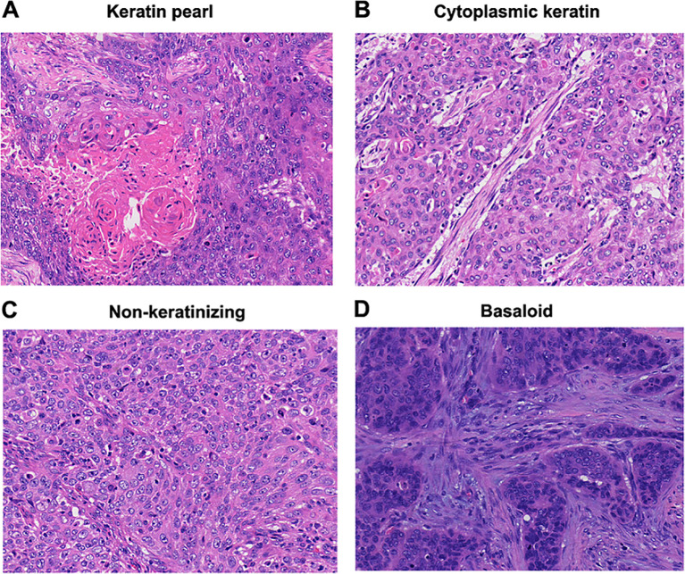
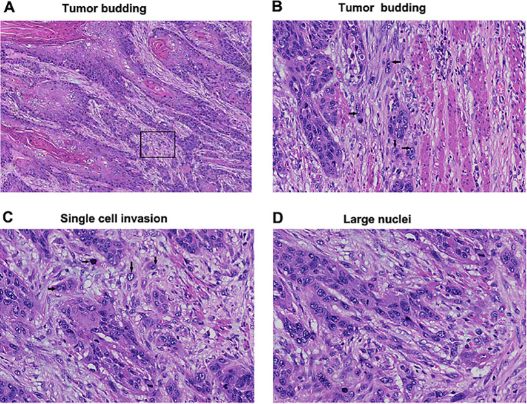
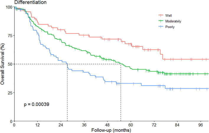
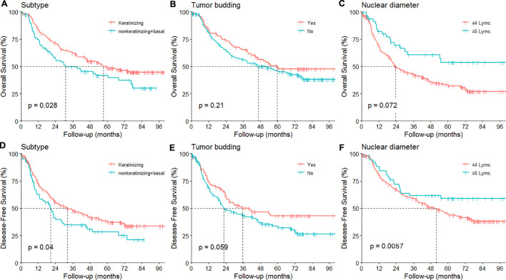
Results: The mean values of tumor budding invasion margins in well, moderately, and poorly differentiated groups were 25.3%, 30.7%, and 36.3%, respectively. Mean tumor budding/10HPFs were 8.0, 10.3, and 13.0, respectively. Well-differentiated tumors showed more keratinizing subtypes, smaller tumor budding invasion margins, more Grade 1 tumor budding (0-4 cells), absence of single-cell invasion, larger nuclear diameter (≥5 lymphocytes), higher mitotic counts, more submucosal invasion, and less lymphovascular invasion. Conversely, poorly differentiated tumors exhibited opposite characteristics. Multivariate analyses identified the nuclear diameter as independent prognostic factors for OS and DFS.
Conclusions: Pathological features can stratify the differentiation landscape in ESCC patients. The nuclear diameter (4 lymphocytes) can help predict prognosis in ESCC than other pathological features.
Implications for practice: We first time report the mean values of tumor budding invasion margins and tumor budding/10HPF in well, moderately, and poorly differentiated groups for esophageal squamous cell carcinoma. The landscape of well differentiation was depicted with more keratinizing subtypes, smaller tumor budding invasion margins, more Grade 1 tumor budding (0-4 cells), absence of single-cell invasion, larger nuclear diameter (≥5 lymphocytes), higher mitotic counts, and less lymphovascular invasion. The nuclear diameter as independent prognostic factors for prognosis. The findings indicate that pathological features can stratify the differentiation landscape in ESCC patients and offer novel insight into definition of well or moderately differentiation.
Keywords: differentiation; esophageal squamous cell carcinoma; nuclear diameter; pathological features; prognosis.
Copyright © 2024 Deng, Zhang, Wang, Li, Xiang, Ma, Zhu, Li and Zhao.
Conflict of interest statement
The authors declare that the research was conducted in the absence of any commercial or financial relationships that could be construed as a potential conflict of interest.
Figures
References
-
- Kadota K, Nitadori J, Woo KM, Sima CS, Finley DJ, Rusch VW, et al. . Comprehensive pathological analyses in lung squamous cell carcinoma: single cell invasion, nuclear diameter, and tumor budding are independent prognostic factors for worse outcomes. J Thorac Oncol. (2014) 9:1126–39. doi: 10.1097/JTO.0000000000000253 - DOI - PMC - PubMed
-
- Zombori-Toth N, Hegedus F, Almasi S, Sejben A, Tiszlavicz L, Furak J, et al. . Proposal of a grading system for squamous cell carcinoma of the lung - the prognostic importance of tumour budding, single cell invasion, and nuclear diameter. Virchows Arch. (2023) 483:393–404. doi: 10.1007/s00428-023-03612-8 - DOI - PMC - PubMed
LinkOut - more resources
Full Text Sources