. 2024 Dec 6:3:1504959.
doi: 10.3389/frtra.2024.1504959. eCollection 2024.

Autologous porcine VRAM flap model for VCA research

Affiliations

Autologous porcine VRAM flap model for VCA research

Caitlin M Blades et al. Front Transplant. .

Abstract

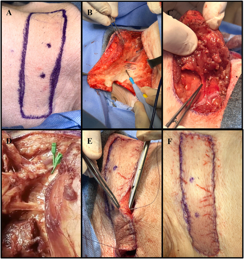
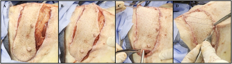
Introduction: As research advances in vascularized composite allotransplantation (VCA), large animal models are essential for translational studies related to immune rejection and graft survival. However, procurement of large flaps can cause significant defects, complicating wound closure and increasing postoperative risks. This study details the surgical techniques and outcomes of autologous vertical rectus abdominis myocutaneous (VRAM) flap transplantation and neck flap isolation with induced ischemia in a swine model. The purpose of this study was to identify the most effective control procedure for use in future VRAM flap allotransplantation research.

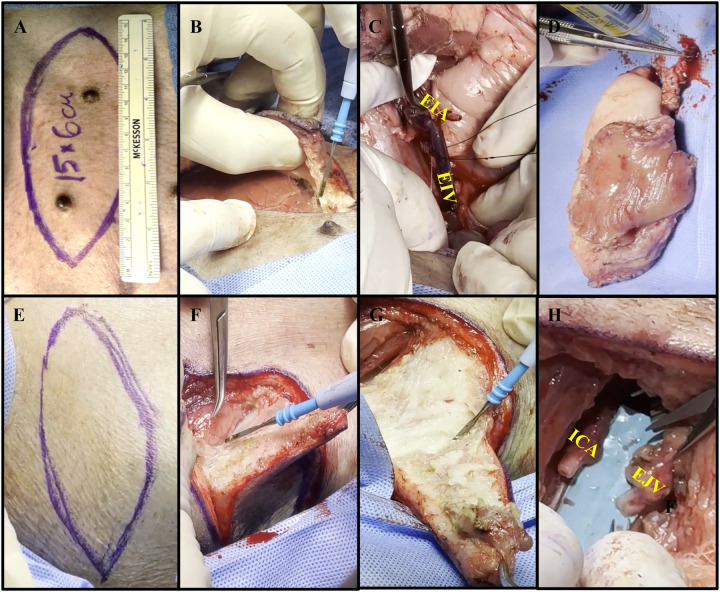
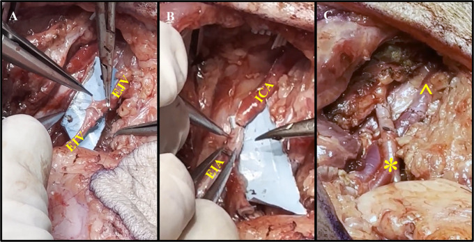
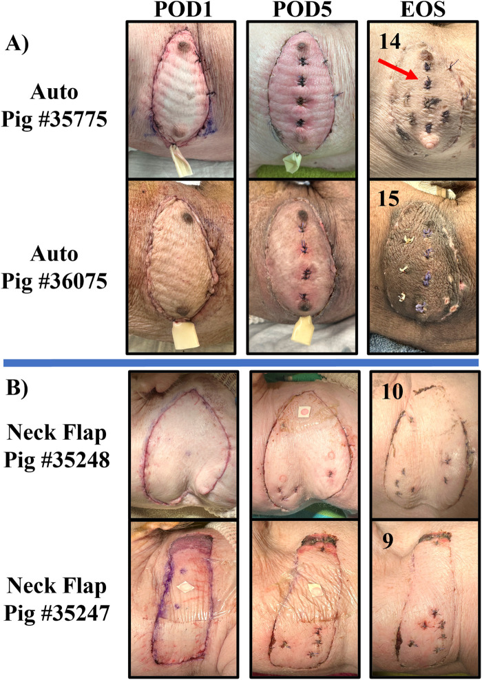
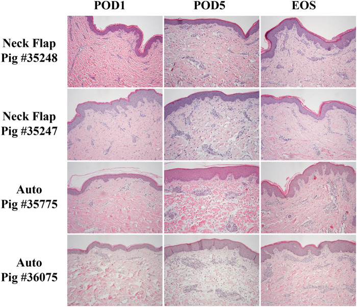
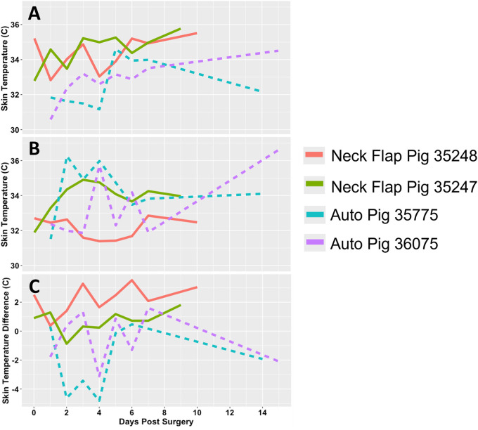
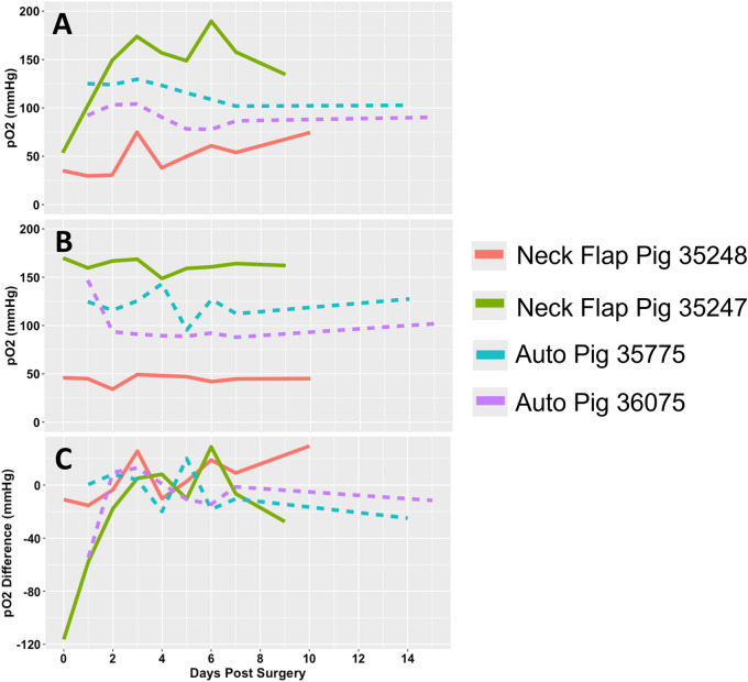
Methods: We performed two left heterotopic autologous VRAM flap transplants and two right anterolateral neck flap isolations using female Yucatan pigs. Postoperatively, animals were monitored for complications and flap healing, with punch biopsies taken on POD1, 5, and at the end of the study for histological analysis. Transcutaneous oxygen and temperature were also recorded.

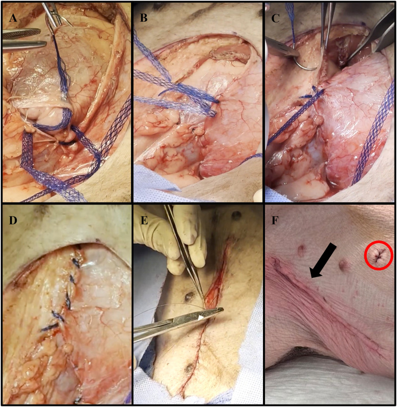
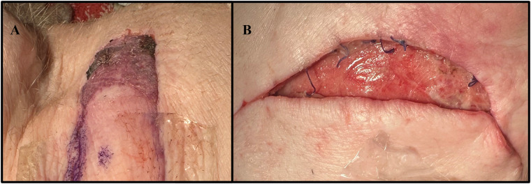
Results: Both autologous flaps survived after vessel anastomosis, with effective closure of abdominal defects using suturable mesh, and no postoperative complications were observed. Histology revealed mild dermal edema and perivascular inflammation on POD5. In the neck flap group, both flaps survived temporary ischemia, however, postoperative complications included dorsal flap necrosis and wound dehiscence, requiring reoperation. No gross inflammation or edema was observed following surgery and histologically there was only mild dermal edema on POD5.

Discussion: We have developed a low-risk, technically feasible porcine autologous VRAM flap transplantation model and our findings support its use in future VCA studies.

Keywords: autologous; ischemia; neck flap; swine; vertical rectus abdominis myocutaneous flap.


Conflict of interest statement

The authors declare that the research was conducted in the absence of any commercial or financial relationships that could be construed as a potential conflict of interest.

Figures

Figure 1
Figure 1
Collective images of the neck flap isolation and induced ischemia procedure. (A) Preoperative flap markings on the right anterolateral neck of pig #35247. (B) Electrocautery dissection of the subcutaneous tissue portion of the flap. (C) Isolation of the main perforator vessels branching from the internal carotid artery. (D) Clamping of the two main perforator vessels using Acland clamps. (E) Placement of deep dermal sutures to secure the flap into the defect following induced ischemia. (F) Fully inset flap at the end of surgery.
Figure 2
Figure 2
Collective images of the autologous VRAM flap transplant. (A) Preoperative markings on pig #35775's left lower abdominal quadrant with a paper ruler indicating flap size. (B) Electrocautery dissection through the superficial ventral abdominal muscles, down to the rectus fascia. (C) Use of silk ties to divide and harvest the external iliac vein (EIV), to serve as the main vein of the pedicle. External iliac artery (EIA) dissected to the right. (D) Procured VRAM flap undergoing ex-vivo heparinized saline flush via the EIA. (E) Biconvex surgical marking, slightly smaller than the dimensions of the abdominal flap, on the right anterolateral neck. (F) Creation of a soft tissue defect using electrocautery to carefully dissect the subcutaneous tissue. (G) Further dissection of the neck flap using electrocautery. (H) Isolation of the internal carotid artery (ICA) and external jugular vein (EJV) over 5 cm, with proximal clamping.
Figure 3
Figure 3
Representative images of abdominal wall defect closure using DURAMESH™ Mesh Suture (Chicago, IL) following autologous VRAM flap procurement. (A) Lateral to medial closure of the parietal peritoneum defect using DURAMESH™. (B) Placement of continuous over and over sutures within the Scarpa fascia in a caudal to cranial fashion. (C) Successful re-approximation of the Scarpa fascia. (D) Complete low-tension closure of the Scarpa fascia. (E) Closure of the abdominal skin using a continuous nonabsorbable 4-0 Vicryl running subcuticular suture. (F) Postoperative image of closed left lower abdominal wound (black arrow). The sutured area (red circle) represents where biopsies were taken for histological analysis.
Figure 4
Figure 4
Representative process of vessel anastomoses. (A) Anastomosis of the external jugular vein (EJV) to the external iliac vein (EIV) using end-to-end suturing. (B) Anastomosis of the internal carotid artery (ICA) and external iliac artery (EIA) using end-to-end suturing. (C) Completion of the vascular (*) and venous (^) anastomoses without signs of anastomotic leakage.
Figure 5
Figure 5
Representative images of the autologous VRAM flap transplantation to the anterolateral neck defect. (A) Autologous flap inset on the right neck of pig #35775. (B) Approximation of the autograft skin to the neck skin by placing 3-0 interrupted simple sutures at multiple tension points. (C) Placement of circumferential 3-0 interrupted simple sutures. (D) Complete closure using a continuous nonabsorbable 4-0 Vicryl running subcuticular suture.
Figure 6
Figure 6
Transcutaneous oxygen and temperature sensor placed flush to a representative graft skin. The sensor is comprised of both a reader head (^) and body (*). The head is connected to the body by a ribbon cable (X).
Figure 7
Figure 7
Representative collection of graft images taken on POD 1, 5, and at the end of the study (EOS) for the autologous VRAM flap transplant (Auto) group (A) and the neck flap isolation and induced ischemia (Neck Flap) group (B). The EOS POD is indicated in the top left corner of each image. The neck flap group did not require the placement of Penrose drains due to a lack of postoperative edema. The sutured areas (red arrow) represent places where prior biopsies were taken for histological analysis.
Figure 8
Figure 8
Complications experienced by the neck flap isolation with induced ischemia cohort. (A) Dorsal flap necrosis in Pig #35247 on POD2. (B) Caudal wound dehiscence in Pig #35248 on POD2.
Figure 9
Figure 9
Representative histology comparing the neck flap isolation and induced ischemia (Neck Flap) group to the autologous transplants (Auto) on POD1 and 5 and at the end of the study (EOS). Both neck flaps showed only mild dermal edema on POD5, resolving by the EOS. Both autologous transplants showed mild to moderate dermal edema and mild dermal perivascular inflammation on POD5, with resolution of the edema and partial resolution of the inflammation by the EOS. The EOS POD is indicated in the top left corner of each image. All Hematoxylin & Eosin images are at 100x and equally white-balanced.
Figure 10
Figure 10
Transcutaneous temperature readings. (A) Temperature (°C) measured from the flap of each experimental pig. (B) Temperature (°C) measured from the abdominal control skin of each experimental pig. (C) Difference in temperature (°C) between each graft and their respective control site. Each animal is represented by a different line color with the autologous grafts illustrated by dotted lines and the neck flaps with induced ischemia represented by solid lines. Neck Flap; neck flap isolation with induced ischemia, Auto; autologous VRAM flap transplantation to the neck.
Figure 11
Figure 11
Transcutaneous oxygen tension over time. (A) Partial pressure of oxygen (pO2) readings measured from the flap of each pig. (B) pO2 readings measured from the abdominal control skin of each pig. (C) pO2 difference between each graft and their respective control site. Each animal is represented by a different line color with the autologous grafts illustrated by dotted lines and the neck flaps with induced ischemia represented by solid lines. Neck Flap; neck flap isolation with induced ischemia, Auto; autologous VRAM flap transplantation to the neck.

Similar articles

References

    1. Wahyudi M, Satria O, Aprilya D, Nong I. Vertical rectus abdominis myocutaneous flap for reconstruction of forequarter amputation defect after shoulder soft tissue sarcoma resection: technical consideration. Plast Reconstr Surg Glob Open. (2023) 11(6):e5077. 10.1097/GOX.0000000000005077 - DOI - PMC - PubMed
    1. Salo J, Tukiainen E. Flap reconstruction of the chest wall after oncologic resection. Curr Chall Thorac Surg. (2020) 2:5. 10.21037/ccts.2019.12.05 - DOI
    1. Coombs DM, Patel NB, Zeiderman MR, Wong MS. The vertical rectus abdominis musculocutaneous flap as a versatile and viable option for perineal reconstruction. Eplasty. (2017) 17:ic2. - PMC - PubMed
    1. Küntscher MV, Mansouri S, Noack N, Hartmann B. Versatility of vertical rectus abdominis musculocutaneous flaps. Microsurgery. (2006) 26(5):363–9. 10.1002/micr.20253 - DOI - PubMed
    1. Pomahac B, Gobble RM, Schneeberger S. Facial and hand allotransplantation. Cold Spring Harb Perspect Med. (2014) 4(3):a015651. 10.1101/cshperspect.a015651 - DOI - PMC - PubMed

LinkOut - more resources