. 2024 Nov 19;4(6):342-352.
doi: 10.1021/acsbiomedchemau.4c00088. eCollection 2024 Dec 18.

Bioinformatics-Facilitated Identification of Novel Bacterial Sulfoglycosidases That Hydrolyze 6-Sulfo- N-acetylglucosamine

Affiliations

Bioinformatics-Facilitated Identification of Novel Bacterial Sulfoglycosidases That Hydrolyze 6-Sulfo- N-acetylglucosamine

Mochen Dong et al. ACS Bio Med Chem Au. .

Abstract

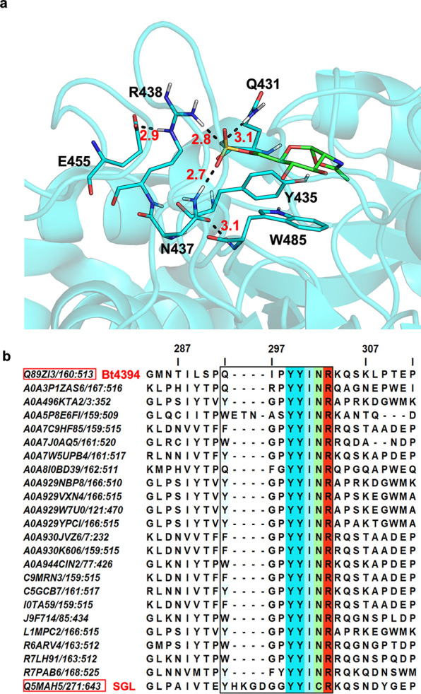
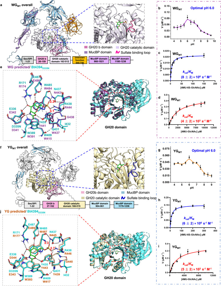
Glycan sulfation is a widespread postglycosylation modification crucial for modulating biological functions including cellular adhesion, signaling, and bacterial colonization. 6-Sulfo-β-GlcNAcases are a class of enzyme that alters sulfation patterns. Such changes in sulfation patterns are linked to diseases such as bowel inflammation, colitis, and cancer. Despite their significance, 6-sulfo-β-GlcNAcases, which cleave β-linked 6-sulfo-N-acetylglucosamine (6S-GlcNAc), have been but rarely identified. This scarcity results mainly from the short, diverse, and distinctive sulfate-binding motifs required for recognition of the 6-sulfate group in 6S-GlcNAc in addition to the conserved GH20 family features. In this study, we discovered 6-sulfo-β-GlcNAcases and assigned two novel sulfate-binding motifs by the use of comparative genomics, structural predictions, and activity-based screening. Our findings expand the known microbiota capable of degrading sulfated glycans and add significant enzymes to the tool kit for analysis and synthesis of sulfated oligosaccharides.


Conflict of interest statement

The authors declare no competing financial interest.

Figures

Figure 1
Figure 1
AlphaFold 3-predicted unrelaxed structure of F3-ORF26WT and its biochemical characterization. (a) Unrelaxed apo F3-ORF26 structure predicted by AlphaFold 3 with its multiple domains highlighted in color and with the GH20 catalytic domain presenting a (β/α)8 barrel fold aligned to Bt4394D335N-6S-NAG-oxazoline structure (PDB: 7DVB, the 6S-NAG-oxazoline is shown as green sticks for clarity; the sulfate-binding loop is highlighted in orange). (b) pH-rate profile for the hydrolysis of 4MU-6S-GlcNAc substrate (500 μM) by F3-ORF26WT (2 nM) across a pH range of 4.0–9.5, with an optimal pH of 6.0. (c) Superposition of the Bt4394D335N-6S-NAG-oxazoline structure and that of the GH20 catalytic domain of F3-ORF26. The inset shows residues critical for catalysis and binding of 6S-GlcNAc in the active site. Michaelis–Menten plots for (d) hydrolysis of 4MU-6S-GlcNAc (5 to 1000 μM at pH 6.0) by F3-ORF26WT (1 nM). (e) Hydrolysis of 4MU-GlcNAc (100 to 8000 μM at pH 6.0) by F3-ORF26WT (0.1 μM).
Figure 2
Figure 2
F3-ORF26 6-sulfo-β-GlcNAcases (highlighted as a yellow dot) mapped on the Uniport SSN created for the GH20 domain. The AST was increased to 250 to finally separate the subclusters that correspond to differences in the sequence similarity.
Figure 3
Figure 3
(a) Structure of Bt4394D335N-6S-NAG-oxazoline intermediate complex (PDB: 7DVB, cyan), showing that the side chain of Q431 appears more exposed to the solvent and coordinated with one sulfate oxygen. H-bond distances are for donor-to-acceptor in Å. (b) The multiple sequence alignment (MSA) generated by Clustal Omega shows the portion of sequences around the sulfate-binding motif (boxed) from the SSN cluster containing SGL. The alignment indicates that NR residues are more conserved. Each sequence is identified by its UniProt ID from the GH20 domain (PF00728). The color scheme is based on JalView Clustal coloring, with a default conservation setting of 30. The top sequence, Q89ZI3, corresponds to Bt4394 and is used as a reference, while the bottom sequence, Q5MAH5, corresponds to SGL.
Figure 4
Figure 4
AlphaFold 3-predicted unrelaxed structure of WG and YG enzymes and their biochemical characterization. (a) Predicted unrelaxed structure of full-length WGWT, with its multiple domains highlighted in color and with the GH20 catalytic domain aligned to the Bt4394D335N-6S-NAG-oxazoline structure (the sulfate-binding loop highlighted in magenta). (b) pH-rate profile for the hydrolysis of 4MU-6S-GlcNAc (50 μM) substrate by WGWT (2 nM) over a pH range of 4.0–9.5, with an optimal pH of 6.0. Michaelis–Menten plots for (c) the hydrolysis of 4MU-6S-GlcNAc (1–150 μM) by WGWT (0.5 nM) and (d) the hydrolysis of 4MU-GlcNAc (100–10000 μM) by WGWT (40 nM) at pH 6.0. H-bond distances are for donor-to-acceptor in Å. (e) Superposition of the predicted GH20 domain structure of WGWT (purple) and the structure of the Bt4394D335N-6S-NAG-oxazoline intermediate (cyan). (f) Predicted unrelaxed structure of full-length YGWT, with the GH20 catalytic domains aligned with the Bt4394D335N-6S-NAG-oxazoline intermediate structure (PDB: 7DVB, only the 6S-NAG-oxazoline is drawn as green sticks for clarity). The sulfate-binding loop is highlighted in dark blue. (g) pH-rate profile for the hydrolysis of 4MU-6S-GlcNAc (100 μM) substrate by YGWT (1.1 nM) over pH range of 4.0–9.0 with an optimal pH of 6.0. Michaelis–Menten plots for (h) the hydrolysis of 4MU-6S-GlcNAc (2.5 μM to 300 μM) by YGWT (2 nM) and (i) the hydrolysis of 4MU-GlcNAc (100 μM to 10000 μM) by YGWT (31.8 nM) at pH 6.0. (j) Superposition of the predicted unrelaxed GH20 domain structures of apoYGWT (salmon) and the Bt4394D335N-6S-NAG-oxazoline intermediate complex (cyan). The inset emphasizes essential residues critical to the catalysis and binding of 6S-GlcNAc in the active site.
Figure 5
Figure 5
Comparison of the sulfate-binding motif in WGWT and YGWT structures predicted by two versions of AlphaFold, with their active sites aligned to the Bt4394D335N-6S-NAG-oxazoline intermediate structure (PDB: 7DVB, only the 6S-NAG-oxazoline ligand in green is shown for clarity). The residues around the sulfate binding motif of unrelaxed WGWT structures predicted by (a) AlphaFold 2 and AlphaFold 3 are shown to highlight differences in conformations for the W437-G438-P439 region. Similarly, the residues around the sulfate binding motif of unrelaxed YGWT structures predicted by (b) AlphaFold 2 and AlphaFold 3 show differences in the conformations for the Y439-G440-P441 region. H-bond distances are for donor-to-acceptor in Å.

Similar articles

References

    1. Muthana S. M.; Campbell C. T.; Gildersleeve J. C. Modifications of glycans: biological significance and therapeutic opportunities. ACS Chem. Biol. 2012, 7 (1), 31–43. 10.1021/cb2004466. - DOI - PMC - PubMed
    1. Whitelock J. M.; Iozzo R. V. Heparan sulfate: a complex polymer charged with biological activity. Chem. Rev. 2005, 105 (7), 2745–2764. 10.1021/cr010213m. - DOI - PubMed
    1. She Y.-M.; Li X.; Cyr T. D. Remarkable Structural Diversity of N-Glycan Sulfation on Influenza Vaccines. Anal. Chem. 2019, 91 (8), 5083–5090. 10.1021/acs.analchem.8b05372. - DOI - PubMed
    1. Xia B.; Royall J. A.; Damera G.; Sachdev G. P.; Cummings R. D. Altered O-glycosylation and sulfation of airway mucins associated with cystic fibrosis. Glycobiology 2005, 15 (8), 747–775. 10.1093/glycob/cwi061. - DOI - PubMed
    1. Lo-Guidice J. M.; Wieruszeski J. M.; Lemoine J.; Verbert A.; Roussel P.; Lamblin G. Sialylation and sulfation of the carbohydrate chains in respiratory mucins from a patient with cystic fibrosis. J. Biol. Chem. 1994, 269 (29), 18794–18813. 10.1016/S0021-9258(17)32238-X. - DOI - PubMed

LinkOut - more resources