. 2021 Dec 17:7:100308.
doi: 10.1016/j.ijcchd.2021.100308. eCollection 2022 Mar.

What can we learn from systematic segmental analysis of fetal heart by postmortem micro-CT: Is it time to change approach?

Affiliations

What can we learn from systematic segmental analysis of fetal heart by postmortem micro-CT: Is it time to change approach?

C Sandrini et al. Int J Cardiol Congenit Heart Dis. .

Abstract

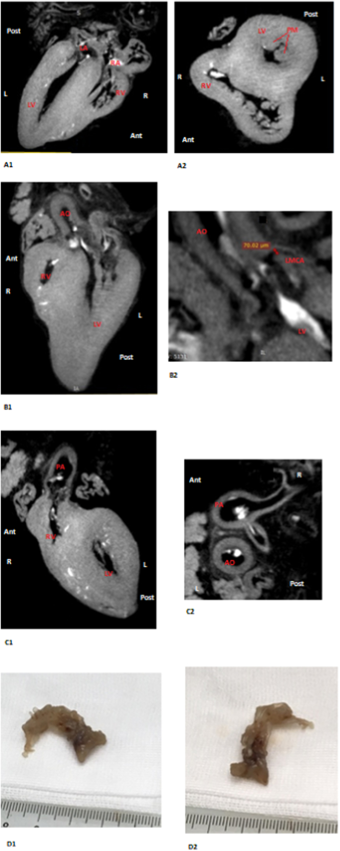
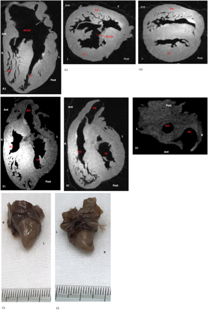
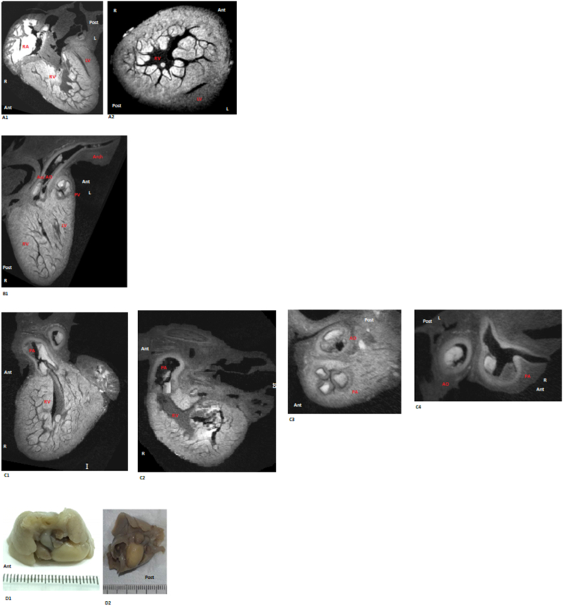
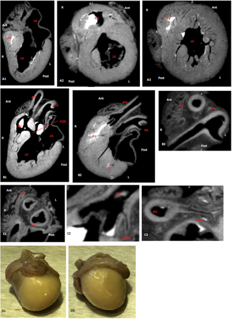
Background: Postmortem analysis of fetal heart is crucial but challenging. Alternative techniques to autopsy have been studied. We investigate micro-CT diagnostic accuracy in the analysis of fetal heart coming from early and late termination of pregnancy.

Methods: Micro-CT and autopsy analyzed human fetal heart using twenty-six indices of cardiac anatomy derived from segmental approach. Sub-analysis of smaller specimens was performed. Primary outcome is the agreement between techniques. Secondary outcomes are sensitivity, specificity, positive and negative predictive value. Indices are defined "visible" or "non-visible" (primary end point) and "normal", "abnormal" or "non-diagnostic" (secondary end point).

Results: Fifty-five cases were included. Agreement was 74.5% and 65.2% for primary end point and 93.3% and 91.8% for secondary end point in general population and in the group of smaller specimens, respectively. Sensitivity, specificity, positive and negative predictive value were 92.8%, 51.8%, 67.1%, 87.2% in the general population and 92.8%, 47.8%, 54.3%, 90.9% in the subgroup of smaller specimens for primary end point. They were 97.3%, 68.3%, 95.1%, 80.0% in the general population and 95.7%, 75.6%, 94.2%, 81.0% in the subgroup of smaller specimens for secondary end point. 86.1% and 91.5% of indices defined as "non-diagnostic" at autopsy would be visible by micro-CT in the two studied populations.

Conclusion: Micro-CT represents a valid alternative to autopsy for postmortem evaluation of human fetal heart. We proposed to use micro-CT for initial analysis of fetal heart and to perform autopsy only in unsolved cases or if histological analysis is needed.

Keywords: Congenital heart disease; Fetal heart; Micro-CT; Postmortem study; Virtual autopsy.


Conflict of interest statement

The authors declare that they have no known competing financial interests or personal relationships that could have appeared to influence the work reported in this paper.

Figures

Fig. 1
Fig. 1
Case 19, normal heart, GA at TOP 12 ​+ ​6. A1: Normal four chamber view; A2: normal short axis view at the level of papillary muscles; B1: normal left ventricular outflow tract; B2: origin of the left coronary artery from aorta, note the dimension of the left main coronary artery (70 ​μm); C1: normal right ventricular outflow tract; C2: short axis view at the level of the great arteries, showing the bifurcation of the pulmonary trunk and the ascending aorta; D1 and D2: external view of the heart at macroscopic dissection. LV: left ventricle; RV: right ventricle; AO: aorta; LMCA: left main coronary artery; PA: pulmonary artery; Ant: anterior; Post: posterior; R: right; L: left.
Fig. 2
Fig. 2
Case 10, atrioventricular septal defect, GA at TOP 13 ​+ ​4. A1: four chamber view showing balanced right and left ventricle and an atrioventricular septal defect; A2: short axis view at the level of atrioventricular valve showing balanced right and left ventricle, septal component of the atrioventricular septal defect and the leaflets of the common atrioventricular valve; A3: short axis view at the level of papillary muscles; B1: left ventricular outflow tract; B2: right ventricular outflow tract; B3: short axis view at the level of great arteries showing the bifurcation of the pulmonary artery and the aorta; C1: anterior view of the sample; C2: posterior view of the sample. AVSD: atrioventricular septal defect; LV: left ventricle; RV: right ventricle; AO: aorta; PA: pulmonary artery; Ant: anterior; Post: posterior; R: right; L: left.
Fig. 3
Fig. 3
Case 5, hypoplastic left heart syndrome, GA at TOP 21 ​+ ​6. A1: four chamber view, note the hypoplastic left ventricle; A2: short axis view at the level of the ventricle, note the hypoplastic left ventricle; B1: left ventricular outflow tract, note developed ascending aorta and the hypoplastic aortic arch; C1 and C2: right ventricular outflow tract; C3: short axis view at the level of semilunar valve; C4: short axis view at the level of pulmonary artery bifurcation; D1: anterior view of the sample showing the normal right ventricle; D2: posterior view of the sample showing the hypoplastic left ventricle. LV: left ventricle; RA: right atrium; RV: right ventricle; AO: aorta; Asc AO: ascending aorta; PA: pulmonary artery; Ant: anterior; Post: posterior; R: right; L: left.
Fig. 4
Fig. 4
case 40, tricuspid atresia (associated to ventricular septal defect and transposed great arteries), GA at TOP 18 ​+ ​4. A1: Four chamber view, note the hypoplastic right ventricle; A2 and A3: basal and apical short axis view, note the hypoplastic right ventricle; B1: outflow tracts view, note the outlet ventricular septal defect and the transposed great arteries; B2: outflow tract showing the transposed great arteries; B3: short axis view at the level of the great arteries showing the anterior aorta and the posterior pulmonary artery with its bifurcation; C1: short axis view at the level of great arteries; C2: note the origin of the left main coronary artery from the aorta; C3: hypoplastic aortic arch; D1: anterior view of the sample, note the prevalence of the left ventricle; D2: posterior view of the sample showing the left ventricle. LV: left ventricle; LA: left atrium; RV: right ventricle; AO: aorta; VSD: ventricular septal defect; PA: pulmonary artery; LMCA: left main coronary artery; Ant: anterior; Post: posterior; R: right; L: left.
Fig. 5
Fig. 5
case 6, pulmonary atresia/ventricular septal defect, GA at TOP 16 ​+ ​3. A1: five chamber view showing the balanced ventricles, the ventricular septal defect and the overriding aorta; A2 and A3: apical and basal short axis view at the level of the ventricle; A4: short axis view under the origin of the great arteries showing the ventricular septal defect; B1: left ventricular outflow tract, note the rotation to the right of the aorta (that seems to originate from the right ventricle from this section) and the presence of a second anterior great artery; B2: short axis view at the level of the origin of the great arteries showing two great vessels in an antero-posterior orientation; B3: bifurcation of the pulmonary artery; C1:cutted sample across the left ventricular outflow tract showing the origin of the ascending aorta and the presence of a second anterior great artery. LV: left ventricle; RV: right ventricle; AO: aorta; VSD: ventricular septal defect; PA: pulmonary artery; LMCA: left main coronary artery; Ant: anterior; Post: posterior; R: right; L: left.

Similar articles

References

    1. Sonek J. First trimester ultrasonography in screening and detection of fetal anomalies. Am J Med Genet C Semin Med Genet. 2007;145C:45–61. doi: 10.1002/ajmg.c.30120. - DOI - PubMed
    1. Hyett J., Perdu M., Sharland G., Snijders R., Nicolaides K.H. Using fetal nuchal translucency to screen for major congenital cardiac defects at 1014 weeks of gestation: population based cohort study. BMJ. 1999;318:81–85. doi: 10.1136/bmj.318.7176.81. - DOI - PMC - PubMed
    1. Ghi T., Huggon I.C., Zosmer N., Nicolaides K.H. Incidence of major structural cardiac defects associated with increased nuchal translucency but normal karyotype. Ultrasound Obstet Gynecol. 2001;18:610–614. doi: 10.1046/j.0960-7692.2001.00584.x. - DOI - PubMed
    1. Atzei A., Gajewska K., Huggon I.C., Allan L., Nicolaides K.H. Relationship between nuchal translucency thickness and prevalence of major cardiac defects in fetuses with normal karyotype. Ultrasound Obstet Gynecol. 2005;26:154–157. doi: 10.1002/uog.1936. - DOI - PubMed
    1. Minnella G.P., Crupano F.M., Syngelaki A., Zidere V., Akolekar R., Nicolaides K.H. Diagnosis of major heart defects by routine first-trimester ultrasound examination: association with increased nuchal translucency, tricuspid regurgitation and abnormal flow in ductus venosus. Ultrasound Obstet Gynecol. 2020;55:637–644. doi: 10.1002/uog.21956. - DOI - PubMed

LinkOut - more resources