

Subtalar Arthroereisis for Symptomatic Flexible Flatfoot in Adolescents: A Prospective Study of 26 Feet
- PMID: 39712326
- PMCID: PMC11663004
- DOI: 10.5455/msm.2024.36.131-136
Subtalar Arthroereisis for Symptomatic Flexible Flatfoot in Adolescents: A Prospective Study of 26 Feet
Abstract
Background: Flexible flatfoot is a normal finding in infants and the arch is shaped spontaneously in most children before the age of 10 years. Flexible flatfoot is a common deformity in both adolescent and adult populations.
Objective: This prospective study aims to assess the functional and radiological outcomes of subtalar arthroereisis in adolescent patients with symptomatic flexible flatfoot.
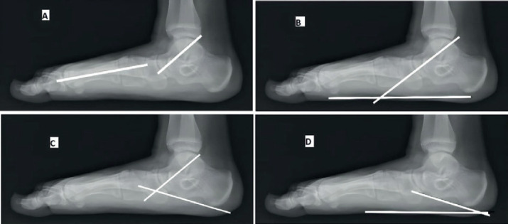
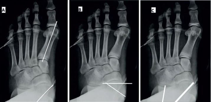
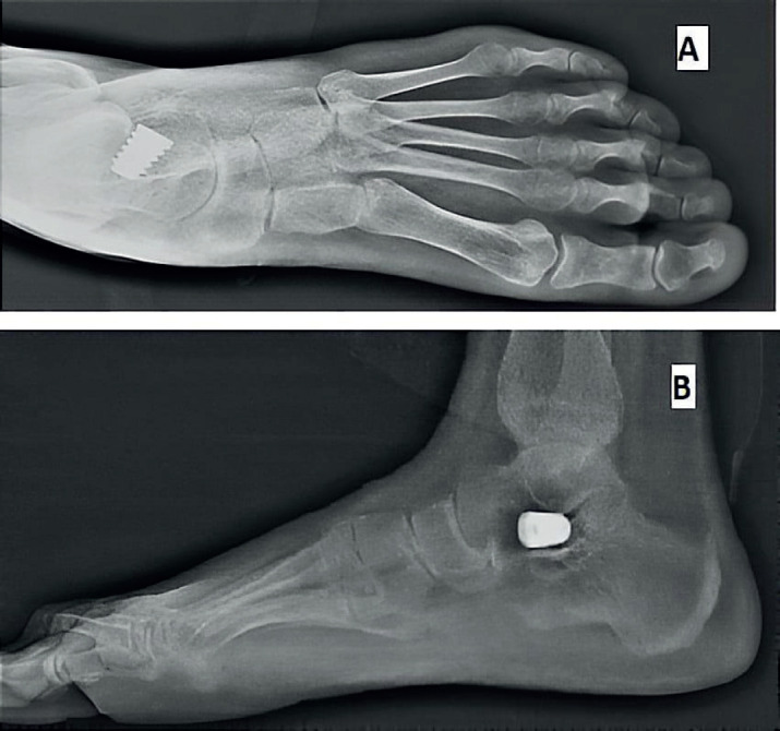
Methods: This is a prospective study and included 26 feet in 19 patients who underwent subtalar arthroereisis for symptomatic flexible flatfeet deformity. Preoperative and postoperative functional assessment based on the American Orthopedic Foot and Ankle Society (AOFAS) hindfoot scale. Radiographic parameters included preoperative and postoperative Kite`s angle, talonavicular coverage angle, Anterior-Posterior talo-1st metatarsal angle, Meary`s angle, talar declination angle, calcaneal inclination angle and lateral talocalcaneal angle.
Results: The mean follow-up period was 22.5±9.4 months and the mean preoperative AOFAS score was 54.6±6.0, while the mean AOFAS score at the last follow-up visit was 86.3±3.9 (P<0.001).The mean preoperative and postoperative radiological measurements were 19.0°±8.2° and 7.4°±3.9° for the AP Talo-1st metatarsal angle (P<0.001); 23.6°±9.1° and 8.0°±4.0° for talonavicular coverage angle (P<0.001); 35.4°±3.7° and 24.1°±3.4° for Kite`s angle (P<0.003); 22.4°±6.1° and 7.5°±3.7° for Meary`s angle (P<0.001); 41.0°±4.4° and 25.2°±7.1° for talar declination angle (P<0.001); 13.5°±3.7° and 21.3°±3.6° for calcaneal inclination angle (P<0.001) and 52.4°±7.2° and 42.9°±4.8° for lateral talocalcaneal angle (P<0.041) respectively.
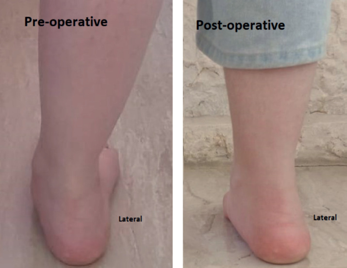
Conclusion: Subtalar arthroereisis is an effective and minimally invasive procedure that showed clinical and radiological improvement for symptomatic flexible flatfoot in our study group.
Keywords: Arthroereisis; flexible Flatfoot; pesplanovalgus; sinus tarsi implant.
© 2024 Mohammad Alkhatatba, Suhaib Bani Essa, Moawiah Khatatbeh, Ahmad Radaideh, Hamzeh Ziad Audat, Ahmad Bani Younes, Mutaz Alrawashdeh, Jehad Abualadas, Naser Obeidat, Jamal Al-Omari, Yazan Anaqreh.
Conflict of interest statement
The authors declare that they have no competing interests.
Figures
References
-
- Michaudet C, Edenfield KM, Nicolette GW, Carek PJ. Foot and Ankle Conditions: Pes Planus. FP Essent. 2018;465:18–23. - PubMed
-
- Staheli LT, Chew DE, Corbett M. The longitudinal arch. A survey of eight hundred and eighty-two feet in normal children and adults. J Bone Joint Surg Am. 1987;69(3):426–428. - PubMed
LinkOut - more resources
Full Text Sources