Review
. 2024 Dec 20;6(1):e70020.
doi: 10.1002/mco2.70020. eCollection 2025 Jan.

Metastatic brain tumors: from development to cutting-edge treatment

Affiliations
Review

Metastatic brain tumors: from development to cutting-edge treatment

Guilong Tanzhu et al. MedComm (2020). .

Abstract

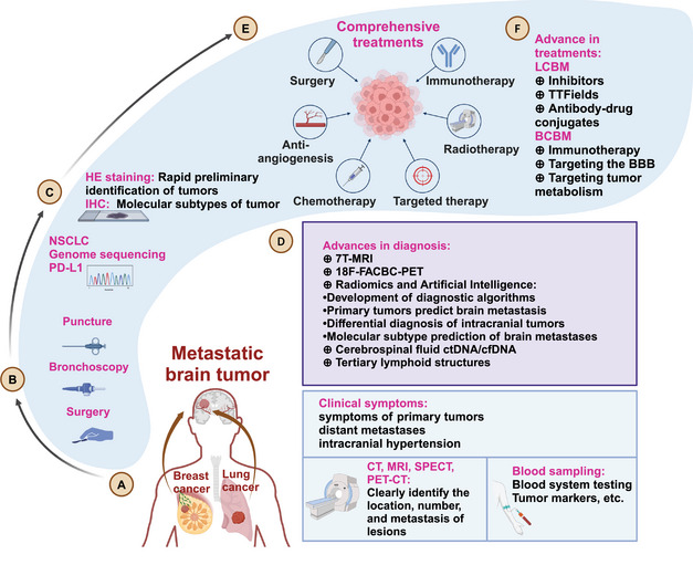
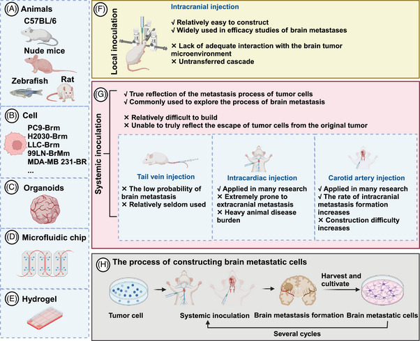
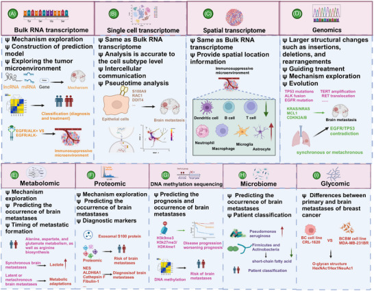
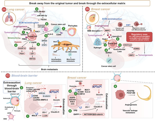
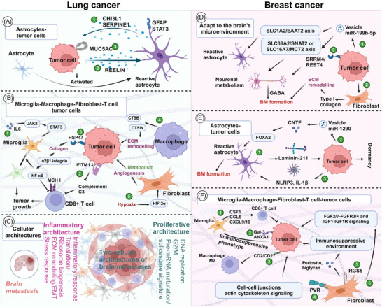
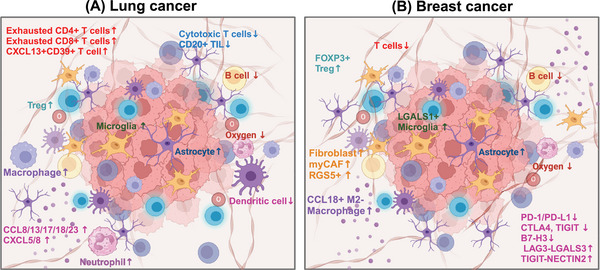
Metastatic brain tumors, also called brain metastasis (BM), represent a challenging complication of advanced tumors. Tumors that commonly metastasize to the brain include lung cancer and breast cancer. In recent years, the prognosis for BM patients has improved, and significant advancements have been made in both clinical and preclinical research. This review focuses on BM originating from lung cancer and breast cancer. We briefly overview the history and epidemiology of BM, as well as the current diagnostic and treatment paradigms. Additionally, we summarize multiomics evidence on the mechanisms of tumor occurrence and development in the era of artificial intelligence and discuss the role of the tumor microenvironment. Preclinically, we introduce the establishment of BM models, detailed molecular mechanisms, and cutting-edge treatment methods. BM is primarily treated with a comprehensive approach, including local treatments such as surgery and radiotherapy. For lung cancer, targeted therapy and immunotherapy have shown efficacy, while in breast cancer, monoclonal antibodies, tyrosine kinase inhibitors, and antibody-drug conjugates are effective in BM. Multiomics approaches assist in clinical diagnosis and treatment, revealing the complex mechanisms of BM. Moreover, preclinical agents often need to cross the blood-brain barrier to achieve high intracranial concentrations, including small-molecule inhibitors, nanoparticles, and peptide drugs. Addressing BM is imperative.

Keywords: diagnosis and treatment; metastatic brain tumors; molecular mechanisms; multiomics; tumor microenvironment.


Conflict of interest statement

The authors declare that there is no conflict of interest in any aspect.

Figures

FIGURE 1
FIGURE 1
Diagnosis and treatment of metastatic brain tumors. (A) Brain metastasis from lung cancer and breast cancer can manifest as clinical symptoms, distant metastasis, and intracranial hypertension; laboratory tests such as blood tumor markers and imaging examinations can be used for auxiliary diagnosis and characterization of metastatic lesions. (B) The diagnostic approach for brain metastasis includes surgery, fiberoptic bronchoscopy, and puncture biopsy. (C) HE staining and IHC are often used to confirm the primary tumor source and classify brain metastasis. For brain metastasis from NSCLC, genomic sequencing and PD‐L1 testing guide the treatment. (D) Advances in the diagnosis of brain metastasis have been driven by technologies such as 7T‐MRI, 18‐FACBC PET, imaging genomics, artificial intelligence, tertiary lymphoid structures, and liquid biopsy like ctDNA/cfDNA. (E) Treatments of brain metastasis typically involve a combination of surgical treatment, immunotherapy, antiangiogenic therapy, radiotherapy, chemotherapy, and targeted therapy. (F) Advances in the treatments include inhibitors, TTFields, antibody–drug conjugates for lung cancer brain metastasis and immunotherapy, strategies like targeting the BBB or tumor metabolism for breast cancer brain metastasis.
FIGURE 2
FIGURE 2
Common preclinical models in brain metastasis research. (A–E) Common models include animals (such as mice (most common), rats, zebrafish), brain‐tropism cells (often labeled with the end of Brm or BR), organoids, and microfluidic chips. (F and G) The advantages and disadvantages of local and systemic inoculation are considered to construct animal models of brain metastasis. (H) Describes the process of constructing brain‐tropism cells (also called brain metastatic cells).
FIGURE 3
FIGURE 3
The role and the latest progress of various omics in brain metastasis. Transcriptome (bulk RNA transcriptome, single‐cell transcriptome, and spatial transcriptome), genomics is the most common omics in brain metastasis. In addition, metabolomics revealed the mechanism and timing of occurrence of breast cancer brain metastasis. Proteomics, DNA methylation sequencing, microbiome, and glycomic have also gradually developed. Although the functions of each omics are generally similar, they complement each other's strengths and weaknesses. Integrating multiple omics data are the current trend.
FIGURE 4
FIGURE 4
Mechanism of metastatic brain metastasis. (A‐B) Break away from the original tumor and break through the extracellular matrix. (A) Lung cancer. (A①) Genes such as RAC1, PMS2, MUC5AC regulate ECM, metabolism, tumorigenesis, and angiogenesis to promote brain metastasis. (A②) Hypoxia stimulates the RASSF1A/kinase Hippo pathway, exacerbating the formation of brain metastasis. (A③) EGFR mutation promotes brain metastasis through ERK1/2–E2F1–WNT5A. (A④) LGALS8–AS1 targets miR‐885‐3p to mediate the expression of FSCN1, ultimately leading to the formation of brain metastasis. (A⑤) HLA‐G expression is increased, and it acts on adjacent tumor cells through SPAG9/STAT3 to promote their self‐renewal ability and growth. (A⑥) CD44+ lung cancer stem cells derive vascular pericytes, form metastatic niches, and migrate across the endothelium by mediated GPR124. (A⑦) Nicotine induces neutrophils to produce exosomal miR‐4466, promoting stemness and invasion properties of lung cancer cells. (B) Breast cancer. (B①) Hyal‐1 produces hydrogel to wrap and promote tumor cells growth. (B②) Lipocalin‐2 regulates MMP9 and exerts various functions such as angiogenesis, ECM reconstruction, BBB disruption, and tumorigenesis. (B③) The loss of Kmt2c/Kmt2d promotes tumor growth through the H3K27me3–KDM6A–MMP3 axis. (B④) High expression of NDRG1 is associated with stemness and promotes tumor growth. (C) Extravasation through blood–brain barrier. (C①‐②) Lung cancer. (C①) MSLN‐overexpressing cells achieve brain metastasis by enhancing the ability to penetrate the BBB. Mechanistically, MSLN facilitates the expression and activation of MET through the c‐Jun N‐terminal kinase (JNK) signaling pathway. (C②) Exosomal LncRNA LINC01356 and miR‐375‐3p derived from brain metastatic cells, as well as Lnc–MMP2‐2‐overexpressing or CXCR4+ tumor cells, promote the occurrence of brain metastasis by inhibiting the tight junctions of the BBB (including Claudin‐5, Occulin, and ZO‐1). (C③‐⑤) Breast cancer. (C③‐⑤) Tumor cells enhance BBB adhesion through RET, MUC1, VCAM1, VLA‐4, ICAM1/2, and secretion of exosomes. Meanwhile, the EGFR–DOCK4–RAC1 axis regulates cell morphology and facilitates their passage through the BBB. (C⑥) Tumor cells and pericytes promote vascular leakage and metastasis by mimicking angiogenesis through PDGFRβ and PDGF–BB.
FIGURE 5
FIGURE 5
The colonization mechanism of tumor cells in the intracranial microenvironment. (A–C) Lung cancer. (A) Astrocytes–tumor cells interactions. (A①) The aggressive proliferation of metastatic brain tumor is linked to the release of CHI3L1 from p‐STAT3+ astrocytes. (A②) In SCLC, reelin is secreted to stimulate the recruitment of astrocytes, which subsequently leads to the secretion of SERPINE1 and other proteins by astrocytes, thereby promoting the growth of SCLC. (A③) Tumor cells interact with astrocytes, inducing an upregulation of MUC5AC expression in tumor cells and enhancing brain colonization. (B) Interactions between tumor cells and microglia, macrophage, fibroblast, and T cells. (B①) Increased IL6 regulates the JAK2/STAT3 signaling pathway to induce the anti‐inflammatory effect of microglia, thereby promoting the colonization of tumor cells. (B②) Low expression or loss of IFITM1 results in a decrease in complement component 3 and MHC I molecules, ultimately inhibiting the killing effect of CD8+ T cells. (B③) The HSP47–collagen axis achieves M2 polarization through the α2β1 integrin/NF‐κB pathway, leading to the upregulation of anti‐inflammatory cytokines and inhibiting the antitumor response of CD8+ T cell. (B④) High expression of cathepsins CTSB and CTSW in macrophages contributes to multiple tumor‐promoting processes including invasion and metastasis. (B⑤) Hypoxia mediates HIF‐2α, leading to CAFs undergoing a unique lineage transition. The transformed CAFs exhibit angiogenesis, trigger metabolic program rearrangement, and promote tumor cells growth. (C) Cellular architectures The proliferative BM functional archetypes are prominent in DNA replication, G2/M, and pre‐mRNA maturation as well as the spliceosome signature while the inflammatory BM functional archetypes are prominent in inflammatory response, ECM remodeling, and stress response ability. (D–F) Breast cancer. (D)Tumor cells adapt to the brain microenvironment. (D①) Tumor cells secrete vesicles rich in miR‐199b‐5p, which act on SLC1A2/EAAT2 axis to activate astrocytes and alter neuronal metabolism through SLC38A2/SNAT2 or SLC16A7/MCT2 axis. (D②) Tumor cells stimulate fibroblast secretion of type I collagen to reconstruct ECM and promote tumor growth. (D③) Tumor cells overexpress SRRM4/EST4, promoting GABA production and leading to tumorigenesis. (E) Astrocytes–tumor cells interactions. (E①) Astrocytes secrete Laminin‐211 to promote tumor cell dormancy. (E②) Tumor cells secrete vesicles rich in miR‐1290 to activate the CNTF–FOXA2 axis. (E③) Meanwhile, tumor cells increase NLRP3 and IL‐1β in astrocytes, which leads to the activation of astrocytes and promotes tumor metastasis. (F) Microglia–macrophage–fibroblast‐T cell‐tumor cells. (F①) Microglia secrete CSF1, CCL5, CXCL9/10 to activate CD8+T cells and kill tumor cells. (F②) Tumor cells overexpress Gal‐3 and ANXA1 to activate the immunosuppressive phenotype of microglia. (F③) Meanwhile, tumor cells expressing CD2/CD27 inhibit M2 type macrophage. (F④‐⑤) Fibrocytes activate cell junctions actin cytoskeleton signaling through PVR, and shape an immunosuppressive microenvironment by secreting periostin and biglycan through RGS5. (F⑥) Fibrocytes promote tumor growth through FGF2/7–FGFR3/4 and IGF1‐IGF1R signaling.
FIGURE 6
FIGURE 6
A brief summary of the tumor microenvironment of brain metastasis from lung cancer and breast cancer. Overall, brain metastases from both breast and lung cancers display a is more immunosuppressive and fibrotic microenvironment compared with their primary lesions. (A) Lung cancer. Exhausted T cells, Treg cells, macrophages, neutrophil, microglia, astrocyte, cytokines and chemokines increased. However, dendritic cells, cytotoxic T cells, and B cells decreased. (B) Breast cancer. CCL18+M2 macrophage, RGS5+ fibroblast, myCAF, FOXP3+Treg cells, LGALS1+ microglia, astrocyte increased. And dendritic cells, T cells, and B cells decreased. PD‐1/PD‐L1, CTLA4, TIGIT, B7‐H3 decreased, and the immune checkpoint that mainly mediates immune escape may be related to LAG3–LGALS3 and TIGIT–NECTIN2.

Similar articles

Cited by

  • Current Bioinformatics Tools in Precision Oncology.
    Wolde T, Bhardwaj V, Pandey V. Wolde T, et al. MedComm (2020). 2025 Jul 9;6(7):e70243. doi: 10.1002/mco2.70243. eCollection 2025 Jul. MedComm (2020). 2025. PMID: 40636286 Free PMC article. Review.
  • Medulloblastoma: biology and immunotherapy.
    Poggi A, Reggiani F, Azevedo HS, Raffaghello L, Pereira RC. Poggi A, et al. Front Immunol. 2025 Jul 3;16:1602930. doi: 10.3389/fimmu.2025.1602930. eCollection 2025. Front Immunol. 2025. PMID: 40677711 Free PMC article. Review.

References

    1. Boire A, Brastianos PK, Garzia L, Valiente M. Brain metastasis. Nat Rev Cancer. 2020;20(1):4‐11. - PubMed
    1. Miccio JA, Tian Z, Mahase SS, et al. Estimating the risk of brain metastasis for patients newly diagnosed with cancer. Commun Med (Lond). 2024;4(1):27. - PMC - PubMed
    1. Relli V, Trerotola M, Guerra E, Alberti S. Abandoning the notion of non‐small cell lung cancer. Trends Mol Med. 2019;25(7):585‐594. - PubMed
    1. Gillespie CS, Mustafa MA, Richardson GE, et al. Genomic alterations and the incidence of brain metastases in advanced and metastatic NSCLC: a systematic review and meta‐analysis. J Thorac Oncol. 2023;18(12):1703‐1713. - PubMed
    1. Scandurra G, Lombardo V, Scibilia G, et al. New frontiers in the treatment of patients with HER2+ cancer and brain metastases: is radiotherapy always useful? Cancers (Basel). 2024;16(13):2466. - PMC - PubMed

LinkOut - more resources