. 2025 Jan;12(1):015001.
doi: 10.1117/1.NPh.12.1.015001. Epub 2024 Dec 19.

Distribution of spine classes shows intra-neuronal dendritic heterogeneity in mouse cortex

Affiliations

Distribution of spine classes shows intra-neuronal dendritic heterogeneity in mouse cortex

Carina C Theobald et al. Neurophotonics. 2025 Jan.

Abstract

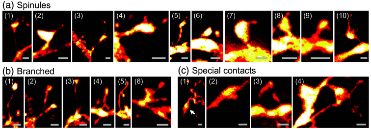
Significance: Neuronal dendritic spines are central elements for memory and learning. Their morphology correlates with synaptic strength and is a proxy for function. Classic light microscopy cannot resolve spine morphology well, and techniques with higher resolution (electron microscopy and super-resolution light microscopy) typically do not provide spine data in large fields of view, e.g., along entire dendrites. Therefore, it remains unclear if spine types are organized on mesoscopic scales, despite their undisputed importance for understanding the brain.

Aim: Recently, it was shown that the distribution of spine type is dendrite-specific in the turtle cortex, suggesting a mesoscopic organization, but leaving the question open if such a dendrite specificity also exists in mammals. Here, we determine if such a difference in spine-type distribution among dendrites also exists in the mouse brain.

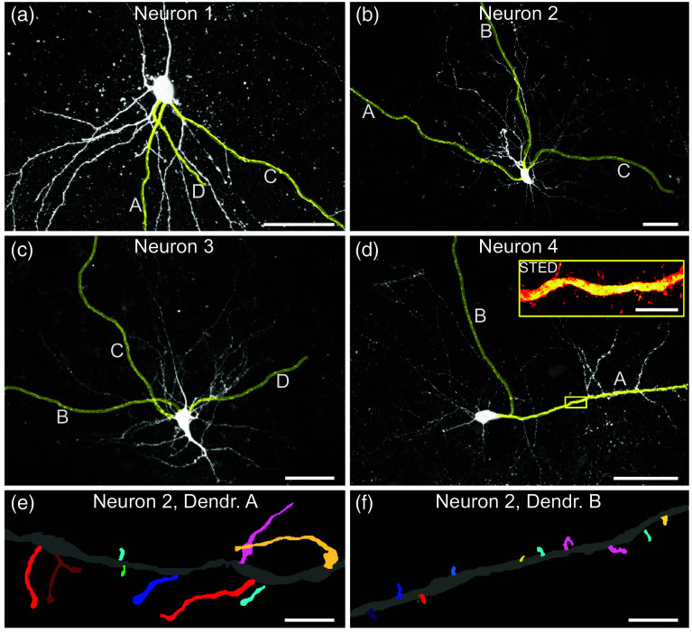
Approach: We used super-resolution stimulated emission depletion microscopy of complete dendrites and advanced morphological analysis in three dimensions to decipher morphological differences of spines on different dendrites.

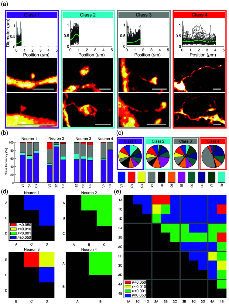
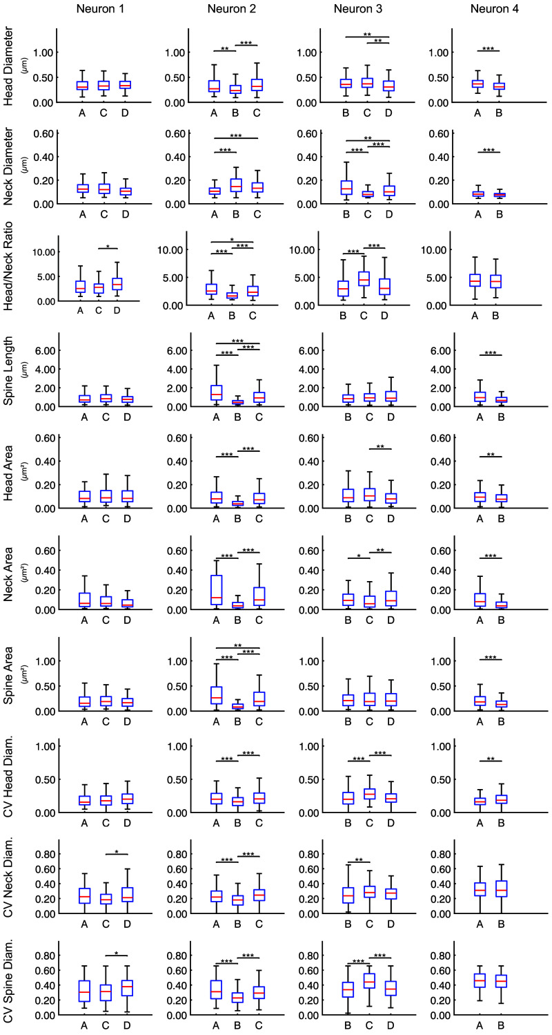
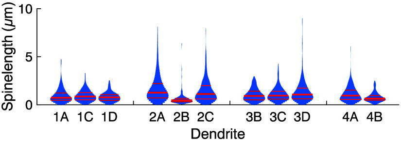
Results: We found that spines of different shapes decorate different dendrites of the same neuron to a varying extent. Significant differences among the dendrites are apparent, based on spine classes as well as based on quantitative descriptors, such as spine length or head size.

Conclusions: Our findings may indicate that it is an evolutionarily conserved principle that individual dendrites have distinct distributions of spine types hinting at individual roles.

Keywords: dendrite-specific; dendritic spine; spine morphology; spine shape; super-resolution stimulated emission depletion microscopy; three-dimensional analysis.


Figures

Fig. 1
Fig. 1
Cortical neurons. (a)–(d) Confocal overview images of the perisomatic region of the neurons filled with markers in the mouse cortex (maximum-intensity projections). Letters and yellow markings identify the individual dendrites, which were then imaged with STED microscopy [inset in panel (d)], eventually also beyond the field of view shown here. Individual neurons were filled with biocytin and revealed with the streptavidin-coupled fluorophore Atto 647N. Scale bars, 50  μm and 5  μm in the inset. (e) and (f) Segmentation of individual spines from two dendrites of the same neuron highlights differences (maximum-intensity projections shown). Scale bars, 2  μm.
Fig. 2
Fig. 2
Spine classes are inhomogeneously distributed on the dendrites. (a) Spines were categorized into four classes by hierarchical clustering of shape and length: upper panels—individual diameter profiles (gray) and average profile of each class (green). The total lengths of the profiles were scaled to the average spine length of the respective class; lower panels—representative spine examples. Scale bars, 1  μm. (b) Relative abundance of each spine class on the dendrites, the distribution differs among dendrites. Numbers 1 to 4 refer to the four neurons, letters to the dendrites (see Fig. 1). Color code as in panel (a). (c) Pie charts showing which fraction of a specific spine class is found on each dendrite. (d) Pairwise Pearson’s chi-square tests confirm significant differences among the repartition of the classes on the dendrites within three of the four neurons. p values color coded: p0.05 red, p0.01 yellow, p0.001 green, and p>0.05 blue. All tests have been corrected for multiple comparisons. (e) Pairwise Pearson’s chi-square tests for all dendrites of all cells.
Fig. 3
Fig. 3
Spine descriptors are distinct among dendrites. Quantitative spine descriptors show significant differences in dendrites of the same neuron. Box plots show the median and quartiles; the whiskers extend to the most extreme data points not considered outliers (outliers not depicted). *p0.05, **p0.01, and ***p0.001. All tests have been corrected for multiple comparisons (Kruskal–Wallis tests). Each column represents one neuron. All tested descriptors differ significantly among several dendrites, with the fewest differences in neuron 1. Width variability within spines given as coefficient of variation (CV).
Fig. 4
Fig. 4
Spine length distributions vary among dendrites. The spine lengths show distinct distributions on different dendrites. Violin plots: red lines show the median and quartiles.
Fig. 5
Fig. 5
Spinules, branches, and special contacts. (a) Spines with spinules. (b) Branched spines. (c) Spines with unusual contacts. Arrow in (1) points to a synapse. Scale bars, 500 nm.

Similar articles

Cited by

References

    1. DeFelipe J., Fariñas I., “The pyramidal neuron of the cerebral cortex: morphological and chemical characteristics of the synaptic inputs,” Progr. Neurobiol. 39, 563–607 (1992).PGNBA510.1016/0301-0082(92)90015-7 - DOI - PubMed
    1. Araya R., Vogels T. P., Yuste R., “Activity-dependent dendritic spine neck changes are correlated with synaptic strength,” Proc. Natl. Acad. Sci. U. S. A. 111, E2895–E2904 (2014).10.1073/pnas.1321869111 - DOI - PMC - PubMed
    1. Harris K., Stevens J., “Dendritic spines of rat cerebellar Purkinje cells: serial electron microscopy with reference to their biophysical characteristics,” J. Neurosci. 8, 4455–4469 (1988).JNRSDS10.1523/JNEUROSCI.08-12-04455.1988 - DOI - PMC - PubMed
    1. Yuste R., Denk W., “Dendritic spines as basic functional units of neuronal integration,” Nature 375, 682–684 (1995).10.1038/375682a0 - DOI - PubMed
    1. Yuste R., Majewska A., Holthoff K., “From form to function: calcium compartmentalization in dendritic spines,” Nat. Neurosci. 3, 653–659 (2000).NANEFN10.1038/76609 - DOI - PubMed