A Rare Case of Situs Inversus Partialis With Levocardia in a 73-Year-Old Patient
- PMID: 39712821
- PMCID: PMC11661905
- DOI: 10.7759/cureus.74061
A Rare Case of Situs Inversus Partialis With Levocardia in a 73-Year-Old Patient
Abstract
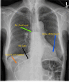
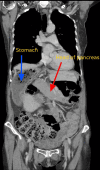
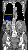
Situs inversus partialis (SIP) is an extremely rare congenital disorder in which most of the visceral organs are located on the opposite side of their usual anatomical locations. The condition is usually associated with levocardia, in which the apex of the heart is directed toward the left side. In our case study, a female patient with a history of dysphagia and weight loss presented to the outpatient clinic under the urgent two-week wait pathway. She underwent a CT scan of the thorax, abdomen, and pelvis, which revealed that she has SIP in which some of the abdominal organs are reversed and others are in their normal anatomical locations. The patient was diagnosed with esophageal carcinoma. However, because of her rare condition, it was challenging to insert a nasogastric tube into her reversed stomach and to explore other feeding options, e.g., jejunostomy. This report discusses a rare case of SIP, incidentally discovered in a patient with suspected esophageal malignancy, and its associated surgical challenges in her management.
Keywords: congenital birth defect; dysphagia; esophageal carcinoma; situs inversus with levocardia; weight loss.
Copyright © 2024, Mahrous et al.
Conflict of interest statement
Human subjects: Consent for treatment and open access publication was obtained or waived by all participants in this study. Conflicts of interest: In compliance with the ICMJE uniform disclosure form, all authors declare the following: Payment/services info: All authors have declared that no financial support was received from any organization for the submitted work. Financial relationships: All authors have declared that they have no financial relationships at present or within the previous three years with any organizations that might have an interest in the submitted work. Other relationships: All authors have declared that there are no other relationships or activities that could appear to have influenced the submitted work.
Figures


Similar articles
-
Situs inversus with levocardia in a 15-year-old male adolescent: a case report.J Med Case Rep. 2023 Dec 3;17(1):499. doi: 10.1186/s13256-023-04254-9. J Med Case Rep. 2023. PMID: 38042875 Free PMC article.
-
Robot-assisted distal gastrectomy with lymph node dissection for gastric cancer in a patient with situs inversus partialis: a case report with video file.Surg Case Rep. 2018 Feb 13;4(1):16. doi: 10.1186/s40792-018-0422-7. Surg Case Rep. 2018. PMID: 29441475 Free PMC article.
-
Incidental finding of levocardia with situs inversus: case report.Radiol Case Rep. 2024 Dec 17;20(3):1386-1388. doi: 10.1016/j.radcr.2024.11.042. eCollection 2025 Mar. Radiol Case Rep. 2024. PMID: 39801529 Free PMC article.
-
Neonatal intestinal obstruction associated with situs inversus totalis: two case reports and a review of the literature.J Med Case Rep. 2017 Sep 18;11(1):264. doi: 10.1186/s13256-017-1423-z. J Med Case Rep. 2017. PMID: 28918753 Free PMC article. Review.
-
Prolonged survival with isolated levocardia and situs inversus.Cleve Clin J Med. 1991 May-Jun;58(3):243-7. doi: 10.3949/ccjm.58.3.243. Cleve Clin J Med. 1991. PMID: 1893555 Review.
References
-
- Abdominal manifestations of situs anomalies in adults. Fulcher AS, Turner MA. Radiographics. 2002;22:1439–1456. - PubMed
-
- Situs inversus partialis (levocardia): an incidental discovery of an extremely rare case during the autopsy of a female with suicidal poisoning. Kanani J, Sheikh MI. Surg Exp Pathol. 2024;7:10.
Publication types
LinkOut - more resources
Full Text Sources
Miscellaneous
