. 2024 Dec 17:13:789-803.
doi: 10.2147/ITT.S495095. eCollection 2024.

Lipopolysaccharide in Bile Promotes the Neutrophil Extracellular Traps-Induced Gallstone Formation by Activating the Gallbladder Immune Barrier

Affiliations

Lipopolysaccharide in Bile Promotes the Neutrophil Extracellular Traps-Induced Gallstone Formation by Activating the Gallbladder Immune Barrier

Jingjing Yu et al. Immunotargets Ther. .

Abstract

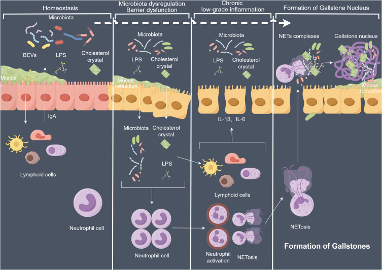
Background: Cholelithiasis areis a common digestive system disorder, with cholesterol gallstones being the most prevalent type. Gallstones lead to many severe complications, posing a significant burden on global healthcare systems. Many studies have shown associations between biliary microbiota, gallbladder immune microenvironment, and gallstone formation. However, the specific immune mechanisms underlying the cholesterol gallstone formation have not been fully elucidated.

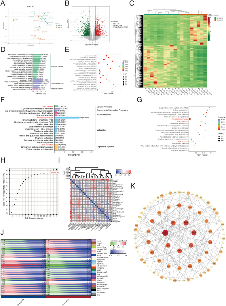
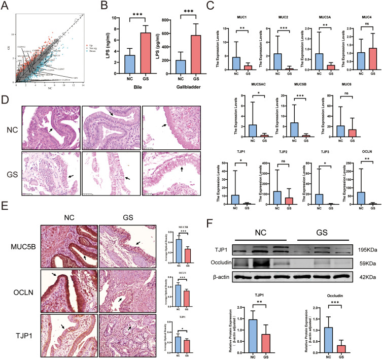
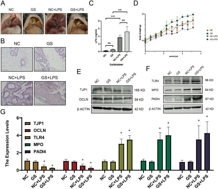
Methods: In this study, gallbladderand bile samples from 8 asymptomatic patients with cholelithiasis undergoing cholecystectomy and 11 healthy liver transplant donors were collected for tissue transcriptome sequencing and differential analysis. Male C57BL/6J mice were fed a normal or lithogenic diet for 6 weeks. Starting from the third week, lipopolysaccharide (LPS) or specific regulators were injected intraperitoneally once a week for a total of 3 times. Enzyme-linked immunosorbent assay, quantitative polymerase chain reaction, Western blot, immunohistochemistry, and immunofluorescence were employed for quantitative, qualitative or localization analysis of LPS, neutrophil extracellular traps (NETs), inflammatory factors, proteins, and mRNAs using samples collected from mice.

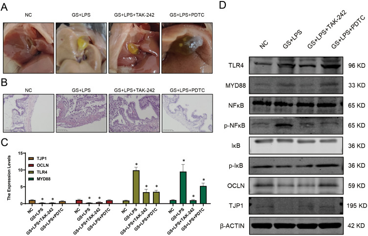
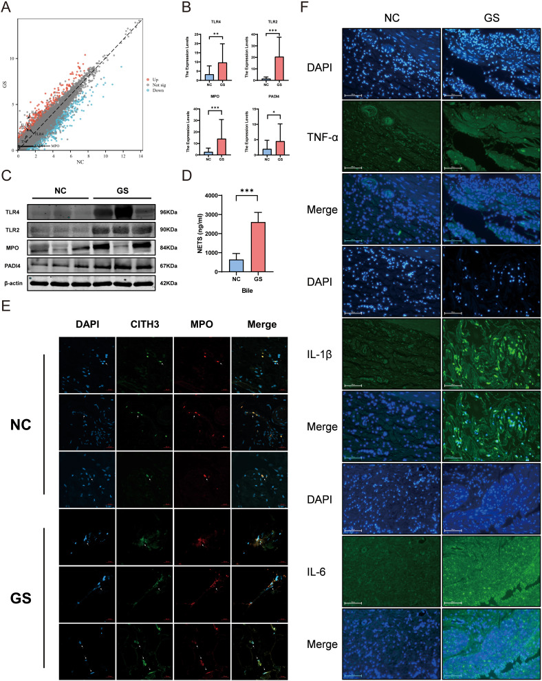
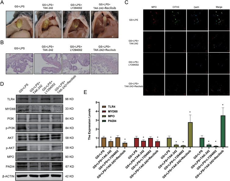
Results: In patients with cholelithiasis, the gallbladder mechanical barrier is impaired, resulting in an immune-activated state. LPS induces damage to the gallbladder mucosal mechanical barrier through the Toll-like receptor 4 (TLR4)/myeloid differentiation factor 88 (MyD88)/nuclear factor kappa-B (NF-κB) signaling pathway. Furthermore, it stimulates the continuous production of NETs through the TLR4/Phosphoinositide 3-kinase (PI3K)/Protein kinase B (Akt) signaling pathway, aggravating the formation of gallstones.

Conclusion: With the biliary dysbiosis, excessive LPS can invade the submucosa of the gallbladder, leading to chronic inflammation that recruits neutrophils to form NETs, which are ultimately expelled into bile, thereby promoting the formation of gallstones.

Keywords: gallstone; immune barrier; lipopolysaccharide; neutrophil extracellular traps.


Conflict of interest statement

The authors declare no conflicts of interest in this work.

Figures

None
Graphical abstract
Figure 1
Figure 1
Transcriptome analysis of patients with cholelithiasis. (A-C) DEGs between the GS and the NC group. (A: PCA; B: Volcano plot; C: Heatmap) (D-G) Classification and enrichment results of differentially expressed metabolites between the GS and the NC group. (D and E: GO; F and G: KEGG) (H) The nature of the network topology constructed using unique Power values. (I and J) Correlation between different modules and the proportion of normal and cholecystolithiasis samples. (K) Venn plot of DEGs.
Figure 2
Figure 2
Impairment of gallbladder mucosal barrier in patients with cholelithiasis. (A) Volcano plot of gene expression related to the mucosal barrier from RNA sequencing results. (B) LPS levels in gallbladder tissues and bile detected by ELISA. (C) qPCR analysis of the expression levels of mucosal barrier-related genes (GS vs NC, outliers were excluded based on the mean ± 3*standard deviation criterion). (D) H&E staining of the gallbladder mucosal barrier (GS vs NC). (E) IHC detection of MUC5B, OCLN, and TJP1 expression (GS vs NC), the gallbladder mucosal epithelial cells have been indicated by arrows. (F) Protein expression levels of TJP1 and OCLN (GS vs NC). (mean ± SD, *p <0.05, **p <0.01, ***p <0.001).
Figure 3
Figure 3
LPS as a potential key factor in gallbladder mechanical barrier impairment and NETs formation promotion. (A) Gallstone formation in mice. (B) H&E staining of the gallbladder in mice. (C) LPS concentration in bile from mice measured by ELISA. (D) Changes in body weight of mice over time. (E) Expression profiles of genes related to the gallbladder mucosal barrier. (F) Expression profiles of genes associated with NETs formation. (G) Transcriptional levels of relevant genes in model mice. (mean ± SD, *p <0.05, **p<0.01).
Figure 4
Figure 4
LPS induces gallbladder mucosal mechanical barrier impairment via the TLR4/MyD88/NF-κB signaling pathway. (A) Gallstone formation in mice from different groups. (B) H&E staining of the gallbladder in mice from different groups. (C) Transcriptional levels of TLR4, OCLN, and MyD88 in each group. (D) Expression profiles of genes related to the TLR4/MyD88/NF-κB signaling pathway in different groups (mean ± SD, *p <0.05).
Figure 5
Figure 5
Increased formation of NETs in the gallbladder and bile of patients with cholelithiasis, indicating chronic inflammation in the gallbladder. (A) Volcano plot of gene expression related to NETs formation in gallbladder tissues of patients with cholelithiasis based on RNA sequencing. (B) Expression levels of key genes involved in NETs formation in GS and NC group determined by qPCR. (C) Protein expression levels of key genes involved in NETs formation. (GS vs NC). (D) Quantification of NETs content in bile samples from GS and NC groups using ELISA. (E) Formation of NETs in gallbladder tissues of GS and NC groups. (F) Immunofluorescence results for inflammatory cytokines TNF-α, IL-1β, and IL-6 in gallbladder tissues of GS and NC groups. (mean ± SD, *p <0.05, **p <0.01, ***p <0.001).
Figure 6
Figure 6
LPS promotes NETs formation in the gallbladder via the TLR4/PI3K/Akt signaling pathway. (A) Formation of gallstones in mice. (B) H&E staining of gallbladder tissues from mice. (C) NETs formation in different groups. (D) Protein expression levels of genes related to the PI3K/Akt signaling pathway. (E) Transcription levels of NETs-related genes (mean ± SD, *p <0.05).

Similar articles

Cited by

References

    1. Shaffer EA. Gallstone disease: epidemiology of gallbladder stone disease. Best Pract Res Clin Gastroenterol. 2006;20(6):981–996. doi:10.1016/j.bpg.2006.05.004 - DOI - PubMed
    1. Stinton LM, Shaffer EA. Epidemiology of gallbladder disease: cholelithiasis and cancer. Gut Liver. 2012;6(2):172–187. doi:10.5009/gnl.2012.6.2.172 - DOI - PMC - PubMed
    1. Everhart JE, Ruhl CE. Burden of digestive diseases in the United States Part III: liver, biliary tract, and pancreas. Gastroenterology. 2009;136(4):1134–1144. doi:10.1053/j.gastro.2009.02.038 - DOI - PubMed
    1. Di Ciaula A, Wang DQ, Portincasa P. An update on the pathogenesis of cholesterol gallstone disease. Curr Opin Gastroenterol. 2018;34(2):71–80. doi:10.1097/MOG.0000000000000423 - DOI - PMC - PubMed
    1. Boyer JL. Bile formation and secretion. Compr Physiol. 2013;3(3):1035–1078. doi:10.1002/cphy.c120027 - DOI - PMC - PubMed

LinkOut - more resources