. 2024 Sep 28:15:100399.
doi: 10.1016/j.jpi.2024.100399. eCollection 2024 Dec.

Digital mapping of resected cancer specimens: The visual pathology report

Affiliations

Digital mapping of resected cancer specimens: The visual pathology report

Carly Fassler et al. J Pathol Inform. .

Abstract

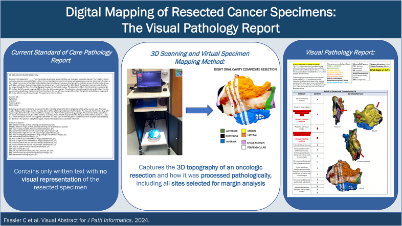
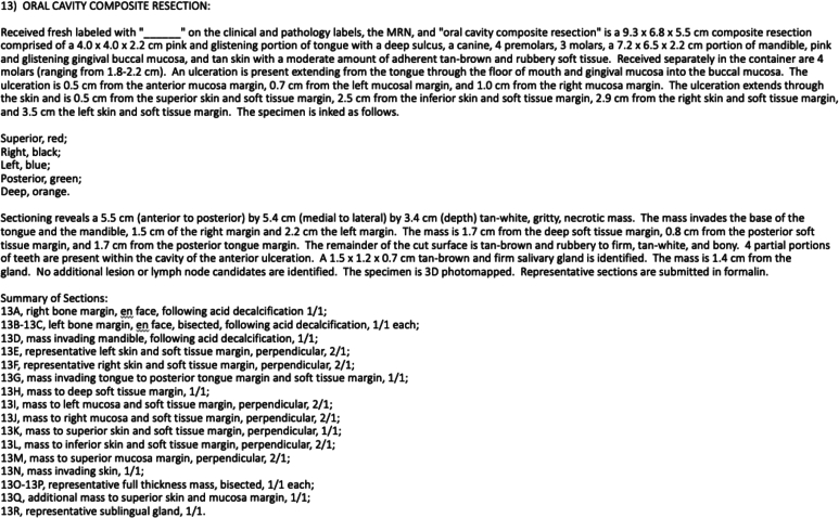
Background: The current standard-of-care pathology report relies only on lengthy written text descriptions without a visual representation of the resected cancer specimen. This study demonstrates the feasibility of incorporating virtual, three-dimensional (3D) visual pathology reports to improve communication of final pathology reporting.

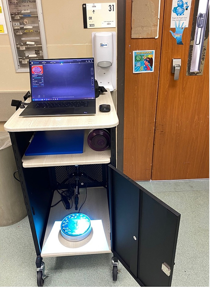
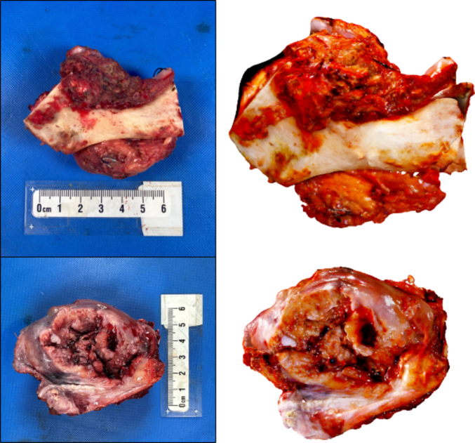
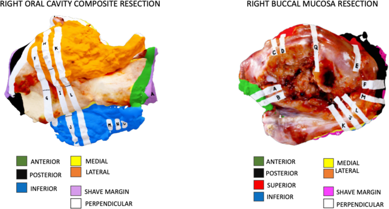
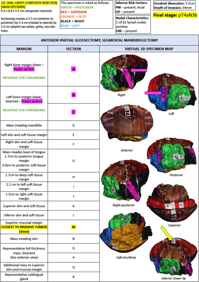
Materials and methods: Surgical specimens are 3D scanned and virtually mapped alongside the pathology team to replicate grossing. The 3D specimen maps are incorporated into a hybrid visual pathology report which displays the resected specimen and sampled margins alongside gross measurements, tumor characteristics, and microscopic diagnoses.

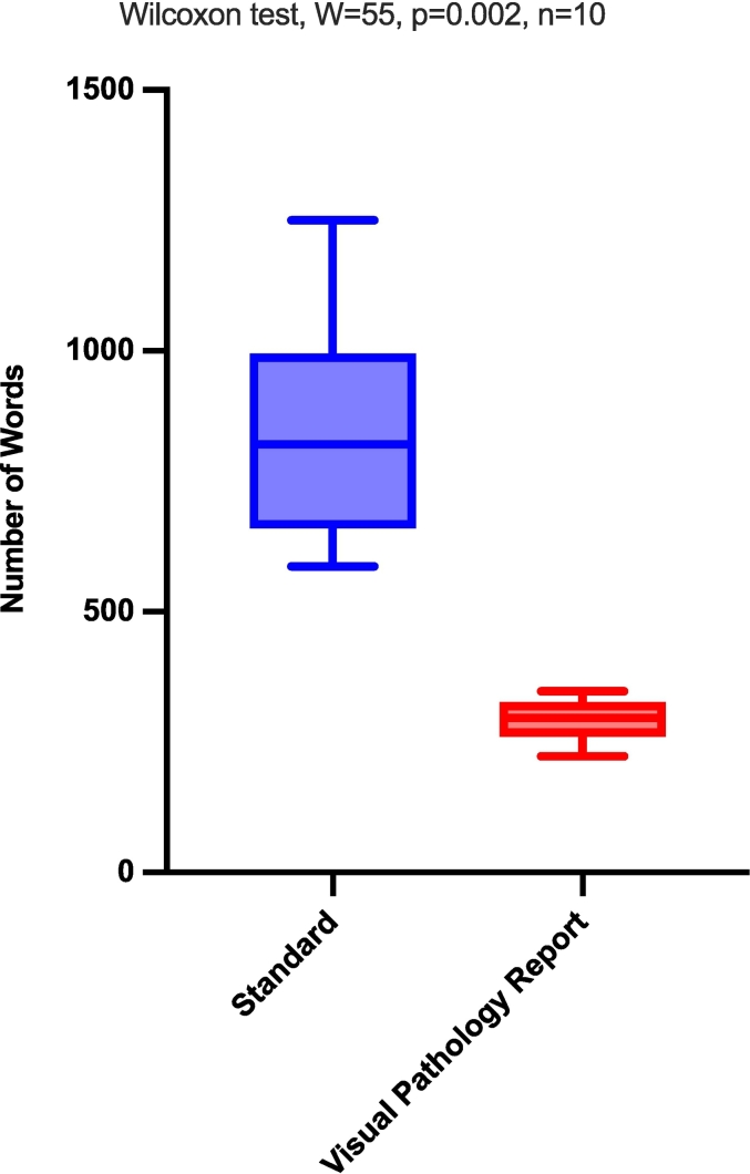
Results: Visual pathology reports were created for 10 head and neck cancer cases. Each report concisely communicated information from the final pathology report in a single page and contained significantly fewer words (293.4 words) than standard written pathology reports (850.1 words, p < 0.01).

Conclusions: We establish the feasibility of a novel visual pathology report that includes an annotated visual model of the resected cancer specimen in place of lengthy written text of standard of care head and neck cancer pathology reports.

Keywords: 3D scanning; Head and neck cancer; Margin status; Oral cavity cancer; Pathology communication; Virtual mapping.


Figures

Unlabelled Image
Graphical abstract
Fig. 1
Fig. 1
Mobile 3D scanning cart that may be set up outside of the OR (shown here) or pathology lab. Scanning occurs in the lower compartment of the cart and the door is closed to ensure optimal dark conditions for 3D scanning.
Fig. 2
Fig. 2
Two-dimensional (2D) images of a right oral cavity composite resection (top) and right buccal mucosa (bottom) resections (left) alongside corresponding screen captures from the three-dimensional (3D) scan of each resection (right).
Fig. 3
Fig. 3
Virtual 3D specimen maps (from Fig. 2 right oral cavity composite and right buccal resection) created at the time of grossing using computer-aided design (CAD) software. Corresponding key to ink colors seen below each map. White sections denote perpendicular sections, pink/purple sections denote shave margins. Each section is marked with the corresponding letter to the histology cassettes each section is submitted in. (For interpretation of the references to color in this figure legend, the reader is referred to the web version of this article.)
Fig. 4
Fig. 4
Sample page from the current standard of care final pathology report comprised of only written text with no visual representation of the resected cancer specimen.
Fig. 5
Fig. 5
Pathology report of the future for the case described in the text in Fig. 4. Pink arrows demonstrate the sites of frozen section margin sampling and their subsequent reporting. The yellow arrow demonstrates the close permanent margin identified in section K. (For interpretation of the references to color in this figure legend, the reader is referred to the web version of this article.)
Fig. 6
Fig. 6
Comparison of word count between standard pathology reports and visual pathology reports.

Similar articles

References

    1. Ettl T., El-Gindi A., Hautmann M., et al. Positive frozen section margins predict local recurrence in R0-resected squamous cell carcinoma of the head and neck. Oral Oncol. Apr 2016;55:17–23. doi: 10.1016/j.oraloncology.2016.02.012. - DOI - PubMed
    1. Binahmed A., Nason R.W., Abdoh A.A. The clinical significance of the positive surgical margin in oral cancer. Oral Oncol. Sep 2007;43(8):780–784. doi: 10.1016/j.oraloncology.2006.10.001. - DOI - PubMed
    1. Network N.C.C. Head and Neck Cancers (Version 2.2024) https://www.nccn.org/professionals/physician_gls/pdf/head-and-neck.pdf Updated 01/09/2024.
    1. Cooper J.S., Pajak T.F., Forastiere A.A., et al. Postoperative concurrent radiotherapy and chemotherapy for high-risk squamous-cell carcinoma of the head and neck. N Engl J Med. May 6 2004;350(19):1937–1944. doi: 10.1056/NEJMoa032646. - DOI - PubMed
    1. Bernier J., Domenge C., Ozsahin M., et al. Postoperative irradiation with or without concomitant chemotherapy for locally advanced head and neck cancer. N Engl J Med. May 6 2004;350(19):1945–1952. doi: 10.1056/NEJMoa032641. - DOI - PubMed

LinkOut - more resources