

Perforated anterior tenia coli-type appendicitis in a case of vermiform appendix duplex in a toddler: a case report
- PMID: 39713058
- PMCID: PMC11660923
- DOI: 10.1093/jscr/rjae784
Perforated anterior tenia coli-type appendicitis in a case of vermiform appendix duplex in a toddler: a case report
Erratum in
-
Correction to: Perforated anterior tenia coli-type appendicitis in a case of vermiform appendix duplex in a toddler: a case report.J Surg Case Rep. 2025 Jul 24;2025(7):rjaf521. doi: 10.1093/jscr/rjaf521. eCollection 2025 Jul. J Surg Case Rep. 2025. PMID: 40709353 Free PMC article.
Abstract
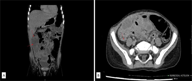
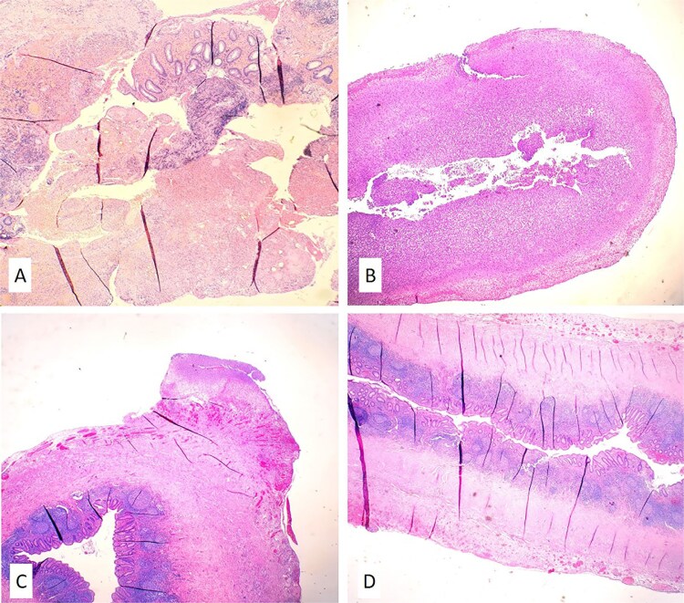
Duplication of the vermiform appendix is a rare anomaly observed in patients undergoing appendectomy. A 27-month-old male toddler presented with a 9-day history of abdominal pain, vomiting, and diarrhea, progressing to an acute abdomen with signs of severe peritonitis. Intraoperative findings revealed a periappendicular infiltrate from a perforated vermiform appendix of the tenia coli type. A second, inflamed appendix was incidentally discovered in its typical location during the procedure. Vermiform appendix duplication presents a clinical challenge due to its rarity and potential for complications. According to the Cave-Wallbridge classification, this case represents Type B2, or the tenia coli variant, characterized by a perforated appendix originating at the tenia coli convergence and a smaller, secondary appendix in a retrocecal position. This case emphasizes the importance of thorough distal and proximal exploration during initial appendectomy when this anomaly is suspected, particularly in cases of Type B2.
Keywords: appendix vermiform duplication; case report; perforated appendicitis; small child.
Published by Oxford University Press and JSCR Publishing Ltd. © The Author(s) 2024.
Conflict of interest statement
None declared.
Figures

References
Publication types
LinkOut - more resources
Full Text Sources
