. 2024 Dec 6:12:1407729.
doi: 10.3389/fbioe.2024.1407729. eCollection 2024.

Collagen scaffold-seeded iTenocytes accelerate the healing and functional recovery of Achilles tendon defects in a rat model

Affiliations

Collagen scaffold-seeded iTenocytes accelerate the healing and functional recovery of Achilles tendon defects in a rat model

Thomas Später et al. Front Bioeng Biotechnol. .

Abstract

Introduction: Tendon injuries represent an ongoing challenge in clinical practice due to poor regenerative capacity, structure, and biomechanical function recovery of ruptured tendons. This study is focused on the assessment of a novel strategy to repair ruptured Achilles tendons in a Nude rat model using stem cell-seeded biomaterial.

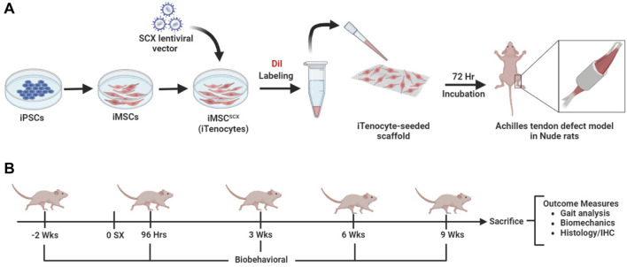
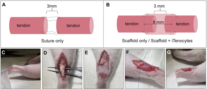
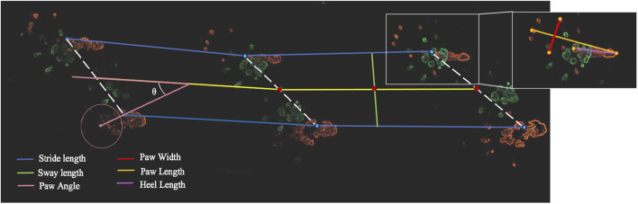
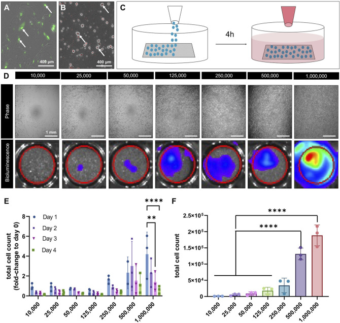
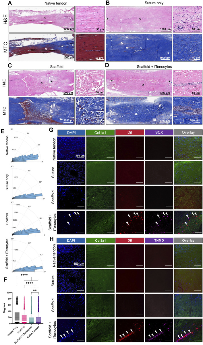
Methods: Specifically, we have used induced pluripotent stem cell (iPSC)-derived mesenchymal stem cells (iMSCs) overexpressing the early tendon marker Scleraxis (SCX, iMSCSCX+, iTenocytes) in combination with an elastic collagen scaffold. Achilles tendon defects in Nude rat models were created by isolating the tendon and excising 3 mm of the midsection. The Achilles tendon defects were then repaired with iTenocyte-seeded scaffolds, unseeded scaffolds, or suture only and compared to native Nude rat tendon tissue using gait analyses, biomechanical testing, histology, and immunohistochemistry.

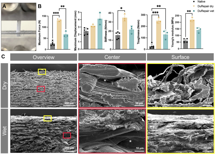
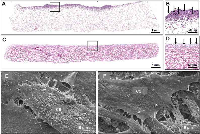
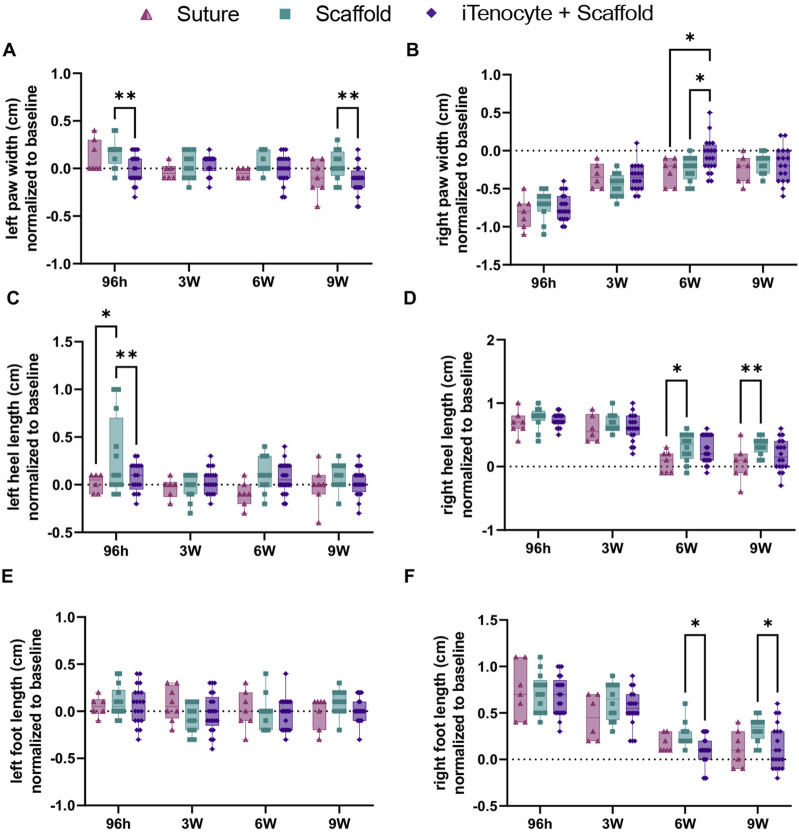
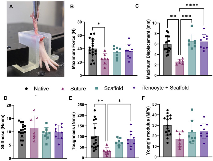
Results: The results show faster functional recovery of gait in iTenocyte-seeded scaffold group comparing to scaffold only and suture only groups. Both iTenocyte-seeded scaffold and scaffold only treatment groups had improved biomechanical properties when compared to suture only treatment group, however no statistically significant difference was found in comparing the cell seeding scaffold an scaffold only group in terms of biomechanical properties. Immunohistochemistry staining further demonstrated that iTenocytes successfully populated the collagen scaffolds and survived 9 weeks after implantation in vivo. Additionally, the repaired tissue of iTenocyte-treated injuries exhibited a more organized structure when compared to tendon defects that were repaired only with suturing or unseeded scaffolds.

Conclusion: We suggest that iTenocyte-seeded DuRepair™ collagen scaffold can be used as potential treatment to regenerate the tendon tissue biomechanically and functionally.

Keywords: Achilles tendon rupture repair; collagen scaffold; stem cells; tissue engineering; tissue regeneration.


Conflict of interest statement

The authors declare that the research was conducted in the absence of any commercial or financial relationships that could be construed as a potential conflict of interest.

Figures

None
iPSCs were differentiated into iMSCs and transduced with SCX-GFP+ lentiviral vector to create iMSC-SCXGFP+ (A). The cells were DiI labeled and seeded onto the scaffolds for 72 h and implanted into an Achilles tendon defect model in Nude rats. Rats underwent gait testing before injury, 96 h, 3-, 6- and 9-weeks post injury. After 9 weeks, biomechanical testing and histological analysis was performed to evaluate healing efficacy (B).
FIGURE 1
FIGURE 1
Surgical procedure of Achilles tendon resection and defect treatment. Schematic illustration of defect repair by suture only. After tendon tissue removal, the two ends of tendon were sutured together while maintaining a 3 mm defect (A). Schematic illustration of defect treatment with unseeded or seeded scaffolds. After the creation of a 3 mm defect in Achilles tendon tissue, scaffolds were wrapped around the remaining tendon ends with an overlap of 2.5 mm on each side, leaving a 3 mm gap in the center of the tube-like scaffold structure (B). Images of the in vivo procedure (C–G). Right foot of the animal, fixed to the underlaying surface to prevent unwanted moving of the rat (C). After opening skin and muscle layers, a forceps was passed through the tissue underneath the Achilles tendon (D). The Achilles tendon was cut to create a standardized 3 mm tissue defect and the scaffold was placed underneath the defect, with the cell-seeded side of seeded scaffolds towards the tendon (E). Both ends of the scaffolds were sutured to the ends of the tendon and maintained the 3 mm standardized gap, resulting in a tube-like structure of the scaffold, wrapping it around the defect (F, G).
FIGURE 2
FIGURE 2
Functional recovery gait test of Achilles tendon defects in Nude rats. Gait testing sheet with hind paws in orange and fore paws in green. Stride length (blue), sway length (green), paw width (red), paw length (orange), paw angle (pink) and heel length (purple) were measured using the hind paws.
FIGURE 3
FIGURE 3
Material characterization DuRepair™ collagen scaffolds. Representative image of a dry DuRepair™ scaffold fixed with two mechanical clamps for biomechanical testing (A) Biomechanical outcome measures (maximum force, maximum displacement, stiffness, toughness, and young’s modulus) of native tendon tissue (●), DuRepair™ dry (▼), DuRepair™ wet (■). (B) n = 3 *p < 0.05 **p < 0.01 ***p < 0.001. Representative scanning electron microscopic cross section images of dry and wet DuRepair™ collagen scaffolds. Surface (red outlines) and center (yellow outlines) zones represent higher magnifications of wet and dry overview images (C).
FIGURE 4
FIGURE 4
Viability of pBM-MSCLuc+ seeded collagen scaffolds in vitro. Representative confocal microscopy image of iTenocytes in culture, in which the nucleus is expressing SCX-GFP fluorescence (A). Representative image of DiI-labeled pBM-MSC-Luc+ in culture (B). Schematic illustration of the cellular seeding process. pBM-MSC-Luc cells were homogeneously seeded on top of collagen scaffolds and incubated for 4 h before medium was added to assure proper cellular attachment (C). Light microscopic images of cell-seeded scaffolds on non-adherent Poly(2-hydroxyethyl methacrylate)-coated wells (top) and bioluminescence images showing increasing bioluminescent signals of scaffolds seeded (bottom) ranging from 10,000, to 1,000,000 cells (D). Total cell count of seeded scaffolds from days 1 (●), day 2 (), day 3 (▼), and day 4 (■) given as fold-change to day 0 (right after seeding) (E). Total cells count of attached cells after 4 days of seeding via cell viability assay (F). **p < 0.01, ****p < 0.0001, n = 3.
FIGURE 5
FIGURE 5
Cellular attachment and surface topography of cell-seeded collagen scaffolds. Representative H&E-stained section of collagen scaffold 4 h after seeding with 500,000 pBM-MSC-Luc+ (A). Higher magnification of A, showing cellular presence in exclusively the superficial zone of the scaffold (B). Representative H&E-stained section of a collagen scaffold 3 days after seeding with 500,000 pBM-MSCLuc+ (C). Higher magnification of C, showing cellular presence in exclusively the superficial zone of the scaffold (D). Two representative scanning electron microscopic images of a collagen scaffold seeded with 500,000 pBM-MSCLuc+. Arrows point to the attached cells (E, F).
FIGURE 6
FIGURE 6
Functional recovery of Achilles tendon defects treated with iTenocyte-seeded collagen scaffolds in vivo. Paw widths of left (A) and right (B) paws. Heel lengths of left (C) and right (D) paws. Foot lengths of left (E) and right (F) paws 96 h, 3-,6- and 9- weeks after injury and treatment of suture (▲), scaffold (■), and iTenocyte-seeded scaffold (). Baseline is represented as the dotted line. *p < 0.05, **p < 0.01, ***p < 0.001, ****p < 0.0001. Suture only n = 11; scaffold only n = 12, scaffold with iTenocytes n = 12.
FIGURE 7
FIGURE 7
Biomechanical characterization of repaired defects. Representative image of a rat foot mechanically fixed onto MTS machine, while continuous mechanical stretching is applied by upward movement onto the treated Achilles tendon tissue (A). Biomechanical outcome measures maximum force (B), maximum displacement (C), stiffness (D), toughness (E), and young’s modulus (F) of native tendon tissue (●) suture (▲), scaffold (■), and iTenocyte-seeded scaffold (♦). *p < 0.05, **p < 0.01, ***p < 0.001, ****p < 0.0001.
FIGURE 8
FIGURE 8
Histology and immunofluorescence of Achilles tendon defects in vivo. Representative H&E and MTC-stained sections of native tendon tissue (A), tendon defects treated with suture only (B), scaffolds only (C), or iTenocyte-seeded scaffolds (D). Rose plots of the nuclei orientation (E). Statistical analysis of the variability of the nuclei orientations (F) Representative images of native tissue as well as tendon defects treated with suture only scaffold, or iTenocyte-seeded scaffold. DAPI (blue) served as a nuclear staining and Cy2-conjugated antibodies against collagen 1 and 3 was used to visualize structural collagen within native tissue, and suture-treated animals as well as implanted scaffolds (green). DiI labeled cells (red) as well as Cy5-conjugated tenocyte markers SCX and TNMD (pink) were only observed in tendon defects treated with iTenocyte-seeded scaffold (G, H). Scale bars = 150 µm **p < 0.01, ****p < 0.0001.

Similar articles

References

    1. Andarawis-Puri N., Flatow E. L., Soslowsky L. J. (2015). Tendon basic science: development, repair, regeneration, and healing. J. Orthop. Res. 33 (6), 780–784. 10.1002/jor.22869 - DOI - PMC - PubMed
    1. Baldwin M., Snelling S., Dakin S., Carr A. (2018). Augmenting endogenous repair of soft tissues with nanofibre scaffolds. J. R. Soc. Interface 15 (141), 20180019. 10.1098/rsif.2018.0019 - DOI - PMC - PubMed
    1. Bottagisio M., Lovati A. B. (2017). A review on animal models and treatments for the reconstruction of Achilles and flexor tendons. J. Mater Sci. Mater Med. 28 (3), 45. 10.1007/s10856-017-5858-y - DOI - PubMed
    1. Brown J. P., Galassi T. V., Stoppato M., Schiele N. R., Kuo C. K. (2015). Comparative analysis of mesenchymal stem cell and embryonic tendon progenitor cell response to embryonic tendon biochemical and mechanical factors. Stem Cell Res. Ther. 6 (1), 89–98. 10.1186/s13287-015-0043-z - DOI - PMC - PubMed
    1. Cai J., Xie X., Li D., Wang L., Jiang J., Mo X., et al. (2020). A novel knitted scaffold made of microfiber/nanofiber core-sheath yarns for tendon tissue engineering. Biomater. Sci. 8 (16), 4413–4425. 10.1039/d0bm00816h - DOI - PubMed

LinkOut - more resources