. 2024 Dec 21;30(47):5032-5054.
doi: 10.3748/wjg.v30.i47.5032.

Macrophage-derived cathepsin L promotes epithelial-mesenchymal transition and M2 polarization in gastric cancer

Affiliations

Macrophage-derived cathepsin L promotes epithelial-mesenchymal transition and M2 polarization in gastric cancer

Lu-Xi Xiao et al. World J Gastroenterol. .

Abstract

Background: Advanced gastric tumors are extremely prone to metastasize the in 20%-30% of gastric cancer, and patients have a poor prognosis despite systemic chemotherapy. Peritoneal metastases from gastric cancer usually indicate the end stage of the disease without curative treatment.

Aim: To peritoneal metastasis for facilitating clinical therapy are urgently needed.

Methods: Immunohistochemical staining and immunofluorescence staining were used to demonstrate the high expression of cathepsin L (CTSL) in human gastric cancer tissues and its localization in cells. Lentivirus transfection was used to construct stable cell lines. Transwell invasion assays, wound healing assays, and animal tests were used to determine the relationships between CTSL and epithelial-mesenchymal transition (EMT) and tumorigenic potential in vivo.

Results: We observed that macrophage-derived CTSL promoted gastric cancer cell migration and metastasis via the EMT pathway in vitro and in vivo, which involved macrophage polarization. Our findings suggest that macrophages improve extracellular matrix remodeling and hence facilitate tumor metastasis. Ablation of CTSL in macrophages within the tumor microenvironment may improve tumor therapy and the prognosis of patients with gastric cancer peritoneal metastasis.

Conclusion: In consideration of our findings, tumor-associated macrophage-derived CTSL is an important factor that promotes the metastasis and invasion of gastric cancer cells, and the targeting of CTSL may potentially improve the prognosis of patients with gastric cancer with peritoneal metastasis.

Keywords: Cancer prevention; Epithelial-mesenchymal transition; Gastric cancer; Immunology; Inflammation; Invasion and metastasis; Tumor-associated macrophages.


Conflict of interest statement

Conflict-of-interest statement: The authors declare that they have no competing interests.

Figures

Figure 1
Figure 1
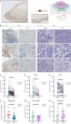
Upregulated expression of cathepsin L in gastric cancer patients with peritoneal metastasis. A: The general level of cathepsin L (CTSL) expression among different cancers from the Gene Expression Profiling Interactive Analysis platform in The Cancer Genome Atlas database; B and C: CTSL expression in gastric tumors (n = 408) was significantly greater than that in normal gastric tissues (n = 211) and was associated with tumor stage in the STAD dataset. The error bars represent the SD, aP < 0.05 according to Student’s t test; D: As a differentially expressed gene, CTSL was upregulated in GSE33651 and GSE11896 (log2fc = 1.786, log2fc = 2.162) via GEO2R analysis; E and F: The qPCR analysis of mRNAs extracted from 64 paired gastric tissues from patients at Nanfang Hospital revealed a significant increase in CTSL expression in tumor tissues (n = 64), P = 0.0412, especially in the group with metastasis (n = 12), P = 0.0434. The data points are presented as the means ± SD. The error bars represent the SD. aP < 0.05 according to Student’s t test; bP < 0.01 according to Student’s t test; G and H: Representative immunohistochemistry (IHC) images of tumor and paracancerous normal sections stained for CTSL; scale bar, 100 μm. The quantification of positively stained cells in the sections via Image J was performed via the geometric mean of 3 representative views from each section; the 53 sections from Nanfang Hospital were used in total. The error bars represent the SD; cP < 0.001 according to Student’s t test; I-K: Representative IHC images of tumor sections from patients with or without peritoneal metastasis stained for CTSL; the error bars represent the SD; aP < 0.05; cP < 0.001 according to Student’s t test. The quantification of positively stained cells in the sections was the same as above for 53 sections from Nanfang Hospital (with metastasis: 18 cases; without metastasis: 35 cases) and 39 sections from the Central Hospital of Wuhan (with metastasis: 9 cases; without metastasis: 30 cases) were used for each group. T: Tumor; N: Normal; PM: Peritoneal metastasis; CTSL: Cathepsin L.
Figure 2
Figure 2
Cathepsin L is more localized in the gastric tumor margin than in the bulk. A: Example diagram of the method for classifying tumor sections stained for cytokeratin, which defines the 2-mm-wide area away from the edge of the tumor as the tumor margin; scale bar, 500 μm; B-E: Representative immunohistochemistry images of the margin and bulk of each tumor section from patients with or without peritoneal metastasis stained for cathepsin L; scale bar, 50 μm. The number of positive cells in the tumor sections was quantified via Image J via the geometric mean of 3 representative views from each section. The data points are presented as the means ± SD. The error bars represent the SD; cP < 0.001; bP < 0.01 according to Student’s t test; F-H: Significant analysis of positive cell counts according to the metastasis state in the tumor margin or bulk region by Image J via the geometric mean of 3 representative views from each section. The data points are presented as the means ± SD. The error bars represent the SD; NS: Not significant, P > 0.05; aP < 0.05; cP < 0.001 according to Student’s t test. CTSL: Cathepsin L; PM: Peritoneal metastasis.
Figure 3
Figure 3
Cathepsin L is critical for macrophages to promote gastric tumor invasion and migration via epithelial-mesenchymal transition. A: Representative immunohistochemistry images of multinucleated giant cells stained for cathepsin L (CTSL) in gastric tumor sections. Magnification, 40 ×; B: Immunofluorescence analysis of CTSL and CD68 in the tumor tissue of gastric cancer patients; scale bar, 20 mm; C: The qPCR and Western blot analysis of shCTSL-treated THP-1 cells. The error bars represent the SD; NS: P > 0.05; cP < 0.001; bP < 0.01 according to one-way analysis of variance (ANOVA); D: Invasion assay of the gastric cancer cell lines MKN45 and MGC803 cocultured with shCTSL or shNC THP-1 cells. Magnification, 20 ×. The chemotactic cells that migrated through the Matrigel in the five views of each group were quantified manually. The error bars represent the SD; bP < 0.01; cP < 0.001; dP < 0.0001 according to one-way ANOVA; E: Wound healing assay of the gastric cancer cell lines MKN45 and MGC803 cocultured with shCTSL or shNC THP-1 cells for 48 hours. Magnification, 4 ×. The wound closure area was quantified via Image J via data from 3 independent experiments. The error bars represent the SD; bP < 0.01; according to Student’s t test; F: Western blot analysis of E-cadherin levels in various gastric cancer cell lines; G: Western blot analysis of epithelial-mesenchymal transition-related proteins (E-cadherin, β-catenin, N-cadherin, and Snail) in MKN45 and MGC803 cells cocultured with shCTSL or shNC-transfected THP-1 cells for 48 hours. Blots from three independent experiments were quantified via Image J. The error bars represent the SD; cP < 0.001; bP < 0.01; aP < 0.05 according to one-way ANOVA. CTSL: Cathepsin L; DAPI: 4',6-diamidino-2-phenylindole.
Figure 4
Figure 4
Cathepsin L + macrophages exert orienting effects on polarization. A: Multiplied immunohistochemistry (mIHC) analysis of cathepsin L (CTSL), CD68, and CDD163 in human gastric tumor sections; scale bar, 20 μm; B: Correlation analysis between CTSL and macrophage markers (CD68 or CD163) via Pearson’s R value measured by Image J; |R| ≤ 1; C and D: Representative mIHC images stained for CTSL, CD68, and CD163 in the tumor margin and bulk of human gastric tissues; magnification, 20 ×, 80 ×; E: Immunofluorescence analysis of CD163 and CD86 expression in shCTSL-transfected or shNC-transfected THP-1 cells; scale bar, 20 μm; F: Quantitative real-time polymerase chain reaction analysis of mRNAs extracted from shCTSL-treated or shNC-treated macrophages induced into the M0/M1/M2 state in vitro; the results revealed increased M1 and decreased M2 marker mRNA levels in shCTSL-treated macrophages. The error bars represent the SD; cP < 0.001; bP < 0.01; aP < 0.05 according to one-way analysis of variance. CTSL: Cathepsin L; DAPI: 4',6-diamidino-2-phenylindole; TNF-α: Tumor necrosis factor-α; IL: Interleukin; Arg-1: Arginase 1.
Figure 5
Figure 5
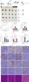
Cathepsin L knockdown impairs macrophage-induced gastric cancer tumorigenesis in vivo. A: Experimental design of the animal study. Wild type male BALB/c nude mice had MKN45 cells implanted into the subcutaneous space and were mixed with either sh cathepsin L (CTSL) or shNC tumor-associated macrophage (TAM); B: Morphological characteristics of tumors in the MKN45 + shNC TAM, MKN45 + shCTSL TAM, MKN45 alone, and shNC TAM groups; C: Volume of tumor growth at the indicated time points over 3 days. The error bars represent the SD; D: Tumor weight and volume ex vivo. The error bars represent the SD. bP < 0.01; aP < 0.05 according to one-way analysis of variance (ANOVA); E: Immunohistochemistry analysis of mouse tumor sections from different groups stained for CTSL, E-cadherin, CD68, and Ki67 proteins. Magnification, 40 ×. Quantification of E-cadherin, CTSL, and CD68 protein expression was performed via Image J. The error bars represent the SD. NS: Not significant, P > 0.05; aP < 0.05; bP < 0.01; cP < 0.001 according to one-way ANOVA. TAM: Tumor-associated macrophage; HE: Haematoxylin and eosin; CTSL: Cathepsin L.

Similar articles

Cited by

References

    1. Cortés-Guiral D, Hübner M, Alyami M, Bhatt A, Ceelen W, Glehen O, Lordick F, Ramsay R, Sgarbura O, Van Der Speeten K, Turaga KK, Chand M. Primary and metastatic peritoneal surface malignancies. Nat Rev Dis Primers. 2021;7:91. - PubMed
    1. Koemans WJ, Luijten JCHBM, van der Kaaij RT, Grootscholten C, Snaebjornsson P, Verhoeven RHA, van Sandick JW. The metastatic pattern of intestinal and diffuse type gastric carcinoma - A Dutch national cohort study. Cancer Epidemiol. 2020;69:101846. - PubMed
    1. Garg M. Emerging roles of epithelial-mesenchymal plasticity in invasion-metastasis cascade and therapy resistance. Cancer Metastasis Rev. 2022;41:131–145. - PubMed
    1. Lankelma JM, Voorend DM, Barwari T, Koetsveld J, Van der Spek AH, De Porto AP, Van Rooijen G, Van Noorden CJ. Cathepsin L, target in cancer treatment? Life Sci. 2010;86:225–233. - PubMed
    1. Chung SM. Variant cathepsin L activity from gastric cancer tissue. Jpn J Cancer Res. 1990;81:813–819. - PMC - PubMed

MeSH terms