Ankle and foot deformities and malformations in proximal femoral focal deficiency
- PMID: 39713180
- PMCID: PMC11656460
- DOI: 10.1177/18632521241301942
Ankle and foot deformities and malformations in proximal femoral focal deficiency
Abstract
Purpose: To describe foot abnormalities in proximal femoral focal deficiency and their correlation to the severity.
Methods: Eighty-nine extremities in 87 patients were evaluated between 1996 and 2020 clinically and radiologically. Fibula length, ankle shape, tarsal coalitions, and the number of foot rays were recorded. Extremities with proximal femoral focal deficiency were classified according to Pappas and divided into severe (classes II and V), medium severe (classes III and IV), and mild groups (classes VII, VIII, and IX).
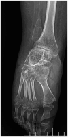
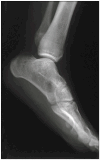
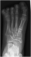
Results: The fibula was short in 89% and absent in 11% of cases. An absent fibula occurred mostly in severe class III and only in 4% of mild grades (statistically significant, p = 0.004). The valgus ankle joint prevailed in 82% of cases. Spherical ankle joints (18% of cases) were associated in all cases with a tarsal coalition. Tarsal coalitions occurred in 14.6% and were present in all classes except class IV. Five ray feet were found in 83% of cases, four ray feet were found in 16%, and three ray feet in one extremity. Reduction in the number of foot rays occurred more commonly in association with fibular aplasia (30%).
Conclusions: Abnormalities of the fibula and ankle joint represent a constant part of proximal femoral focal deficiency, whereas tarsal coalition and a reduction of foot rays do not. The severity of foot abnormalities does not correlate to the severity of proximal femoral focal deficiency but does with fibular aplasia.
Keywords: Foot; fibula deficiency; lateral ray deficiency; proximal femoral focal deficiency; subtalar synostosis.
© The Author(s) 2024.
Conflict of interest statement
The author(s) declared no potential conflicts of interest with respect to the research, authorship, and/or publication of this article.
Figures


Similar articles
-
[Synostosis and tarsal coalitions in children. A study of 68 cases in 47 patients].Rev Chir Orthop Reparatrice Appar Mot. 1994;80(3):252-60. Rev Chir Orthop Reparatrice Appar Mot. 1994. PMID: 7899645 French.
-
Talocalcaneal coalition in patients who have fibular hemimelia or proximal femoral focal deficiency. A comparison of the radiographic and pathological findings.J Bone Joint Surg Am. 1994 Sep;76(9):1363-70. doi: 10.2106/00004623-199409000-00011. J Bone Joint Surg Am. 1994. PMID: 8077266
-
The Nature of Foot Ray Deficiency in Congenital Fibular Deficiency.J Pediatr Orthop. 2017 Jul/Aug;37(5):332-337. doi: 10.1097/BPO.0000000000000646. J Pediatr Orthop. 2017. PMID: 26356313
-
Management of Complex Tarsal Coalition in Children.Foot Ankle Clin. 2021 Dec;26(4):941-954. doi: 10.1016/j.fcl.2021.07.012. Epub 2021 Aug 30. Foot Ankle Clin. 2021. PMID: 34752245 Review.
-
[The treatment of congenital foot abnormalities].Z Orthop Ihre Grenzgeb. 1989 Jan-Feb;127(1):3-14. doi: 10.1055/s-2008-1040081. Z Orthop Ihre Grenzgeb. 1989. PMID: 2655332 Review. German.
Cited by
-
The Significance and Limitations of Pre- and Postnatal Imaging in the Diagnosis and Management of Proximal Focal Femoral Deficiency.Diagnostics (Basel). 2025 May 22;15(11):1302. doi: 10.3390/diagnostics15111302. Diagnostics (Basel). 2025. PMID: 40506874 Free PMC article.
References
-
- Hillmann JS, Mesgarzadeh M, Revesz G, et al.. Proximal femoral focal deficiency: radiologic analysis of 49 cases. Radiology 1987; 165: 769–773. - PubMed
-
- Manner HM, Radler C, Ganger R, et al.. Dysplasia of the cruciate ligaments: radiographic assessment and classification. J Bone Joint Surg Am 2006; 88: 130–137. - PubMed
-
- Chomiak J, Podskubka A, Dungl P, et al.. Cruciate ligaments in proximal femoral focal deficiency: arthroscopic assessment. J Pediatr Orthop 2012; 32(1): 21–28. - PubMed
-
- Grogan DP, Holt GR, Ogden JA. Talocalcaneal coalition in patients who have fibular hemimelia or proximal femoral focal deficiency. a comparison of the radiographic and pathological findings. J Bone Joint Surg Am 1994; 76: 1363–1370. - PubMed
-
- Stanitski DF, Stanitski CL. Fibular hemimelia: a new classification system. J Pediatr Orthop 2003; 23(1): 30–34. - PubMed
LinkOut - more resources
Full Text Sources

