An integrated transcriptomic analysis of brain aging and strategies for healthy aging
- PMID: 39713269
- PMCID: PMC11659761
- DOI: 10.3389/fnagi.2024.1450337
An integrated transcriptomic analysis of brain aging and strategies for healthy aging
Abstract
Background: It is been noted that the expression levels of numerous genes undergo changes as individuals age, and aging stands as a primary factor contributing to age-related diseases. Nevertheless, it remains uncertain whether there are common aging genes across organs or tissues, and whether these aging genes play a pivotal role in the development of age-related diseases.
Methods: In this study, we screened for aging genes using RNAseq data of 32 human tissues from GTEx. RNAseq datasets from GEO were used to study whether aging genes drives age-related diseases, or whether anti-aging solutions could reverse aging gene expression.
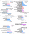
Results: Aging transcriptome alterations showed that brain aging differ significantly from the rest of the body, furthermore, brain tissues were divided into four group according to their aging transcriptome alterations. Numerous genes were downregulated during brain aging, with functions enriched in synaptic function, ubiquitination, mitochondrial translation and autophagy. Transcriptome analysis of age-related diseases and retarding aging solutions showed that downregulated aging genes in the hippocampus further downregulation in Alzheimer's disease but were effectively reversed by high physical activity. Furthermore, the neuron loss observed during aging was reversed by high physical activity.
Conclusion: The downregulation of many genes is a major contributor to brain aging and neurodegeneration. High levels of physical activity have been shown to effectively reactivate these genes, making it a promising strategy to slow brain aging.
Keywords: aging gene; brain aging; neurodegenerative diseases; retard aging; transcriptome.
Copyright © 2024 Liu, Nie, Wang, Chen, Zeng, Shu and Huang.
Conflict of interest statement
Authors XN, PS were employed by Shenzhen Hujia Technology Co., Ltd. The remaining authors declare that the research was conducted in the absence of any commercial or financial relationships that could be construed as a potential conflict of interest.
Figures


Similar articles
-
The Transcriptional Landscape of Microglial Genes in Aging and Neurodegenerative Disease.Front Immunol. 2019 Jun 4;10:1170. doi: 10.3389/fimmu.2019.01170. eCollection 2019. Front Immunol. 2019. PMID: 31214167 Free PMC article.
-
Transcriptomic Changes Highly Similar to Alzheimer's Disease Are Observed in a Subpopulation of Individuals During Normal Brain Aging.Front Aging Neurosci. 2021 Dec 1;13:711524. doi: 10.3389/fnagi.2021.711524. eCollection 2021. Front Aging Neurosci. 2021. PMID: 34924992 Free PMC article.
-
Downregulation of glial genes involved in synaptic function mitigates Huntington's disease pathogenesis.Elife. 2021 Apr 19;10:e64564. doi: 10.7554/eLife.64564. Elife. 2021. PMID: 33871358 Free PMC article.
-
Aging Brain, Dementia and Impaired Glymphatic Pathway: Causal Relationships.Psychiatr Danub. 2023 Oct;35(Suppl 2):236-244. Psychiatr Danub. 2023. PMID: 37800234 Review.
-
Phosphorylated tau as a toxic agent in synaptic mitochondria: implications in aging and Alzheimer's disease.Neural Regen Res. 2022 Aug;17(8):1645-1651. doi: 10.4103/1673-5374.332125. Neural Regen Res. 2022. PMID: 35017410 Free PMC article. Review.
Cited by
-
Mechanistic insights into the anti-aging effects of Crocus sativus in a D-Gal-induced in vitro neural senescence model.PLoS One. 2025 Jul 16;20(7):e0320572. doi: 10.1371/journal.pone.0320572. eCollection 2025. PLoS One. 2025. PMID: 40668823 Free PMC article.
References
-
- Berchtold N. C., Prieto G. A., Phelan M., Gillen D. L., Baldi P., Bennett D. A., et al. (2019). Hippocampal gene expression patterns linked to late-life physical activity oppose age and AD-related transcriptional decline. Neurobiol. Aging 78 142–154. 10.1016/j.neurobiolaging.2019.02.012 - DOI - PMC - PubMed
LinkOut - more resources
Full Text Sources

