. 2024 Dec 20;12(4):69-82.
doi: 10.14252/foodsafetyfscj.D-24-00010. eCollection 2024 Dec.

Construction of a CYP2J2-Template System and Its Application for Ligand Metabolism Prediction

Affiliations

Construction of a CYP2J2-Template System and Its Application for Ligand Metabolism Prediction

Yasushi Yamazoe et al. Food Saf (Tokyo). .

Abstract

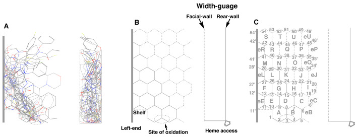
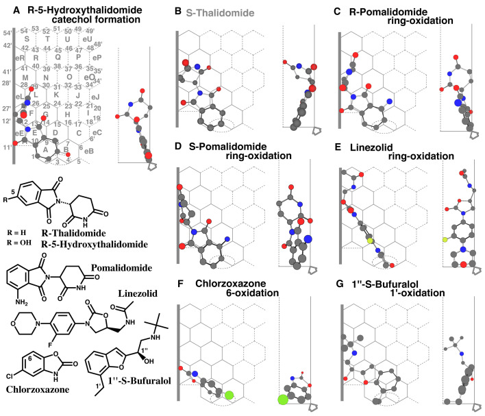
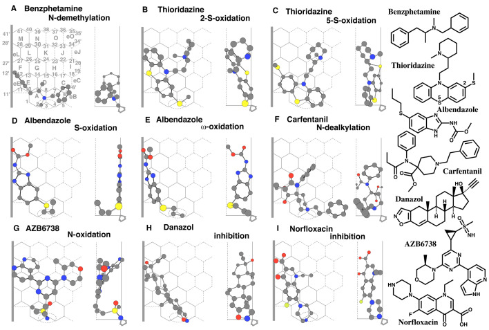
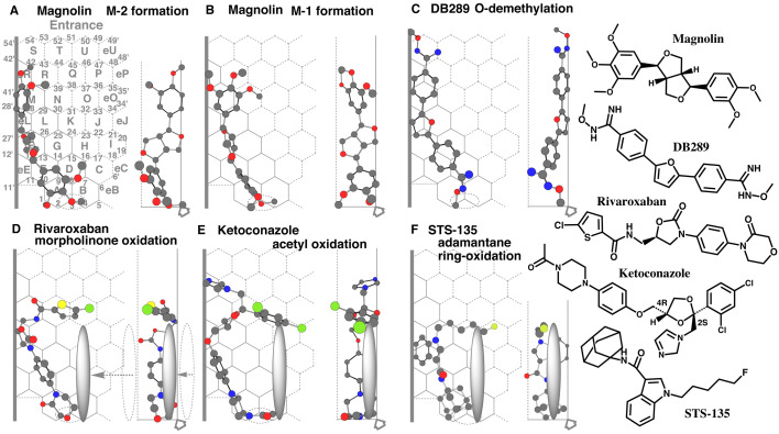
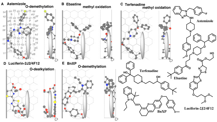
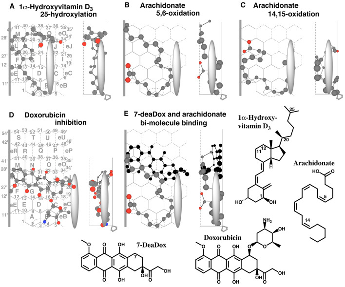
A Template system for the understanding of human CYP2J2-mediated reactions was constructed from the assembly of the ligands with the introduction of ideas of allowable width, Trigger-residue and the residue-initiated movement of ligands in the active site, which were in common with other Template* systems for human CYP1A1, CYP1A2, CYP2B6, CYP2C8, CYP2C9, CYP2C18, CYP2C19, CYP2E1, CYP3A4, CYP3A5, and CYP3A7 (Drug Metab Pharmacokinet 2016, 2017, 2019, 2020, 2021, 2022, 2023, 2024, and in press 2024). CYP2J2 system also includes ideas of bi-molecule binding of ligands on the Template. From their placements on the Template and rules for interaction modes, verifications of good and poor substrates, regio/stereo-selectivity, and inhibitory interaction became available faithfully for these ligands. The refined CYP2J2-Template system will thus offer reliable estimations of this human CYP catalysis toward ligands of diverse structures, together with their deciphering information to lead to judgments.

Keywords: CYP2J2-mediated metabolism; fused-grid Template; ligand immobilization; poor and good substrates; simulation of ligand-interaction on Template..


Conflict of interest statement

Conflict of interest: The authors declare no conflict of interest.

Figures

Fig. 1.
Fig. 1.
Construction of Template 3D structures of CYP2J2 substrates described in Table 1 were assembled considering that their sites of metabolism came close on a plane. The site of metabolism (Site of oxidation) is shown as a dotted oval. Their 90°-rotated structures are shown on the right side and included in a column. A vertical bar was set at the left end (A). An overlapped area was extracted as a hexagonal-grid Template. Site of oxidation is shown as a dotted oval. Two structures, termed Shelf and Left-end are located on the left side borders. The allowable width termed Width-gauge was set between Facial-wall (dotted line) and Rear-wall (solid line). A gray-colored arrow indicated the direction of heme access (B). The Ring names and Position numbers were assigned in alphabetical order (A-U) and from 1 to 54, respectively. Parts of side-end regions were also assigned as eB, eC, eE, eJ, eL, eO, eP, eR, and eU (C).
Fig. 2.
Fig. 2.
Placements of small-sized ligands Placements of R-5-hydroxythalidomide catechol formation (A), of ring oxidations of S-thalidomide (B) and R- (C) and S-pomalidomide (D), of linezolid ring-oxidation (E), of chlorzoxazone 6-oxidation (F), and 1”-S-bufuralol 1’-oxidations (G), are shown as cylindrical shapes of 3D structures on full-size (A) and trimmed Templates (B-G). Ligands are also indicated as 2D structures on the left bottom parts with parts of their chemical positions. Functional and non-functional placements are distinguished with dark- and gray-colored structure names, respectively.
Fig. 3.
Fig. 3.
Ligand interaction with Trigger-residue Placements of benzphetamine N-demethylation (A), of thioridazine 2-S- (B) and 5-S-oxidations (C), of albendazole S- (D) and ω-oxidations (E), of carfentanil N-dealkylation (F), of AZB6738 N-oxidation (G), of danazol inhibition (H), and also of norfloxacin inhibition (I) are shown as cylindrical shapes of 3D structures on trimmed Template. Ligands are also indicated as 2D structures with parts of their chemical positions.
Fig. 4.
Fig. 4.
Interactions of slender shape ligands with Left-end and Shelf Placements of magnolin M-2 (A) and M-1 formations (B), of DB289 O-demethylation (C), of rivaroxaban morpholinone ring-oxidation (D), of ketoconazole acetyl-oxidation (E), of STS-135 adamantane ring-oxidation (F) are shown as cylindrical shapes of 3D structures. Ligands are also indicated as 2D structures on the right. The movement of Trigger-residue from the initial position (dotted pillar symbol) to the ligand contact (gray pillar symbol) is shown in D.
Fig. 5.
Fig. 5.
Placements of diagnostic substrates of CYP2J2 and pillar-shape Trigger-residue Placements of astemizole O-demethylation (A), of ebastine methyl-oxidation (B), of terfenadine methyl-oxidation (C), of luciferin-2j2/4F12 O-dealkylation (D), and of BnXP O-demethylation (E) are shown as cylindrical shapes of 3D structures. Ligands are also indicated as 2D structures on the right. Thick arrow at the bottom of Width-gauge indicates the access direction of heme-oxygen atom.
Fig. 6.
Fig. 6.
Placements of endobiotics Placements of 1α-hydroxyvitamin D3 25-oxidation (A), of arachidonate 5,6- (B) and 14,15-oxidations (C), of doxorubicin inhibition (D), of bi-molecule binding of 7-deaDox and arachidonate for the 5,6-oxidation (E) are shown as cylindrical shapes of 3D structures. Trigger molecule (7-deaDox) is shown in dark color. Ligands are also indicated as 2D structures with parts of their position numbers. Thick arrow at the bottom of Width-gauge indicates the access direction of heme-oxygen atom.

Similar articles

References

    1. Wu S,Moomaw CR,Tomer KB,Falck JR,Zeldin DC. Molecular cloning and expression of CYP2J2, a human cytochrome P450 arachidonic acid epoxygenase highly expressed in heart. J Biol Chem. 1996; 271(7): 3460–3468. 10.1074/jbc.271.7.3460 - DOI - PubMed
    1. Xu M,Ju W,Hao H,Wang G,Li P. Cytochrome P450 2J2: distribution, function, regulation, genetic polymorphisms and clinical significance. Drug Metab Rev. 2013; 45(3): 311–352. 10.3109/03602532.2013.806537 - DOI - PubMed
    1. Enayetallah AE,French RA,Grant DF. Distribution of soluble epoxide hydrolase, cytochrome P450 2C8, 2C9 and 2J2 in human malignant neoplasms. J Mol Histol. 2006; 37(3-4): 133–141. 10.1007/s10735-006-9050-9 - DOI - PubMed
    1. Matsumoto S,Yamazoe Y. Involvement of multiple human cytochromes P450 in the liver microsomal metabolism of astemizole and a comparison with terfenadine. Br J Clin Pharmacol. 2001; 51(2): 133–142. 10.1111/j.1365-2125.2001.01292.x - DOI - PMC - PubMed
    1. Matsumoto S,Hirama T,Matsubara T,Nagata K,Yamazoe Y. Involvement of CYP2J2 on the intestinal first-pass metabolism of antihistamine drug, astemizole. Drug Metab Dispos. 2002; 30(11): 1240–1245. 10.1124/dmd.30.11.1240 - DOI - PubMed