

This is a preprint.
A FRET assay to quantitate levels of the human β-cardiac myosin interacting heads motif based on its near-atomic resolution structure
- PMID: 39713291
- PMCID: PMC11661104
- DOI: 10.1101/2024.12.05.626936
A FRET assay to quantitate levels of the human β-cardiac myosin interacting heads motif based on its near-atomic resolution structure
Abstract
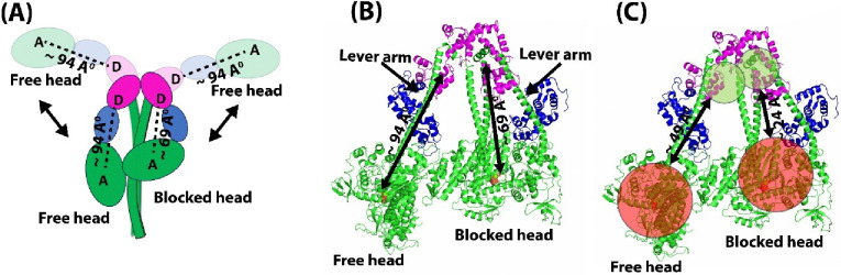
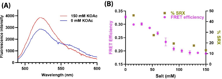
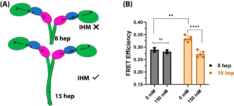
In cardiac muscle, many myosin molecules are in a resting or "OFF" state with their catalytic heads in a folded structure known as the interacting heads motif (IHM). Many mutations in the human β-cardiac myosin gene that cause hypertrophic cardiomyopathy (HCM) are thought to destabilize (decrease the population of) the IHM state. The effects of pathogenic mutations on the IHM structural state are often studied using indirect assays, including a single-ATP turnover assay that detects the super-relaxed (SRX) biochemical state of myosin functionally. Here we develop and use a fluorescence resonance energy transfer (FRET) based sensor for direct quantification of the IHM state in solution. The FRET sensor was able to quantify destabilization of the IHM state in solution, induced by (a) increasing salt concentration, (b) altering proximal S2 tail length, or (c) introducing the HCM mutation P710R, as well as stabilization of the IHM state by introducing a dilated cardiomyopathy-causing mutation (E525K). Our FRET sensor conclusively showed that these perturbations indeed alter the structural IHM state. These results establish that the structural IHM state is one of the structural correlates of the biochemical SRX state in solution.
Conflict of interest statement
COMPETING INTEREST STATEMENT J.A.S. is cofounder and on the Scientific Advisory Board of Cytokinetics, Inc., a company developing small-molecule therapeutics for the treatment of hypertrophic cardiomyopathy. J.A.S. is cofounder and CEO, and K.M.R. is cofounder and Research and Clinical Advisor, of Kainomyx, Inc., a company developing small molecule therapeutics targeting cytoskeletal proteins for a variety of clinical conditions. DDT is a founder and serves as executive officer for Photonic Pharma LLC (PP), a company involved in the early phase of drug discovery, for treatment of diseases involving myosin and other proteins. OR is the sole proprietor of Editing Science LLC, which had no role in this study.
Figures

References
-
- Adhikari A. S., Kooiker K. B., Sarkar S. S., Liu C., Bernstein D., Spudich J. A., & Ruppel K. M. (2016). Early-onset hypertrophic cardiomyopathy mutations significantly increase the velocity, force, and actin-activated ATPase activity of human β-cardiac myosin. Cell Reports, 17(11), 2857–2864. - PMC - PubMed
-
- Adhikari A. S., Trivedi D. V., Sarkar S. S., Song D., Kooiker K. B., Bernstein D., Spudich J. A., & Ruppel K. M. (2019). β-Cardiac myosin hypertrophic cardiomyopathy mutations release sequestered heads and increase enzymatic activity. Nature Communications, 10(1), 1–10. 10.1038/s41467-019-10555-9 - DOI - PMC - PubMed
-
- Anderson R. L., Trivedi D. V., Sarkar S. S., Henze M., Ma W., Gong H., Rogers C. S., Gorham J. M., Wong F. L., Morck M. M., Seidman J. G., Ruppel K. M., Irving T. C., Cooke R., Green E. M., & Spudich J. A. (2018). Deciphering the super relaxed state of human β-cardiac myosin and the mode of action of mavacamten from myosin molecules to muscle fibers. Proceedings of the National Academy of Sciences of the United States of America, 115(35), E8143–E8152. 10.1073/pnas.1809540115 - DOI - PMC - PubMed
Publication types
Grants and funding
LinkOut - more resources
Full Text Sources