[Preprint]. 2024 Dec 11:2024.12.09.627393.
doi: 10.1101/2024.12.09.627393.

The CECR2 bromodomain displays distinct binding modes to select for acetylated histone proteins versus non-histone ligands

Affiliations

The CECR2 bromodomain displays distinct binding modes to select for acetylated histone proteins versus non-histone ligands

Margaret Phillips et al. bioRxiv. .

Abstract

The cat eye syndrome chromosome region candidate 2 (CECR2) protein is an epigenetic regulator involved in chromatin remodeling and transcriptional control. The CECR2 bromodomain (CECR2-BRD) plays a pivotal role in directing the activity of CECR2 through its capacity to recognize and bind acetylated lysine residues on histone proteins. This study elucidates the binding specificity and structural mechanisms of CECR2-BRD interactions with both histone and non-histone ligands, employing techniques such as isothermal titration calorimetry (ITC), nuclear magnetic resonance (NMR) spectroscopy, and a high-throughput peptide assay. The CECR2-BRD selectively binds acetylated histone H3 and H4 ligands, exhibiting a preference for multi-acetylated over mono-acetylated targets. The highest affinity was observed for tetra-acetylated histone H4. Neighboring post-translational modifications, including methylation and phosphorylation, modulate acetyllysine recognition, with significant effects observed for histone H3 ligands. Additionally, this study explored the interaction of the CECR2-BRD with the acetylated RelA subunit of NF-κB, a pivotal transcription factor in inflammatory signaling. Dysregulated NF-κB signaling is implicated in numerous pathologies, including cancer progression, with acetylation of RelA at lysine 310 (K310ac) being critical for its transcriptional activity. Recent evidence linking the CECR2-BRD to RelA suggests it plays a role in inflammatory and metastatic pathways, underscoring the need to understand the molecular basis of this interaction. We found the CECR2-BRD binds to acetylated RelA with micromolar affinity, and uses a distinctive binding mode to recognize this non-histone ligand. These results provide new insight on the role of CECR2 in regulating NF-κB-mediated inflammatory pathways. Functional mutagenesis of critical residues, such as Asn514 and Asp464, highlight their roles in ligand specificity and binding dynamics. Notably, the CECR2-BRD remained monomeric in solution and exhibited differential conformational responses upon ligand binding, suggesting adaptive recognition mechanisms. Furthermore, the CECR2-BRD exclusively interacts with nucleosome substrates containing multi-acetylated histones, emphasizing its role in transcriptional activation within euchromatic regions. These findings position the CECR2-BRD as a key chromatin reader and a promising therapeutic target for modulating transcriptional and inflammatory processes, particularly through the development of selective bromodomain inhibitors.


Conflict of interest statement

COMPETING INTERESTS: EpiCypher (M.R.M. and L.K.) is a commercial developer of the dCypher peptide-binding platform used in this study.

Figures

Figure 1.
Figure 1.. CECR2 bromodomain broadly recognizes acetylated histone peptides.
Using the dCypher assay, a panel of 287 histone peptides was probed with 26 nM GST-CECR2-BRD to assess binding interactions. The X-axis represents the identity and amino acid coverage of each histone peptide, while the Y-axis measures the Alpha Counts indicating binding strength. A) CECR2 shows robust binding across multi-acetylated H2A, H2B, H3, and H4 histone peptides. B-E) Detailed binding profiles of CECR2 with single and multi-acetyl peptides of H2A, H2B, H3, and H4, respectively. F) CECR2 demonstrates tolerance for methylation on H2A, H3, and H4 peptides, maintaining binding strength in the presence of adjacent methylated residues. G) Phosphorylated residues negatively impact certain CECR2 binding to acetylated peptides. All values shown are the averages ± standard deviation (n = 2).
Figure 2.
Figure 2.. CECR2 bromodomain equally binds acetyl, propionyl, and butyryl.
The binding of CECR2-BRD was examined using dCypher and acylated histone peptides on H3 and H4. A) CECR2 shows robust binding to acetyl, propionyl, and butyryl modifications at various lysine residues on the H3 and H4. B) Investigation of multiple acyl modifications at H3K9 reveals CECR2-BRD only binding acetyl (ac, propionyl (pr), and butyryl (bu) residues while no binding was detectable to crotonyl (cr), beta-hydroxybutyryl (bhb), succinyl (su), valeryl (vl), or hexanoyl (hxo). All values shown are the averages ± standard deviation (n = 2).
Figure 3:
Figure 3:. Normalized Chemical Shift Perturbation (CSP) plots for histone H3/H4 interaction with the CECR2 bromodomain.
In the histogram plots above, the CECR2 bromodomain residues are along the x-axis, while their chemical shift (ppm) induced by adding histone ligands in a 1:10 (protein: peptide) molar ratio (except 1:5 for CECR2:H4K5acK8acK12acK16ac), along the y-axis. CSPs induced by different histone ligands are color-coded in the following order: red for H3K9ac, blue for H3K4acK9acK14acK18ac, green for H4K5ac, and orange for H4K5acK8acK12acK16ac ligand respectively. Prolines are marked by an asterisk (*), the conserved asparagine (N514) by an arrow, and the # symbol highlights unassigned residues. The dotted line represents the 1 standard deviation cut-off. The secondary structure containing four alpha-helices and ZA/BC loop regions of the CECR2 bromodomain are shown above the histogram.
Figure 4.
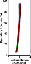
Figure 4.. Sedimentation velocity analysis of the CECR2 bromodomain alone and in complex with acetylated histone H3 and H4 ligands.
The sedimentation velocity distribution van Holde-Weischet overlay plot includes: CECR2-BRD apo control (purple), CECR2-BRD + H3K4acK9acK14acK18ac 1:2 (blue), CECR2-BRD + H3K4acK9acK14acK18ac 1:2 (cyan), CECR2-BRD + H3K4acK9acK14acK18ac 1:0.5 (orange), CECR2-BRD + H3K14ac 1:0.5 (red), CECR2-BRD + H4K5acK8acK12acK16ac 1:0.5 (green), CECR2-BRD + H4K5acK8acK12acK16ac 1:1 (yellow), CECR2-BRD + H4K8ac 1:0.5 (brown).
Figure 5:
Figure 5:. Comparing the Binding Pocket interactions of Acetylated RelA with Acetylated Histones.
A) Normalized Chemical Shift Perturbation (CSP) plot for mono-acetylated RelA versus the tetra-acetylated histone H3/H4 ligands. CECR2-BRD residues are along the x-axis, while their chemical shift induced by adding various ligands is represented along the y-axis (D ppm). CSPs induced by mono-acetylated RelAK310ac (305–315) are shown in purple (1:10 molar ratio of CECR2: RelA), tetra-acetylated H4K5acK8acK12acK16ac (1–24) in orange (1:5 molar ratio of CECR2: H4), and H3K4acK9acK14acK18ac (1–24) in blue (1:10 molar ratio of CECR2: H3). Residue D464 was broadened beyond detection upon RelAK310ac addition and is highlighted in red asterisk. Prolines are marked by black asterisks (*), conserved asparagine N514 by an arrow, and the # symbol highlights unassigned residues. The dotted line represents the standard deviation cut-off of total CSPs. The secondary structure of the CECR2-BRD containing four alpha-helices and ZA/BC loop regions is shown above the histogram. B) Mapping the binding pocket of tetra acetylated H3 and H4 ligands onto the apo crystal structure of the CECR2-BRD (PDB ID: 3NXB). Highlighted in orange are the residues showing CSPs above standard deviation (>1 D ppm) upon ligand addition (1:5 for CECR2:H4K5acK8acK12acK16ac and 1:10 for CECR2:H3K4acK9acK14acK18ac molar ratio). Green residues are assigned, grey unassigned, while black residues denote Prolines. C) Mapping the binding pocket of acetylated RelAK310ac onto the apo crystal structure of the CECR2-BRD (PDB ID: 3NXB). Highlighted in pink are the residues showing CSPs above standard deviation (>1 D ppm) upon ligand addition (1:10 protein to peptide molar ratio).
Figure 6:
Figure 6:. Normalized Chemical Shift Perturbation (CSP) plots illustrating the interaction of DMSO and NVS-CECR2–1 with the CECR2 bromodomain.
Normalized Chemical shifts (Δ ppm) induced by the addition of 3% DMSO are represented in black, while those induced by NVS-CECR2–1 including 3% DMSO) are shown in orange. Prolines are denoted by an asterisk (*), the conserved asparagine N514 by an arrow, and unassigned residues by the # symbol. The dotted line represents the 1 standard deviation cut-off. The secondary structure, comprising four alpha-helices and ZA/BC loop regions of the CECR2-BRD, is delineated above the histogram.
Figure 7.
Figure 7.. CECR2 bromodomain robustly binds multi-acetylated H3 and H4 nucleosomes.
Using the dCypher assay, CECR2 was evaluated against nucleosomes; X-axis represents the concentration of CECR2, while the Y-axis measures the Alpha Counts indicating binding strength. The key identifies each nucleosome. A) CECR2 was assessed against H3K27ac and multi-acetyl H2A and H3 nucleosomes showing strong binding only to H3Tetraac. B) CECR2 interactions to single and multi-acetyl H4 nucleosomes revealed binding only H4Tetraac. All values shown are the averages ± standard deviation (n = 2).

Similar articles

References

    1. Banting G. S.; Barak O.; Ames T. M.; Burnham A. C.; Kardel M. D.; Cooch N. S.; Davidson C. E.; Godbout R.; McDermid H. E.; Shiekhattar R. CECR2, a protein involved in neurulation, forms a novel chromatin remodeling complex with SNF2L. Hum Mol Genet 2005, 14 (4), 513–524. DOI: 10.1093/hmg/ddi048. - DOI - PubMed
    1. Thompson P. J.; Norton K. A.; Niri F. H.; Dawe C. E.; McDermid H. E. CECR2 is involved in spermatogenesis and forms a complex with SNF2H in the testis. J Mol Biol 2012, 415 (5), 793–806. DOI: 10.1016/j.jmb.2011.11.041. - DOI - PubMed
    1. Oppikofer M.; Bai T.; Gan Y.; Haley B.; Liu P.; Sandoval W.; Ciferri C.; Cochran A. G. Expansion of the ISWI chromatin remodeler family with new active complexes. EMBO Rep 2017, 18 (10), 1697–1706. DOI: 10.15252/embr.201744011. - DOI - PMC - PubMed
    1. Wu L.; Zhao G.; Xu S.; Kuang J.; Ming J.; Wu G.; Wang T.; Wang B.; Zhu P.; Pei D.; et al. The nuclear factor CECR2 promotes somatic cell reprogramming by reorganizing the chromatin structure. J Biol Chem 2021, 296, 100022. DOI: 10.1074/jbc.RA120.014598. - DOI - PMC - PubMed
    1. Lee S. K.; Park E. J.; Lee H. S.; Lee Y. S.; Kwon J. Genome-wide screen of human bromodomain-containing proteins identifies Cecr2 as a novel DNA damage response protein. Mol Cells 2012, 34 (1), 85–91. DOI: 10.1007/s10059-012-0112-4. - DOI - PMC - PubMed

Publication types

LinkOut - more resources