[Preprint]. 2024 Dec 12:2024.12.06.627272.
doi: 10.1101/2024.12.06.627272.

Perceptual choice and motor signals in mouse somatosensory cortex

Affiliations

Perceptual choice and motor signals in mouse somatosensory cortex

Genki Minamisawa et al. bioRxiv. .

Abstract

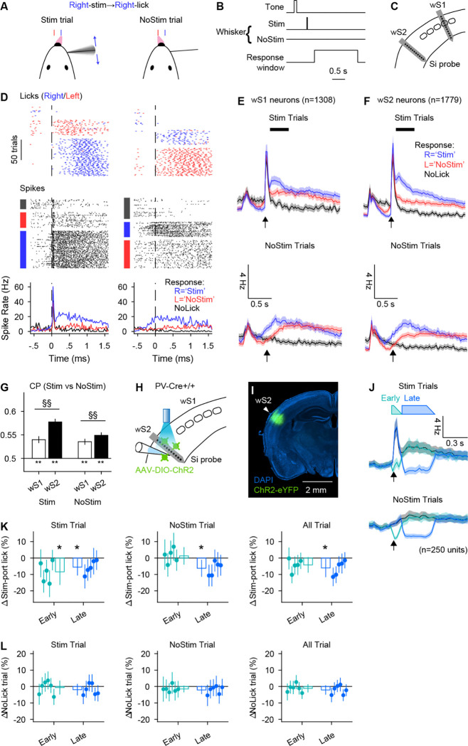
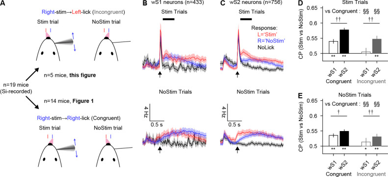
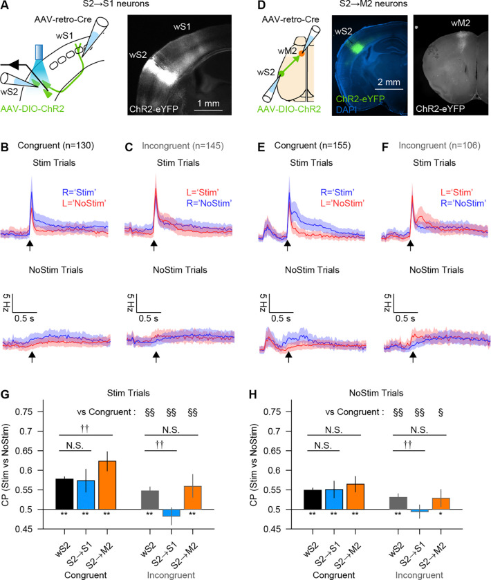
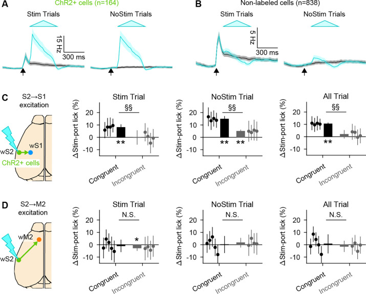
Somatosensory cortex activity relates both to sensation and movement, reflecting their intimate relationship, but the extent and nature of sensory-motor interactions in the somatosensory cortex remain unclear. Here, we investigated perception-related sensory and motor signals in the whisker areas of mouse primary (wS1) and secondary (wS2) somatosensory cortices. We recorded neuronal activity while mice performed a whisker detection task using two alternative lickports, one each to indicate the presence or absence of a whisker deflection on a given trial. One group of mice reported the presence of the whisker stimulus by licking at the port on the same ("congruent") side of the animal as the stimulated whisker, whereas a second group of mice did so by licking at the opposite ("incongruent") side. Activity of single neurons in wS1 and wS2 correlated with perceptual choice. This choice-related activity was enhanced when responding to the congruent side. wS2 neurons projecting along two output pathways-to wS1 or to whisker secondary motor cortex, wM2-also showed choice-related activity, but differed in their dependence on congruence and in the effects of optogenetic manipulation. Thus, somatosensory cortex contains pathway- and action-specific choice-related activity.


Figures

Figure 1.
Figure 1.. Choice- and licking-related activity in wS1 and wS2.
(A) Whisker detection task with Right-stim→Right-lick (congruent) contingency. (B)Time course of the task. (C) Silicon probes were acutely implanted to wS1 and wS2 during the task. (D) Activity of an example neuron in a session. From top to bottom, lick and spike raster plots sorted by trial outcomes, and spike rate histogram triggered by the expected stimulus onset. Left and right columns correspond to data from Stim and NoStim trials, respectively. Lick directions were indicated by colors of the ticks or boxes on the left (Left: red, Right: blue). (E) Spike rate of all silicon probe-recorded wS1 neurons during Stim and NoStim trials. Expected stimulus onsets are indicated by black arrows. Shadings are 95% confidence intervals. Black box above the spike rate plot indicates the analysis window ([0, 500 ms] from the expected stimulus time) for choice probability in panel G. 31 recordings from 13 animals. (F) Same as E for wS2 neurons. 38 recordings from 14 animals. (G) Choice probabilities of Stim- over NoStim-port licking. *, **: p<.05 and <.01, Student’s t-test (null hypothesis: average CP=0); §, §§: p<.05 and 0.01, Two-sample t-test (null hypothesis: CPS1 = CPS2). (H-I) wS2 was inhibited by photo-excitation of PV-positive inhibitory interneurons. (J) Spike rates of wS2 neurons that were not excited by a light stimulation (presumably principal neurons). Expected stimulus onsets are indicated by black arrows. Shadings are 95% confidence intervals. Colored boxes above indicate the time courses of photo-stimuli. (K) Differences in the probability of stim-port licking by wS2 inhibition. Dots and bars correspond to each animal’s data and the average of all animals, respectively. Vertical lines on top of scatter and bar plots indicate 95% CIs. *: p<0.05 and <0.01, student’s t-test (null hypothesis: average=0). N=5 animals. (L) Differences in the probability of NoLick trials by wS2 inhibition. Conventions are the same as panel K. Significant difference from 0 was not observed in any condition. N = 5 animals.
Figure 2.
Figure 2.. Stronger choice-related activity in congruent vs incongruent tasks.
(A) A subset of animals were trained for a whisker detection task with the opposite contingency from Figure 1 (Right-stim→Left-lick, incongruent). (B-C) Spike rate of all silicon probe-recorded wS1 and wS2 neurons under ‘incongruent’ contingency task. 9 and 16 recordings in wS1 and wS2 from 5 animals. (D-E) Choice probabilities of Stim- over NoStim-port licking. Shadings are 95% CI. *, **: p<.05 and <.01, Student’s t-test (null hypothesis: CP=0.5); †, ††: p<.05 and <.01, Two-sample t-test (null hypothesis: CPS1 = CPS2). §, §§: p<.05 and 0.01, Two-sample t-test (null hypothesis: congruent = incongruent in the same neuron and trial types).
Figure 3.
Figure 3.. S2→S1 neurons exhibit choice-related activity specifically in the congruent task.
(A) S2→S1 neurons were labeled with ChR2 by a combination of AAV-retro-Cre and AAV-DIO-ChR2 injections in wS1 and wS2 respectively. (B-C) PSTHs with 95% CI for S2→S1 neurons identified by a reliable response to photo-stimuli. Expected stimulus onset is indicated by an arrow. (D) Same as (A), but for S2→M2 neurons. Axonal projection in wM2 (right panel) indicates a successful labeling of wM2-projecting neurons. (E-F) Same as (B-C), but for S2→M2 neurons. (G-H) Choice probabilities of Stim- over NoStim-port licking. *, **: p<.05 and <.01, student’s t-test (null hypothesis: average=0.5); †, ††: p<.05 and <.01, two-sample t-test (null hypothesis: CP = CPS2 in the same neuron type). §, §§: p<.05 and 0.01, two-sample t-test (null hypothesis: congruent = incongruent in the same neuron type).
Figure 4.
Figure 4.. Excitation of S2→S1 neurons biases choice toward the stimulus-associated port in the congruent task.
(A) PSTHs with 95% CI from identified ChR2+ cells in wS2 (i.e., S2→S1 and S2→M2 neurons). Cyan and black traces correspond to trials with and without a photostimulus. Cyan triangle indicates the timing and amplitude of photo stimulus. 19 sessions from 11 animals. (B) PSTHs of the cells that were not responsive to the photo stimulus in the same recording sessions as (A). (C) Behavioral effect of photo-excitation of S2→S1 neurons during the task. Differences in the probability of Stim-port licking by a photo stimulation for different task contingency (congruent/incongruent) and trial (Stim/NoStim/All) types. Each dot corresponds to each animal’s data, and a bar is the average of all animals in the same conditions. Error bars: 95% CI (for both scatter and bar plots). *, **: p<0.05 and <0.01, student’s t-test (null hypothesis: average=0). §§: p<0.01, two-sample t-test (null hypothesis: congruent = incongruent). (D) Same as (C) but for S2→M2 neurons.

Similar articles

References

    1. Kwon S. E., Yang H., Minamisawa G. & O’Connor D. H. Sensory and decision-related activity propagate in a cortical feedback loop during touch perception. Nat. Neurosci. 19, 1243–1249 (2016). - PMC - PubMed
    1. Sachidhanandam S., Sreenivasan V., Kyriakatos A., Kremer Y. & Petersen C. C. H. Membrane potential correlates of sensory perception in mouse barrel cortex. Nat. Neurosci. 16, 1671–1677 (2013). - PubMed
    1. Yang H., Kwon S. E., Severson K. S. & O’Connor D. H. Origins of choice-related activity in mouse somatosensory cortex. Nat. Neurosci. 19, 127–134 (2016). - PMC - PubMed
    1. Yamashita T. & Petersen C. C. Target-specific membrane potential dynamics of neocortical projection neurons during goal-directed behavior. eLife 5, e15798 (2016). - PMC - PubMed
    1. Oryshchuk A. et al. Distributed and specific encoding of sensory, motor, and decision information in the mouse neocortex during goal-directed behavior. Cell Rep. 43, 113618 (2024). - PubMed

Publication types

LinkOut - more resources