[Preprint]. 2024 Dec 10:2024.12.05.627072.
doi: 10.1101/2024.12.05.627072.

Environmentally-relevant doses of bisphenol A and S exposure in utero disrupt germ cell programming across generations resolved by single nucleus multi-omics

Affiliations

Environmentally-relevant doses of bisphenol A and S exposure in utero disrupt germ cell programming across generations resolved by single nucleus multi-omics

Liang Zhao et al. bioRxiv. .

Update in

Abstract

Background: Exposure to endocrine-disrupting chemicals (EDCs), such as bisphenol A (BPA), disrupts reproduction across generations. Germ cell epigenetic alterations are proposed to bridge transgenerational reproductive defects resulting from EDCs. Previously, we have shown that prenatal exposure to environmentally relevant doses of BPA or its substitute, BPS, caused transgenerationally maintained reproductive impairments associated with neonatal spermatogonial epigenetic changes in male mice. While epigenetic alterations in germ cells can lead to transgenerational phenotypic variations, the mechanisms sustaining these changes across generations remain unclear.

Objectives: This study aimed to systematically elucidate the mechanism of transgenerational inherence by prenatal BPA and BPS exposure in the murine germline from F1 to F3 generations at both transcriptomic and epigenetic levels.

Methods: BPA or BPS with doses of 0 (vehicle control), 0.5, 50, or 1000 μg/kg/b.w./day was orally administered to pregnant CD-1 females (F0) from gestational day 7 to birth. Sperm counts and motility were examined in F1, F2, and F3 adult males. THY1+ germ cells on postnatal day 6 from F1, F2, and F3 males at a dose of 50 μg/kg/b.w./day were used for analysis by single-nucleus (sn) multi-omics (paired snRNA-seq and snATAC-seq on the same nucleus).

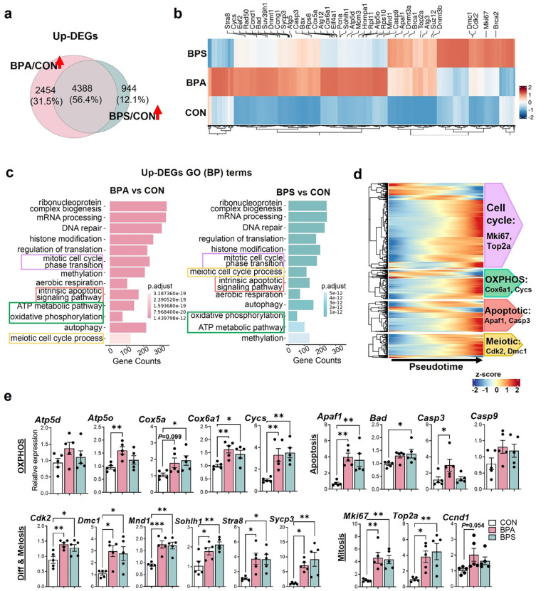
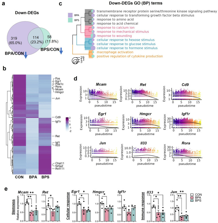
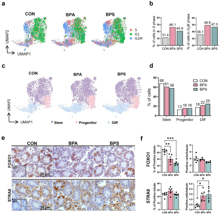
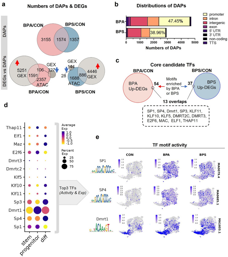
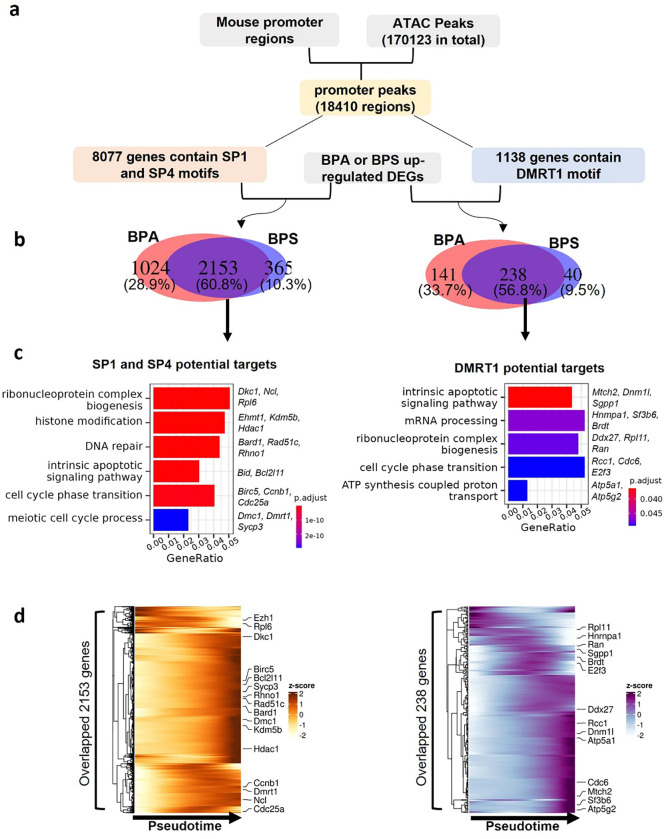
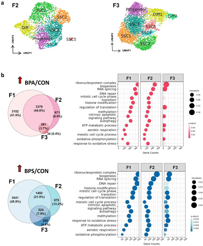
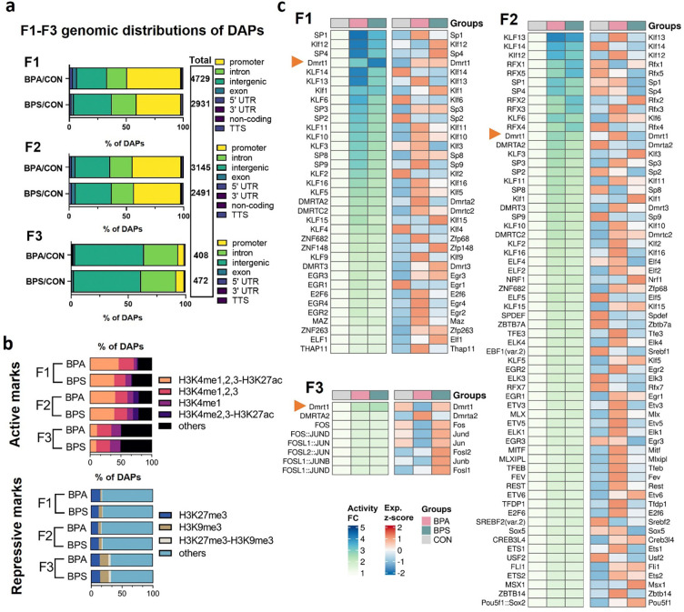
Results: Prenatal exposure to BPA and BPS with 0.5, 50, and 1000 μg/kg/b.w./day reduced sperm counts in mice across F1 to F3 generations. In the F1 neonatal germ cells, ancestral BPA or BPS exposure with 50 μg/kg/b.w./day resulted in increased differentially expressed genes (DEGs) associated with spermatogonial differentiation. It also disrupted the balance between maintaining the undifferentiated and differentiating spermatogonial populations. Differentially accessible peaks (DAPs) by snATAC-seq were primarily located in the promoter regions, with elevated activity of key transcription factors, including SP1, SP4, and DMRT1. Throughout F1-F3 generations, biological processes related to mitosis/meiosis and metabolic pathways were substantially up-regulated in BPA- or BPS-exposed groups. While the quantities of DEGs and DAPs were similar in F1 and F2 spermatogonia, with both showing a significant reduction in F3. Notably, approximately 80% of DAPs in F1 and F2 spermatogonia overlapped with histone post-translational modifications linked to transcription activation, such as H3K4me1/2/3 and H3K27ac. Although BPA exerted more potent effects on gene expression in F1 spermatogonia, BPS induced longer-lasting effects on spermatogonial differentiation across F1 to F3 males. Interestingly, DMRT1 motif activity was persistently elevated across all three generations following ancestral BPA or BPS exposure.

Discussion: Our work provides the first systematic analyses for understanding the transgenerational dynamics of gene expression and chromatin landscape following prenatal exposure to BPA or BPS in neonatal spermatogonia. These results suggest that prenatal exposure to environmentally relevant doses of BPA or BPS alters chromatin accessibility and transcription factor motif activities, consequently contributing to disrupted transcriptional levels in neonatal germ cells, and some are sustained to F3 generations, ultimately leading to the reduction of sperm counts in adults.


Conflict of interest statement

Conflict of interest: The authors declare that the research was conducted without any commercial or financial relationships that could be construed as a potential conflict of interest.

Figures

Figure 1.
Figure 1.
Experimental design and classification of THY1+ testicular cells in the F1 generation. (a) Schematic diagram of the experimental study design. Created with BioRender.com. (b) Sperm counts (left panel) and motility (right panel) across F1 to F3 generations. (c) WNN UMAP visualization of nine major cell types from PND6 testes in the F1 generation. (d) UMAP plot of germ cell subsets defined by clustering analysis. (e) Violin plots of representative marker genes for germ cell subtypes. (f) Monocle pseudotime trajectory analysis of the germ cell subsets defined in (d). Black lines on the UMAP represent the trajectory graph. The root is labeled with a circled 1. (g) Plots showing the expression pattern of representative germ cell marker genes along the pseudotime axis. WNN, “weighted-nearest neighbor” analysis.
Figure 2.
Figure 2.
Genes and biological processes up-regulated by prenatal BPA and BPS exposure in the F1 germ cells. (a) Venn diagram shows the numbers and the overlaps of genes up-regulated by exposure to BPA (BPA/CON) and BPS (BPS/CON). (b) Heatmap shows the up-regulated genes in each treatment group. (c) Enriched GO biological processes in germ cells by prenatal BPA and BPS exposure. (d) Heatmap shows the expression pattern of genes for enriched pathways alongside the pseudo-developmental process of germ cells. (e) Verification of differential gene expression by RT-qPCR. *P < 0.05, **P < 0.01, ***P < 0.001, mean ± SEM, n = 5/group.
Figure 3.
Figure 3.
Down-regulated genes and biological processes by prenatal BPA and BPS exposure in the F1 germ cells. (a) Venn diagram shows the numbers and the overlaps of genes down-regulated by exposure to BPA (BPA/CON) and BPS (BPS/CON). (b) Heatmap shows the down-regulated genes in each treatment group. (c) Treeplot shows the hierarchical clustering of enriched biological processes. (d) Expression pattern of representative down-regulated genes alongside the pseudo-developmental process of germ cells. (e) Verification of differential gene expression by RT-qPCR. *P < 0.05, **P < 0.01, mean ± SEM, n = 5/group.
Figure 4.
Figure 4.
Cell cycle progression and differentiating states of germ cells in F1 germ cells. (a) UMAP distribution of cell cycle phases. (b) The proportions of germ cells in S and G2M phases. (c) UMAP visualization of the distribution of SSCs, progenitors, and differentiating cells. (d) Bar plot shows the cell proportions in three stages of germ cell differentiation. (e) Immunohistochemistry analysis of PND6 testis sections stained with FOXO1 or STRA8. (f) The percentages of FOXO1+ and STRA8+ tubules and positive cells per tubule. *P < 0.05, **P < 0.01, ***P < 0.001, mean ± SEM, n = 5/group. SSC, spermatogonial stem cell.
Figure 5.
Figure 5.
Effects of prenatal exposure to BPA and BPS on chromatin accessibility of germ cells in the F1 generation. (a) Top panel, differentially accessible peaks (DAPs). Bottom panel, the overlapping genes between DAPs (ATAC data) and DEGs (GEX data). (b) The genomic distribution of DAPs caused by gestational BPA and BPS exposure. (c) Core candidate TFs were acquired by intersecting the DEGs with enriched TF motifs. (d) Dot plot shows the gene expression pattern of acquired core TFs in different stages of germ cell differentiation. (e) Motif sequences (left) and UMAP visualization of TF chromVAR deviations (right) of SP1, SP4, and DMRT1 between groups. TF, transcriptional factor.
Figure 6.
Figure 6.
Identification of potential target genes of SP1/SP4 and DMRT1 in the F1 generation. (a) Workflow of the analysis framework. (b) Venn diagrams reveal the numbers and the overlaps of up-regulated genes potentially targeted by SP1/SP4 or DMRT1. (c) GO enrichment analysis of predicated target genes. (d) Heatmap shows the expression pattern of target genes along the pseudotime trajectory.
Figure 7.
Figure 7.
Transcriptomic changes on neonatal germ cells across generations caused by prenatal exposure to BPA and BPS. (a) UMAP plots of germ cell sub-clusters in the F2 and F3 generations. (b) Up-regulated genes and enriched GO terms in F1, F2, and F3 germ cells exposed to BPA or BPS.
Figure 8.
Figure 8.
Changes in chromatin accessibility and histone modifications of neonatal germ cells across 3 generations with F0 prenatal exposure to BPA and BPS. (a) The genomic distribution of DAPs in germ cells of the F1, F2, and F3 generations. (b) Bar plots showing the overlaps of identified DAPs with genomic regions significantly enriched for repressive or active histone marks. (c) Heatmaps showing the activity of TF motifs and their associated gene expression levels between groups in the F1, F2, and F3 generations. FC, fold change. Exp, gene expression.

Similar articles

References

    1. Diamanti-Kandarakis E, Bourguignon JP, Giudice LC, et al. Endocrine-Disrupting Chemicals: An Endocrine Society Scientific Statement. Endocr Rev. 2009;30(4):293–342. doi:10.1210/er.2009-0002 - DOI - PMC - PubMed
    1. Laws MJ, Neff AM, Brehm E, Warner GR, Flaws JA. Endocrine disrupting chemicals and reproductive disorders in women, men, and animal models. Adv Pharmacol San Diego Calif. 2021;92:151–190. doi:10.1016/bs.apha.2021.03.008 - DOI - PMC - PubMed
    1. Sifakis S, Androutsopoulos VP, Tsatsakis AM, Spandidos DA. Human exposure to endocrine disrupting chemicals: effects on the male and female reproductive systems. Environ Toxicol Pharmacol. 2017;51:56–70. doi:10.1016/j.etap.2017.02.024 - DOI - PubMed
    1. Kahn LG, Philippat C, Nakayama SF, Slama R, Trasande L. Endocrine-disrupting chemicals: implications for human health. Lancet Diabetes Endocrinol. 2020;8(8):703–718. doi:10.1016/S2213-8587(20)30129-7 - DOI - PMC - PubMed
    1. Xin F, Susiarjo M, Bartolomei MS. Multigenerational and transgenerational effects of endocrine disrupting chemicals: A role for altered epigenetic regulationt Semin Cell Dev Biol. 2015;43:66–75. doi:10.1016/j.semcdb.2015.05.008 - DOI - PMC - PubMed

Publication types

LinkOut - more resources