[Preprint]. 2025 May 14:2024.12.06.626879.
doi: 10.1101/2024.12.06.626879.

UniFORM: Towards Universal ImmunoFluorescence Normalization for Multiplex Tissue Imaging

Affiliations

UniFORM: Towards Universal ImmunoFluorescence Normalization for Multiplex Tissue Imaging

Kunlun Wang et al. bioRxiv. .

Abstract

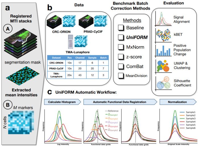
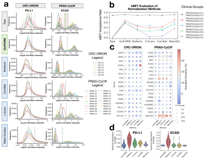
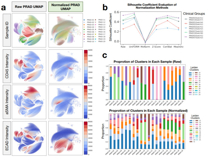
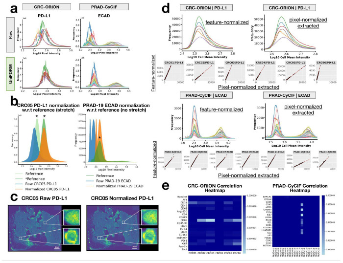
Multiplexed tissue imaging (MTI) technologies enable high-dimensional spatial analysis of tumor microenvironments but face challenges with technical variability in staining intensities. Existing normalization methods, including Z-score, ComBat, and MxNorm, often fail to account for the heterogeneous, right-skewed expression patterns of MTI data, compromising signal alignment and downstream analyses. We present UniFORM, a non-parametric, Python-based pipeline that uses an automated rigid landmark functional data registration approach for normalizing both feature- and pixel-level MTI data. Designed specifically for the distributional characteristics of MTI datasets, UniFORM operates without prior distributional assumptions and performs robustly regardless of distribution modality, including both unimodal and bimodal patterns. It removes technical variation by aligning the biologically invariant component of the signal, typically the negative (non-expressing) population, while preserving biologically meaningful variation in the positive population, thereby maintaining tissue-specific expression patterns essential for downstream analysis. Benchmarking across three distinct MTI platform datasets demonstrates that UniFORM outperforms existing methods in mitigating batch effects while maintaining biological signal fidelity. This is evidenced by improved marker distribution alignment and positive population preservation, enhanced kBET and Silhouette scores, and improved downstream analyses such as UMAP visualizations and Leiden clustering. UniFORM also introduces a novel guided fine-tuning option for complex and heterogeneous datasets. Although optimized for fluorescence-based platforms, UniFORM provides a scalable and robust solution for MTI data normalization, enabling accurate and biologically meaningful interpretations.


Conflict of interest statement

Competing interests The authors declare the following competing interests: G.B.M. is a SAB member or Consultant: for Amphista, Astex, AstraZeneca, BlueDot, Chrysallis Biotechnology, Ellipses Pharma, GSK, ImmunoMET, Infinity, Ionis, Leapfrog Bio, Lilly, Medacorp, Nanostring, Nuvectis, PDX Pharmaceuticals, Qureator, Roche, Signalchem Lifesciences, Tarveda, Turbine, Zentalis Pharmaceuticals. G.B.M. has Stock/Options/Financial relationships with: Bluedot, Catena Pharmaceuticals, ImmunoMet, Nuvectis, SignalChem, Tarveda, and Turbine. G.B.M. has Licensed Technology: HRD assay to Myriad Genetics, DSP patents with Nanostring. G.B.M. has Sponsored research with AstraZeneca. The other authors declare no competing interests.

Figures

Figure 1:
Figure 1:. UniFORM offers universal automatic intensity normalization for both feature-level and pixel-level MTI data.
(a) UniFORM accepts both pixel-level and feature-level data as input. Nucleus/cell segmentation masks are required for pixel-level normalization to exclude non-cell regions. (b) The study workflow includes datasets from two multiplex imaging platforms: CRC-ORION (17 channels, 6 patients, 1 batch), PRAD-CyCIF (20 channels, 20 patients, 7 batches), and TMA-Lunaphore (43 channels, 12 samples, 3 batches). UniFORM was benchmarked against commonly used normalization methods (MxNorm-registration, Z-score, ComBat, Mean Division) and a baseline. The performance of each method was evaluated through signal alignment, kBET analysis, positive population change, and UMAP-Leiden clustering. (c) Overview of the UniFORM automatic normalization workflow. The process begins with histogram calculation, followed by signal alignment using functional data registration, and concludes with normalization. A major novelty and highlight of UniFORM is its pixel-level normalization capability. It undergoes the same pipeline but uses cell/nuclei segmentation masks and a slightly different normalization step.
Figure 2:
Figure 2:. Feature-level normalization evaluation and comparison.
(a) Evaluation of cell mean intensity signal alignment across different normalization methods of unimodal marker PD-L1 and bimodal marker ECAD, with local positive population thresholds (colorful lines) and global threshold (black line) annotated. (b) Evaluation of batch effect correction with kBET with five patient groups, each composed of patient samples with similar cell composition but of different batches, measured by kBET acceptance rate (higher rate indicates better batch correction). (c) Evaluation of positive population change post-normalization across different normalization methods. The size of the bubbles indicates the magnitude of the absolute positive population mean percentage change, and the color of the heatmap indicates the standard deviation of the mean percentage change. (d) Violin plots show the spread and central tendency of the positive percentage change for PD-L1 and ECAD.
Figure 3:
Figure 3:. Impact of UniFORM on downstream analysis.
(a) UMAP of raw and normalized PRAD-CyCIF dataset colored by sample IDs showing mixture after normalization and UMAP of raw and normalized PRAD-CyCIF dataset colored by the intensity value of marker CD45, αSMA, and ECAD showing clustering of higher intensity regimen after normalization. (b) Silhouette coefficient evaluation of mutually exclusive marker separation with five patient groups, a higher Silhouette score indicates better cluster separation. (c) Leiden clustering of raw and normalized PRAD-CyCIF dataset showing enriched cluster proportions across samples after normalization.
Figure 4:
Figure 4:. Pixel-level normalization evaluation.
(a) Evaluation of UniFORM’s pixel intensity signal alignment of unimodal marker PD-L1 and bimodal marker ECAD (b) Demonstration of the sample signal alignment with respect to its reference and the effect of stretching mechanism. (c) Visual inspection of the PD-L1 channel before and after normalization confirms no artifacts are introduced. (d) Features extracted from normalized images achieve good signal alignment and almost perfect Spearman correlation with the feature-level normalized data. (e) Spearman correlation heatmap showing extracted features having almost perfect correlation with the feature-level normalized in both CRC-ORION and PRAD-CyCIF data. Refer to Figure 2a for legend information.

Similar articles

  • Short-Term Memory Impairment.
    Cascella M, Al Khalili Y. Cascella M, et al. 2024 Jun 8. In: StatPearls [Internet]. Treasure Island (FL): StatPearls Publishing; 2025 Jan–. 2024 Jun 8. In: StatPearls [Internet]. Treasure Island (FL): StatPearls Publishing; 2025 Jan–. PMID: 31424720 Free Books & Documents.
  • An image analysis pipeline to quantify the spatial distribution of cell markers in stroma-rich tumors.
    Ruzette AA, Kozlova N, Cruz KA, Muranen T, Nørrelykke SF. Ruzette AA, et al. bioRxiv [Preprint]. 2025 May 1:2025.04.28.650414. doi: 10.1101/2025.04.28.650414. bioRxiv. 2025. PMID: 40654607 Free PMC article. Preprint.
  • Signs and symptoms to determine if a patient presenting in primary care or hospital outpatient settings has COVID-19.
    Struyf T, Deeks JJ, Dinnes J, Takwoingi Y, Davenport C, Leeflang MM, Spijker R, Hooft L, Emperador D, Domen J, Tans A, Janssens S, Wickramasinghe D, Lannoy V, Horn SRA, Van den Bruel A; Cochrane COVID-19 Diagnostic Test Accuracy Group. Struyf T, et al. Cochrane Database Syst Rev. 2022 May 20;5(5):CD013665. doi: 10.1002/14651858.CD013665.pub3. Cochrane Database Syst Rev. 2022. PMID: 35593186 Free PMC article.
  • Leveraging a foundation model zoo for cell similarity search in oncological microscopy across devices.
    Kalweit G, Klett A, Silvestrini P, Rahnfeld J, Naouar M, Vogt Y, Infante D, Berger R, Duque-Afonso J, Hartmann TN, Follo M, Bodurova-Spassova E, Lübbert M, Mertelsmann R, Boedecker J, Ullrich E, Kalweit M. Kalweit G, et al. Front Oncol. 2025 Jun 18;15:1480384. doi: 10.3389/fonc.2025.1480384. eCollection 2025. Front Oncol. 2025. PMID: 40606969 Free PMC article.
  • Management of urinary stones by experts in stone disease (ESD 2025).
    Papatsoris A, Geavlete B, Radavoi GD, Alameedee M, Almusafer M, Ather MH, Budia A, Cumpanas AA, Kiremi MC, Dellis A, Elhowairis M, Galán-Llopis JA, Geavlete P, Guimerà Garcia J, Isern B, Jinga V, Lopez JM, Mainez JA, Mitsogiannis I, Mora Christian J, Moussa M, Multescu R, Oguz Acar Y, Petkova K, Piñero A, Popov E, Ramos Cebrian M, Rascu S, Siener R, Sountoulides P, Stamatelou K, Syed J, Trinchieri A. Papatsoris A, et al. Arch Ital Urol Androl. 2025 Jun 30;97(2):14085. doi: 10.4081/aiua.2025.14085. Epub 2025 Jun 30. Arch Ital Urol Androl. 2025. PMID: 40583613 Review.

References

    1. Lin J.-R., Fallahi-Sichani M., Chen J.-Y. & Sorger P. K. Cyclic Immunofluorescence (CycIF), A Highly Multiplexed Method for Single-cell Imaging. Curr. Protoc. Chem. Biol. 8, 251–264 (2016). - PMC - PubMed
    1. Tan W. C. C. et al. Overview of multiplex immunohistochemistry/immunofluorescence techniques in the era of cancer immunotherapy. Cancer Commun. Lond. Engl. 40, 135–153 (2020). - PMC - PubMed
    1. Hofman P. et al. Multiplexed Immunohistochemistry for Molecular and Immune Profiling in Lung Cancer-Just About Ready for Prime-Time? Cancers 11, 283 (2019). - PMC - PubMed
    1. Harms P. W. et al. Multiplex Immunohistochemistry and Immunofluorescence: A Practical Update for Pathologists. Mod. Pathol. Off. J. U. S. Can. Acad. Pathol. Inc 36, 100197 (2023). - PubMed
    1. Black S. et al. CODEX multiplexed tissue imaging with DNA-conjugated antibodies. Nat. Protoc. 16, 3802–3835 (2021). - PMC - PubMed

Publication types

LinkOut - more resources