[Preprint]. 2024 Dec 11:2024.12.10.627806.
doi: 10.1101/2024.12.10.627806.

Structure of the Staphylococcus aureus bacteriophage 80α neck shows the interactions between DNA, tail completion protein and tape measure protein

Affiliations

Structure of the Staphylococcus aureus bacteriophage 80α neck shows the interactions between DNA, tail completion protein and tape measure protein

James L Kizziah et al. bioRxiv. .

Update in

Abstract

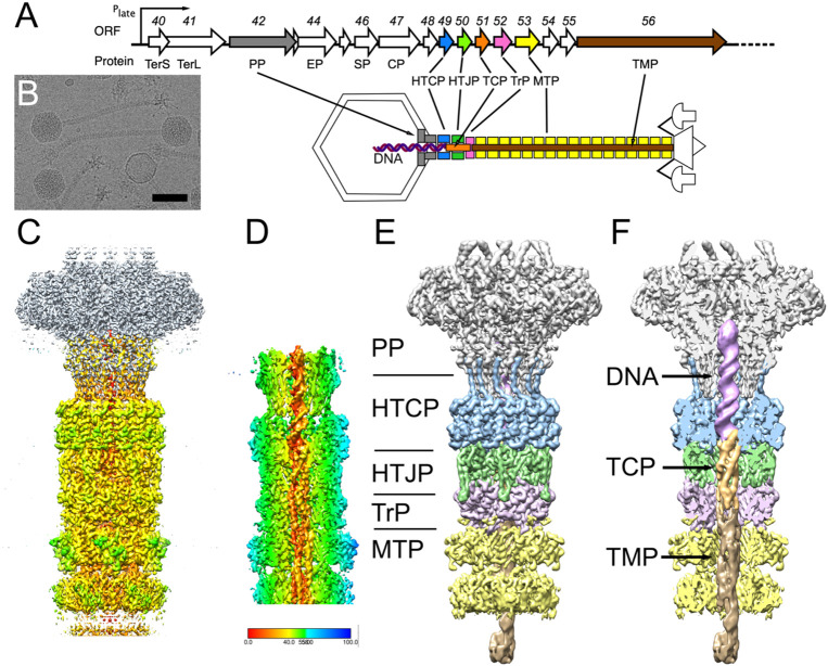
Tailed bacteriophages with double-stranded DNA genomes (class Caudoviricetes) play an important role in the evolution of bacterial pathogenicity, both as carriers of genes encoding virulence factors and as the main means of horizontal transfer of mobile genetic elements (MGEs) in many bacteria, such as Staphylococcus aureus. The S. aureus pathogenicity islands (SaPIs), including SaPI1, are a type of MGEs are that carry a variable complement of genes encoding virulence factors. SaPI1 is mobilized at high frequency by "helper" bacteriophages, such as 80α, leading to packaging of the SaPI1 genome into virions made from structural proteins supplied by the helper. 80α and SaPI1 virions consist of an icosahedral head (capsid) connected via a unique vertex to a long, non-contractile tail. At one end of the tail, proteins associated with the baseplate recognize and bind to the host. At the other end, a connector or "neck" forms the interface between the tail and the head. The neck consists of several specialized proteins with specific roles in DNA packaging, phage assembly, and DNA ejection. Using cryo-electron microscopy and three-dimensional reconstruction, we have determined the high-resolution structure of the neck section of SaPI1 virions made in the presence of phage 80α, including the dodecameric portal (80α gene product (gp) 42) and head-tail-connector (gp49) proteins, the hexameric head-tail joining (gp50) and tail terminator (gp52) proteins, and the major tail protein (gp53) itself. We were also able to resolve the DNA, the tail completion protein (gp51) and the tape measure protein (gp56) inside the tail. This is the first detailed structural description of these features in a bacteriophage, providing insights into the assembly and infection process in this important group of MGEs and their helper bacteriophages.

Keywords: 3D reconstruction; Staphylococcus aureus pathogenicity island 1; bacteriophage tail; connector; cryo-electron microscopy; portal protein; virus capsid.


Figures

Figure 1.
Figure 1.
Structure of the SaPI1 neck. (A) Schematic diagram of part of the late operon of 80α, showing the genes encoding neck proteins. ORF numbers and protein names are shown: TerS, small terminase subunit; TerL, large terminase subunit; PP, portal protein; EP, ejection protein, SP, scaffolding protein; CP, major capsid protein; HTCP, head-tail connector protein; HTJP, head-tail joining protein; TCP, tail completion protein; TrP, tail terminator protein; MTP, major tail protein; TMP, tape measure protein. The schematic diagram indicates the location of the neck proteins in the virion. The DNA is shown as a purple double helix. (B) Cryo-electron micrograph of SaPI1 virions. Scale bar = 50 nm. (C) Isosurface representation of a composite of the C6 neck reconstruction, colored by radius from the central axis, and the previously determined C12 reconstruction of the portal (gray) [30]. (D) Cutaway view of the asymmetric (C1) reconstruction of the neck, showing the DNA, TCP and TMP inside the neck, colored by radius according to the color bar. (E) Segmented reconstruction, colored by protein as in panel A: PP (gp42), gray; HTCP (gp49), blue; HTJP (gp50), green; TrP (gp52), pink; MTP (gp53), yellow. (F) Cutaway view of segmented reconstruction, showing density inside neck, colored as in panel C. In addition, the DNA is purple; TCP (gp51), orange; TMP (gp56), brown.
Figure 2.
Figure 2.
Atomic modeling of the neck. (A) Ribbon representation of the atomic model from the C6 neck reconstruction. The two copies of HTCP (gp49) per asymmetric unit are colored light and dark blue; the HTJP (gp50), TrP (gp52) and MTP (gp53) hexamers are shown in green, pink and yellow, respectively. The C-terminus of HTCP, which was built from the previous portal structure [30] is in cyan. (B) Atomic model of the neck reconstruction with one asymmetric unit colored as in panel A, the rest gray. The previously determined portal protein model is included, with the two copies of PP (gp42) per asymmetric unit colored light and dark brown. (C) An isolated asymmetric unit, colored as in A and B. (D) Slab through the neck model, colored as in panel A. (E) Sections through the neck model at the levels indicated by the arrows in panel D. From left to right: HTCP and HTJP; HTJP and TrP; TrP and MTP. The smallest inner diameters are indicated. (F) Monomers of neck proteins, from left to right: HTCP, HTJP, TrP and MTP. (G) Interaction between HTJP (green surface) and the HTCP (blue) and TrP (pink). (H) Interaction between HTJP (green), HTCP (blue) and TrP (pink) shown as molecular surfaces. (I) Cutaway view showing the electrostatic potential surface of the tail interior. (J) AlphaFold model of HTJP (orange) superimposed on the HTJP model from the neck reconstruction (green), showing the predicted inner ring of α-helices. (K) MTPs from the 80α baseplate reconstruction (orange and tan) superimposed on the MTP from the neck model (yellow), showing the difference in the C-arm.
Figure 3.
Figure 3.
Modeling of the inside of the neck. (A) Detail of internal density with the TCP (gp50, orange), TMP (gp56, shades of green) and DNA (purple) models fitted in. External proteins (HTCP, HTJP, TrP and MTP) are colored as in Fig. 2. (B) Atomic model of the inside of the neck, including the DNA double helix (purple and magenta), TCP (orange) and the three copies of TMP in green, turquoise and aqua, inside the segmented density of the asymmetric reconstruction. (C) Same model as in (B) with the atomic model of the surrounding proteins shown in gray. (D) slabs through the tail model at the levels indicated in (C). (E) Closeup view of the internal proteins and the DNA. (F) Ribbon diagram of the TCP model. (G) Ribbon diagrams of the AlphaFold3 models of the TCPs from phage lambda (gpZ), HK97 (gp10) and SPP1 (gp16.1). (H) The interaction between TCP, TMP and the DNA. The TCP interacting loop is shown in stick representation, colored by element. (I) Detail of the interaction between TCP and the DNA. The 3’ and 5’ ends of the DNA and pertinent residues are indicated. (J) Electrostatic potential surfaces of the inside of the tail. (K) Electrostatic surface of TCP with surrounding proteins and DNA shown in ribbon representation. (L) Top view of the electrostatic surface of TCP with the HTCP shown in ribbon representation. (K) Same view as panel L with TCP and HTCP both shown as electrostatic surfaces.

References

    1. Puxty RJ, Millard AD (2023) Functional ecology of bacteriophages in the environment. Curr Opin Microbiol 71: 102245. - PubMed
    1. Taylor VL, Fitzpatrick AD, Islam Z, Maxwell KL (2019) The Diverse Impacts of Phage Morons on Bacterial Fitness and Virulence. Adv Virus Res 103: 1–31. - PubMed
    1. Schroven K, Aertsen A, Lavigne R (2021) Bacteriophages as drivers of bacterial virulence and their potential for biotechnological exploitation. FEMS Microbiol Rev 45: fuaa041. - PubMed
    1. Rao VB, Fokine A, Fang Q (2021) The remarkable viral portal vertex: structure and a plausible model for mechanism. Curr Opin Virol 51: 65–73. - PMC - PubMed
    1. Dedeo CL, Cingolani G, Teschke CM (2019) Portal Protein: The Orchestrator of Capsid Assembly for the dsDNA Tailed Bacteriophages and Herpesviruses. Annu Rev Virol 6: 141–160. - PMC - PubMed

Publication types

LinkOut - more resources