[Preprint]. 2024 Dec 10:2024.12.09.624203.
doi: 10.1101/2024.12.09.624203.

Modulation of the JAK2-STAT3 pathway promotes expansion and maturation of human iPSCs-derived myogenic progenitor cells

Affiliations

Modulation of the JAK2-STAT3 pathway promotes expansion and maturation of human iPSCs-derived myogenic progenitor cells

Luca Caputo et al. bioRxiv. .

Update in

Abstract

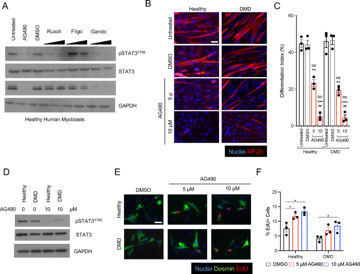
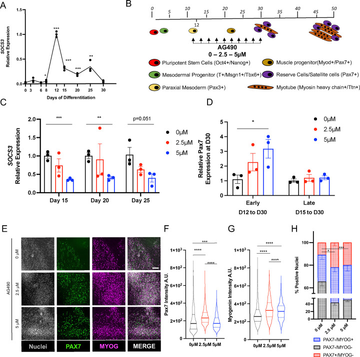
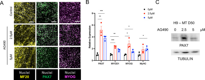
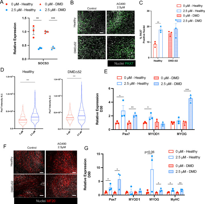
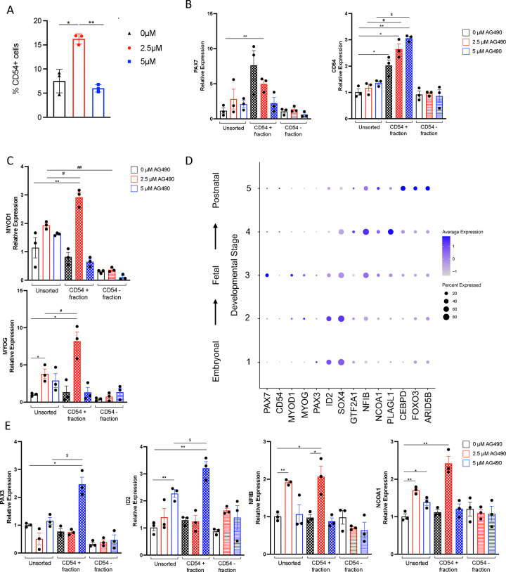
Generation of in vitro induced pluripotent cells (hiPSCs)-derived skeletal muscle progenitor cells (SMPCs) holds great promise for regenerative medicine for skeletal muscle wasting diseases, as for example Duchenne Muscular Dystrophy (DMD). Multiple approaches, involving ectopic expression of key regulatory myogenic genes or small molecules cocktails, have been described by different groups to obtain SMPC towards cell-transplantation in vivo as a therapeutic approach to skeletal muscle diseases. However, hiPSCs-derived SMPC generated using transgene-free protocols are usually obtained in a low amount and resemble a more embryonal/fetal stage of differentiation. Here we demonstrate that modulation of the JAK2/STAT3 signaling pathway during an in vitro skeletal muscle differentiation protocol, increases the yield of PAX7+ and CD54+ SMPCs and drive them to a postnatal maturation stage, in both human ES and patient-derived iPSCs. Importantly, upon removal of the inhibition from the cultures, the obtained SMPCs are able to differentiate into multinucleated myotubes in vitro. These findings reveal that modulation of the JAK2/STAT3 signaling pathway is a potential therapeutic avenue to generate SMPCs in vitro with increase potential for cell-therapy approaches.

Keywords: Duchenne Muscular Dystrophy; STAT3 pathway; hiPSC; myotubes; skeletal muscle progenitor cells.


Conflict of interest statement

Conflict of Interest Statement The authors declare that they have no conflict of interest.

Figures

Figure 1:
Figure 1:. AG490 mediated inhibition of JAK/STAT3 signaling blocks differentiation of human myoblasts
A. Western blot analysis of whole cell lysate of human healthy myoblasts treated with various JAK inhibitors using a-pSTAT3, a-total STAT3, anti-Myogenin and anti-GAPDH antibodies. B. Representative immunofluorescence images of myotubes either from healthy or Duchenne patient-derived myoblasts treated with AG490 or DMSO as vehicle control for MF-20 (red), nuclei are counterstained with Hoechst (blue). Scale bar 100 μm. C. Differentiation index of myotubes for the conditions shown in (B) n=3 biological replicates. D. Western blot analysis of whole cell lysate of human healthy and Duchenne patient-derived myoblasts treated with 10 μM AG490 using a-pSTAT3, a-total STAT3, and anti-GAPDH antibodies. E. Representative immunofluorescence images of myoblasts stained with Desmin (green), EdU (red) nuclei are counterstained with Hoechst (blue). Scale bar 20 μm. F. Quantification of the percentage of proliferating myoblasts treated with DMSO, 5 μM or 10 μM AG490 through EdU staining n=3 biological replicates. ns. p > 0.05 ; *. p≤ 0.05 ; **. p≤ 0.01 ; ***. p≤ 0.001 unpaired Student’s t test. * over untreated control; # over lower concentration of drug; $ over DMSO – vehicle control in C.
Figure 2:
Figure 2:. AG490 treatment leads to expansion of PAX7+ cells in vitro
A. qPCR analysis of SOCS3 expression during myogenic differentiation. B. Schematic representation of the myogenic differentiation protocol, AG490 treatment is between D12 and D30 of differentiation, with refreshment of the media every 48hours. C. qPCR analysis of SOCS3 expression at different days of AG490 treatment. D. qPCR analysis of PAX7 expression at D30 of the differentiation protocol, with AG490 treatment started early (at D12) or late (D15). E. Representative immunofluorescence images of MYOG (magenta) and PAX7 (green) at D30 of differentiation, nuclei are counterstained with Hoechst (gray). Scale bar 50 μm. F. Violin plot or the quantification of the nuclear mean fluorescence intensity (arbitrary unit) (median ± second and third quantile) of the PAX7 IF signal staining. G. Violin plot or the quantification of the nuclear mean fluorescence intensity (arbitrary unit) (median ± second and third quantile) of the MYOG IF signal staining H. percentage of PAX7+ or MYOG+ nuclei at D30 of differentiation. N=3 biological replicates ns. p > 0.05 ; *. p≤ 0.05 ; **. p≤ 0.01 ; ***. p≤ 0.001 unpaired Student’s t test for A - C - H, One-Way ANOVA for F – G.
Figure 3:
Figure 3:. Increased myogenic differentiation upon AG490 removal
A. Representative images of MF-20 (yellow), PAX7 (green) and MYOG (magenta) at D50 of differentiation, nuclei are counterstained with Hoechst (gray). Scale bar 100 μm for MF-20, 50 μm for PAX7 and MYOG. B. qPCR analysis of myogenic factors (PAX7, MYOD1, MYOG, and MyHC) expression at D50 of differentiation. C. Western blot analysis of whole cell lysate of H9-derived myogenic cultures treated with AG490 using a-PAX7, and anti-Tubulin antibodies N=3 biological replicates. ns. p > 0.05 ; *. p≤ 0.05 ; **. p≤ 0.01 ; ***. p≤ 0.001 unpaired Student’s t test.
Figure 4:
Figure 4:. AG490 treatment leads to increased myogenesis in both healthy and DMD myogenic cultures
A. qPCR analysis of SOCS3 expression at D30 of the myogenic differentiation protocol in healthy (circles) and DMDΔ52 (triangles) myogenic cultures. B. Representative immunofluorescence images of PAX7 (green) at D30 of differentiation, nuclei are counterstained with Hoechst (gray). Scale bar 50 μm. C. Percentage of PAX7+ nuclei at D30 of differentiation D. Violin plot or the quantification of the nuclear mean fluorescence intensity (arbitrary unit) (median ± second and third quantile) of the PAX7 IF signal staining in healthy (left panel) and DMDΔ52 (right panel) myogenic cultures. E. qPCR analysis of PAX7, MYOD1 and MYOG expression at D30 of the differentiation protocol F. Representative immunofluorescence images of MF-20 (red) at D50 of differentiation protocol, nuclei are counterstained with Hoechst (gray). Scale bar 50 μm. G. qPCR analysis of myogenic factors (PAX7, MYOD1, MYOG, and MyHC) expression at D50 of differentiation. N=3 biological replicates. ns. p > 0.05 ; *. p≤ 0.05 ; **. p≤ 0.01 ; ***. p≤ 0.001 unpaired Student’s t test for A - C - E - G, One-Way ANOVA for D.
Figure 5:
Figure 5:. AG490 treatment leads to maturation of in vitro derived SMPC
A. Percentage of CD54+ cells obtained from H9-derived myogenic cultures at D30 of differentiation. B. qPCR analysis of PAX7 and CD54/ICAM1 expression in CD54 MACS-purified populations. C. qPCR analysis of MYOD1 and MYOG expression in CD54 MACS-purified populations. D. Dot plot analysis for selected transcription factors differentially expressed in embryonal (STAGE1/2) vs postnatal (STAGE 5) stages. E. qPCR analysis of PAX3, ID2 (STAGE1/2) and NFIB, NCOA1 (STAGE 4) expression in CD54 MACS-purified populations. N=3 biological replicates. ns. p > 0.05 ; *. p≤ 0.05 ; **. p≤ 0.01 ; ***. p≤ 0.001 unpaired Student’s t test.

References

    1. Tedesco F. S. & Cossu G. Stem cell therapies for muscle disorders. Curr. Opin. Neurol. 25, 597–603 (2012). - PubMed
    1. Judson R. N. & Rossi F. M. V. Towards stem cell therapies for skeletal muscle repair. npj Regen. Med. 5, 10 (2020). - PMC - PubMed
    1. Caputo L. et al. Acute conversion of patient-derived Duchenne muscular dystrophy iPSC into myotubes reveals constitutive and inducible over-activation of TGFβ-dependent pro-fibrotic signaling. Skelet Muscle 10, 13 (2020). - PMC - PubMed
    1. Darabi R. et al. Human ES- and iPS-Derived Myogenic Progenitors Restore DYSTROPHIN and Improve Contractility upon Transplantation in Dystrophic Mice. Cell Stem Cell 10, 610–619 (2012). - PMC - PubMed
    1. Darabi R. et al. Functional skeletal muscle regeneration from differentiating embryonic stem cells. Nat. Med. 14, 134–143 (2008). - PubMed

Publication types

LinkOut - more resources