[Preprint]. 2024 Dec 12:2024.12.10.627648.
doi: 10.1101/2024.12.10.627648.

Selective Targeting of a Defined Subpopulation of Corticospinal Neurons using a Novel Klhl14-Cre Mouse Line Enables Molecular and Anatomical Investigations through Development into Maturity

Affiliations

Selective Targeting of a Defined Subpopulation of Corticospinal Neurons using a Novel Klhl14-Cre Mouse Line Enables Molecular and Anatomical Investigations through Development into Maturity

Jake Lustig et al. bioRxiv. .

Update in

Abstract

The corticospinal tract (CST) facilitates skilled, precise movements, which necessitates that subcerebral projection neurons (SCPN) establish segmentally specific connectivity with brainstem and spinal circuits. Developmental molecular delineation enables prospective identification of corticospinal neurons (CSN) projecting to thoraco-lumbar spinal segments; however, it remains unclear whether other SCPN subpopulations in developing sensorimotor cortex can be prospectively identified in this manner. Such molecular tools could enable investigations of SCPN circuitry with precision and specificity. During development, Kelch-like 14 (Klhl14) is specifically expressed by a specific SCPN subpopulation, CSNBC-lat, that reside in lateral sensorimotor cortex with axonal projections exclusively to bulbar-cervical targets. In this study, we generated Klhl14-T2A-Cre knock-in mice to investigate SCPN that are Klhl14+ during development into maturity. Using conditional anterograde and retrograde labeling, we find that Klhl14-Cre is specifically expressed by CSNBC-lat only at specific developmental time points. We establish conditional viral labeling in Klhl14-T2A-Cre mice as a new approach to reliably investigate CSNBC-lat axon targeting and confirm that this identifies known molecular regulators of CSN axon targeting. Therefore, Klhl14-T2A-Cre mice can be used as a novel tool for identifying molecular regulators of CST axon guidance in a relatively high-throughput manner in vivo. Finally, we demonstrate that intersectional viral labeling enables precise targeting of only Klhl14-Cre+ CSNBC-lat in the adult central nervous system. Together, our results establish that developmental molecular delineation of SCPN subpopulations can be used to selectively and specifically investigate their development, as well as anatomical and functional organization into maturity.

Keywords: Brainstem innervation; Cerebellin; Corticospinal; Crim1; Easi-CRISPR; conditional AAV labeling; genomic Cre reporters; segmental axon projection.


Figures

Figure 1.
Figure 1.. Genomic reporters of Cre-dependent recombination do not identify SCPN subpopulation specificity of Cre expression in Klhl14-Cre mice
A-B, Schematic (adapted from Sahni et al., 2021b) showing the locations of distinct CSN subpopulations and their corresponding axonal projections in mouse CNS. CSNBC-lat (green) reside in rostrolateral cortex and extend axons only to bulbar-cervical targets. CSNBC-med (purple) reside in medial cortex and extend axons only to bulbar-cervical targets. CSNTL (red) reside in medial cortex and extend to thoraco-lumbar levels, but extend collaterals across all spinal levels. Klhl14 is only expressed by CSNBC-lat. C, In situ hybridization image on a coronal section of a P4 mouse brain showing Klhl14 expression by CSNBC-lat (from Sahni et. al., 2021b). D, Schematic showing the genomic organization of the WT Klhl14 locus. Klhl14 is encoded by 8 exons. (D’) Magnified view of region boxed in D showing the point of insertion of Cre in Klhl14-Cre mice. The T2A-Cre sequence was targeted to exon 8 immediately 3’ to the STOP codon. The remaining part of the 3’ UTR in exon 8 was used as a homology arm. (D”) Genomic PCR from F2 mice confirmed proper insertion of Cre, with a resultant product of 1703 bp. E, Coronal brain sections collected from a Klhl14-Cre;Emxflpo;Ai65 transgenic mouse. Klhl14-Cre positive cell bodies are labeled via tdTomato reporter expression (in red and in monochrome). TdTomato+ neurons span all layers across both medial (E’) and lateral (E”) cortex. F, Axial spinal sections from the same Klhl14-Cre;Emxflpo;Ai65 transgenic mouse taken at cervical, thoracic, and lumbar levels. TdTomato+ axons are present in the dorsal funiculus at all spinal levels consistent with the widespread recombination in cortex. Scale bars 500 μm for C, 1 mm for E, and 250 μm for E’-E”, and F.
Figure 2.
Figure 2.. Conditional retrograde labeling identifies specificity of Klhl14-Cre expression by CSNBC-lat enabling identification of their cortical location at maturity
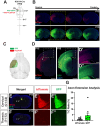
A-A”, Klhl14-Cre heterozygous mice were co-injected with rAAV-FLEX-eGFP and rAAV-tdTomato in the cerebral peduncle at P3 (schematized in A) and the brains collected at P14. A’, Wholemount imaging of the injected brain finds retrogradely labeled tdTomato+ SCPN span the entire sensorimotor cortex while eGFP+ (Klhl14-Cre+) SCPN occupy a smaller area of cortex, specifically residing in lateral cortex. A’, Coronal sections of the same brain reveal that eGFP+ SCPN reside laterally (where Klhl14+ SCPN are known to reside), while tdTomato+ SCPN reside in both medial and lateral cortex. B-B”, Klhl14-Cre heterozygous mice were co-injected with rAAV-FLEX-eGFP and rAAV-tdTomato in cervical spinal cord at P2 (schematized in B) and brains collected at P14. B’, Wholemount imaging finds fewer tdTomato+ CSN labeled than SCPN in A’. eGFP+ CSN occupy a smaller area of cortex than tdTomato+ CSN, indicating specificity of Cre expression. B”, Coronal sections of the same brain find that while tdTomato+ CSN span both medial and lateral cortex, eGFP+ CSN reside laterally (where Klhl14+ SCPN are known to reside). Further, there are fewer eGFP+ CSN than GFP+ SCPN labeled in A”. C, 3D AMaSiNe reconstructions of conditionally labeled neurons from 3 Klhl14-Cre mice each that received retrograde labeling from either the cerebral peduncle (Klhl14-Cre+ SCPN, pink) or cervical spinal cord (Klhl14-Cre+ CSN, blue). SCPN occupy a much larger area of cortex compared to CSN, but both primarily reside in lateral cortex. D, Distribution of Klhl14+ SCPN across different cortical areas (annotations as assigned by the Allen Brain Atlas). Most Klhl14-Cre+ neurons reside in somatosensory cortices. E, Percentage counts of labeled Klhl14-Cre+ neurons in each area, again demonstrating that the majority of Klhl14+ SCPN and CSN reside in sensory cortices. Scale bars are 1mm.
Figure 3.
Figure 3.. Anterograde Labeling in Klhl14-T2A-Cre Mice Confirms that Klhl14+ CSN do not extend axons to the Thoracic Cord
A, Schematic of experimental outline: Klhl14-Cre heterozygous mice were co-injected with AAV-eGFP and AAV-FLEX-tdTomato at P0 in rostrolateral cortex, where Klhl14-Cre+ SCPN reside. Samples were collected at P15. B, Series of coronal brain sections from one injected mouse spanning rostral (left) to caudal (right) showing localization and spread of both AAVs within cortex. C, Sample 3D reconstruction of an injected brain displaying the injection locations and volumes for both eGFP and FLEX-tdTomato. Note that the eGFP (non-Cre dependent) reporter expression spans more medially than tdTomato (Cre-dependent). D, Zoomed in images for the section marked by dotted yellow outline in (B). AAV-FLEX-tdTomato only infects cells in lateral cortex, where Klhl14-Cre SCPN reside (D’). Unlike AAV-FLEX-tdTomato, AAV-eGFP infects cells in both lateral and medial cortex (D”). E-F”, Spinal axial sections from an injected Klhl14-Cre mouse at cervical (E-E”) and thoracic (F-F”) levels. While both tdTomato+ (E’) and eGFP+ axons (E”) are seen in the cervical dorsal funiculus, tdTomato+ axons (F’) are not present in thoracic spinal cord. AAV-eGFP labeled axons (F”) are labeled in the thoracic spinal cord, likely due to nonspecific labeling of CSNmed populations. G, Quantification of thoracic to cervical ratio (T:C ratio) from injected Klhl14-Cre mice at P15. T:C ratio is the percentage of labeled axons in the cervical dorsal funiculus that extend to the thoracic cord. (n=12 for tdTomato+ axons; n=9 mice for GFP+axons). There is much more variability in the T:C ratio for eGFP+ axons than tdTomato+ axons. Scale bars are 1mm for B, D, 100 μm for D’-D”, 250 μm for E, F and 20 μm for insets in E’-E”, F’-F”.
Figure 4.
Figure 4.. Klhl14+ SCPN axons exhibit identical projections in the brainstem as anatomically labeled CSNBC-lat
A, Similar experimental outline as in Figure 3. Klhl14-Cre knock-in mice were co-injected with AAV-eGFP and AAV-FLEX-tdTomato into rostrolateral cortex at P0. eGFP+ axons are from CSNBC-lat. The brainstem from injected mice was analyzed at P15. B, Coronal sections of the brainstem from one such injected mouse spanning the rostrocaudal extent of the brainstem showing axonal collateralization by Klhl14-Cre+ SCPN (magenta) vs. anatomically defined eGFP+ CSNBC-lat (green). C-F, Magnified view of sections highlighted in B showing midbrain (C), pons (D), rostral medulla (E), and caudal medulla (F). Klhl14-Cre+ SCPN axons exhibit nearly identical collateralization across the brainstem as eGFP+ CSNBC-lat axons. Scale bars are 500 μm.
Figure 5.
Figure 5.. Misexpression of Crim1 and Cbln1 in Klhl14-T2A-Cre Mice Redirects Klhl14+ CSN Axons to Thoracic Cord
A, Schematic of experimental outline: P0 Klhl14-Cre heterozygous mice were co-injected with AAV-FLEX-tdTomato and either one of following three AAVs 1) AAV-eGFP (control); AAV-Crim1(Crim1 O/E) or AAV-Cbln1 (Cbln1 O/E) into rostrolateral cortex. B, Graph of average T:C ratios for controls (same results as in Figure 3G) (n=12) compared to Crim1 O/E (n=4) and Cbln1 O/E (n=4). Overexpression of either gene results in an increase in percentage of Klhl14-Cre+ CSN axons that extend into thoracic cord compared to control mice. C-E, Axial spinal sections from P15 Klhl14-Cre mice injected with either control (C-C”), Crim1 O/E (D-D”), or Cbln1 O/E (E-E”) at cervical and thoracic levels. Klhl14-Cre+ (tdTomato+) axons are present in the cervical dorsal funiculus (C’, D’, E’); however, only in Crim1 O/E (D”) and Cbln1 O/E (E”) mice do CSN axons extend to the thoracic dorsal funiculus. F-H, Maximum intensity projection images of thoracic cord horizontal sections (T2-T13) for control (F), Crim1 O/E (G), and Cbln1 O/E (H) groups. Monochrome magnified views of rostral and caudal levels from each cord are shown in F’-H”. In control mice, no CST axons are present within the first few thoracic segments (F’). In contrast, labeled tdTomato+ CSNc axons in both Crim1 O/E (G’) and Cbln1 O/E (H”) mice extend to far caudal levels in the thoracic cord (white arrowheads). Scale bars are 250 μm for C, D, E, 20 μm for insets in C’-C”, D’-D”, E’-E”, 500 μm for F-H, and 50 μm for F’-F”, G’-G”, H’-H”.
Figure 6.
Figure 6.. An Intersectional Approach to target Klhl14+ SCPN at maturity using Klhl14-Cre mice.
A, Schematic of experimental outline: Klhl14-Cre mice received a bilateral retrograde injection of a retro AAV-FLEX-eGFP into the cerebral peduncle at P3. B, Coronal brain sections from an injected mouse at P14 show eGFP+ (Klhl14-Cre+) SCPN in lateral sensorimotor cortex. C, A representative section from the medulla from an injected mouse shows numerous eGFP+ neurons labeled at medial (D, D’) and lateral (D”) levels. Retrograde injection of AAV-FLEX-GFP therefore also labels Klhl14-Cre+ neurons in the brainstem. E, Schematic of experimental outline: Klhl14-Cre mice received bilateral intracortical injections of AAV-Flpo-mCherry into rostrolateral cortex at P2 along with bilateral retrograde injections of AAV-ConFon-eYFP in the cerebral peduncle. F, Coronal brain sections from a P14 injected mouse showing AAV-Flpo-mCherry (red) in lateral cortex. YFP+ (Cre+ and FlpO+) SCPN, i.e., Klhl14-Cre+ SCPN are specifically labeled in lateral cortex. (G) Representative coronal section of the medulla from the same mouse finds YFP+ (Klhl14-Cre+) SCPN axons (H), but no cell bodies are YFP+ (H’, H”). Therefore, this intersectional viral labeling approach labels Klhl14-Cre+ SCPN in cortex, but avoids labeling of Klhl14-Cre+ neurons in the brainstem, highlighting the effectiveness of this intersectional strategy for only targeting Klhl14-Cre+ SCPN at maturity. Scale bars are 1mm in B, F, 500 μm for C, G, and 100 μm for D-D”, H-H”.

Similar articles

Cited by

References

    1. Abe P., Lavalley A., Morassut I., Santinha A.J., Roig-Puiggros S., Javed A., Klingler E., Baumann N., Prados J., Platt R.J., and Jabaudon D. (2024). Molecular programs guiding arealization of descending cortical pathways. Nature 634, 644–651. 10.1038/s41586-024-07895-y. - DOI - PubMed
    1. Botterill J.J., Khlaifia A., Walters B.J., Brimble M.A., Scharfman H.E., and Arruda-Carvalho M. (2021). Off-Target Expression of Cre-Dependent Adeno-Associated Viruses in Wild-Type C57BL/6J Mice. eneuro 8, ENEURO.0363-0321.2021. 10.1523/eneuro.0363-21.2021. - DOI - PMC - PubMed
    1. Daigle T.L., Madisen L., Hage T.A., Valley M.T., Knoblich U., Larsen R.S., Takeno M.M., Huang L., Gu H., Larsen R., et al. (2018). A Suite of Transgenic Driver and Reporter Mouse Lines with Enhanced Brain-Cell-Type Targeting and Functionality. Cell 174, 465–480.e422. 10.1016/j.cell.2018.06.035. - DOI - PMC - PubMed
    1. Di Bella D.J., Habibi E., Stickels R.R., Scalia G., Brown J., Yadollahpour P., Yang S.M., Abbate C., Biancalani T., Macosko E.Z., et al. (2021). Molecular logic of cellular diversification in the mouse cerebral cortex. Nature 595, 554–559. 10.1038/s41586-021-03670-5. - DOI - PMC - PubMed
    1. Fenno L.E., Mattis J., Ramakrishnan C., Hyun M., Lee S.Y., He M., Tucciarone J., Selimbeyoglu A., Berndt A., Grosenick L., et al. (2014). Targeting cells with single vectors using multiple-feature Boolean logic. Nat Methods 11, 763–772. 10.1038/nmeth.2996. - DOI - PMC - PubMed

Publication types