. 2024 Dec 17:16:1813-1822.
doi: 10.2147/CMAR.S482363. eCollection 2024.

Exploratory Research for HIF-1α Overexpression Tumor Antigen in the Activation of Dendritic Cells and the Potent Anti-Tumor Immune Response

Affiliations

Exploratory Research for HIF-1α Overexpression Tumor Antigen in the Activation of Dendritic Cells and the Potent Anti-Tumor Immune Response

Jinjin Zhao et al. Cancer Manag Res. .

Abstract

Background: Tumor-specific antigens play an important role in dendritic cell (DC)-based immunotherapy. The acquisition of tumor-specific antigens, which are essential for DC-based immunotherapy, poses a significant challenge. This study aimed to explore the efficacy of hypoxia inducible factor-1α (HIF-1α) overexpression tumor antigens in DC-based immunotherapy.

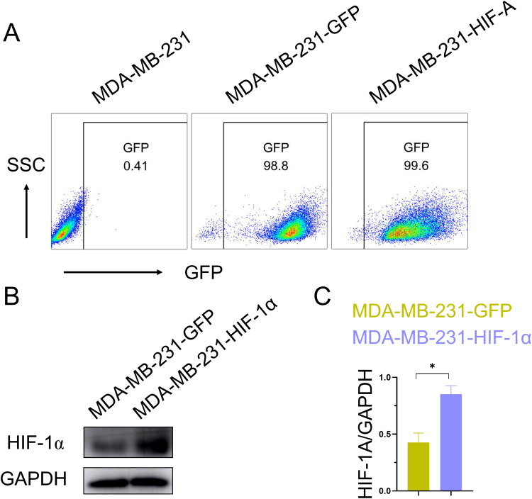
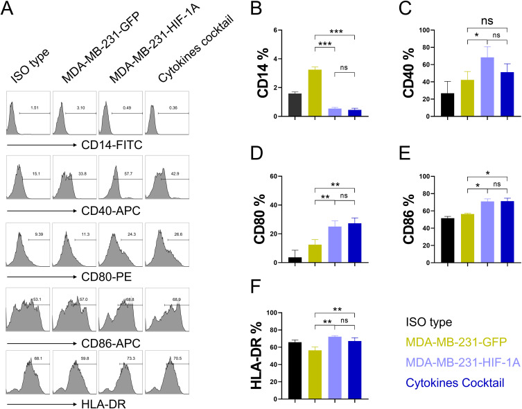
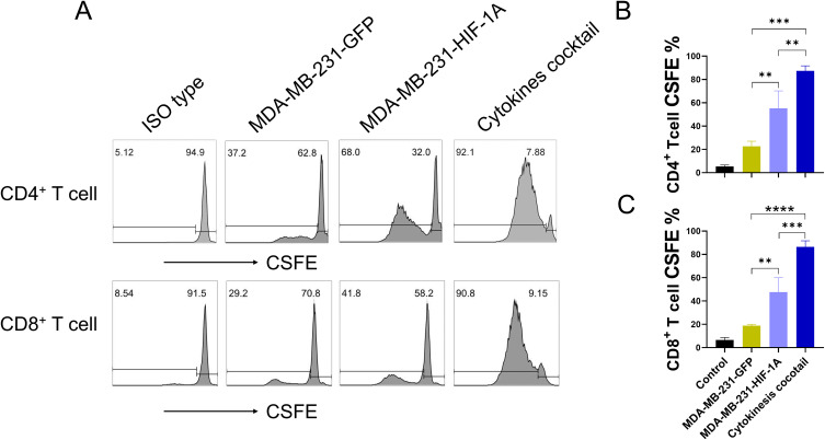
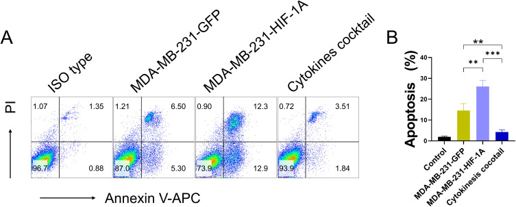
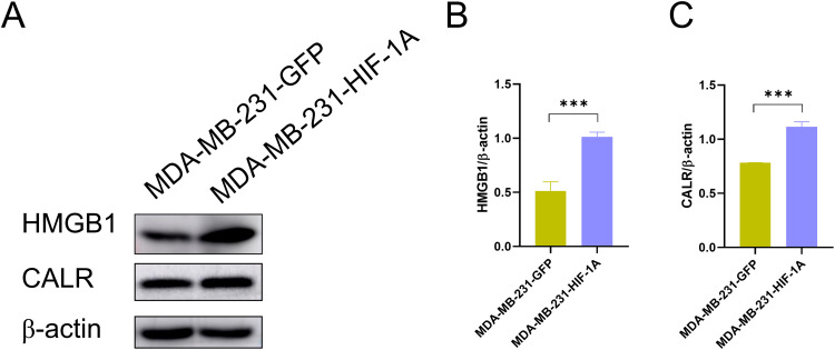
Methods: An HIF-1α over-expression cell line was constructed to prepare HIF-1α overexpression tumor antigens. The expression of CD14, CD40, CD80, CD86, and HLA-DR on the surface of dendritic cells derived from monocytes was assessed using flow cytometry after stimulation with tumor antigens enriched in HIF-1α. T cell proliferation was analyzed by CFSE division following incubation with mature DCs. The apoptotic tumor cells were detected through annexin V/PI staining following coculture with dendritic cells (DCs) stimulated by HIF-1α enriched antigens. The detection of damage-associated molecular pattern molecules (DAMPs) HMGB1 and calreticulin (CALR) was performed using Western blotting.

Results: The results demonstrated that HIF-1α-enriched tumor antigens significantly upregulated the expression of CD40, CD80, CD86, and HLA-DR in DCs compared to normal tumor antigens. Furthermore, co-incubation with HIF-1α-enriched tumor antigen-activated DCs enhanced T cell proliferation and stimulated the T cell-mediated cytotoxicity. Notably, the expression of DAMPs, such as HMGB1 and CALR, was elevated in HIF-1α-enriched tumor antigens.

Conclusion: Our findings demonstrate that tumor antigens enriched with HIF-1α may encompass tumor-specific antigens capable of stimulating DC activation, thereby enhancing T cell proliferation and cytotoxicity. These results provide support for the further advancement of HIF-1α enriched tumor antigens in preclinical and clinical investigations pertaining to tumor treatment.

Keywords: HIF-1α; HMGB1; T cell activation; calreticulin; dendritic cells.


Conflict of interest statement

The authors declare that they have no competing interests in this work.

Figures

Figure 1
Figure 1
HIF-1α High expression MDA-MB-231 cell line was successfully constructed. (A) Flow cytometry results of cells after lentivirus transfection; (B) The level of HIF-1α was measured by Western blot; (C) statistical analysis of protein expression of HIF-1α. The data are expressed as the mean ± SDs. Significant differences are shown by * P< 0.05, **P<0.01, *** P<0.001 by unpaired Student’s t-tests comparing the normal control and HIF-1α high expression group.
Figure 2
Figure 2
The overexpression of HIF-1α tumor antigens induced the maturation of dendritic cells. (A) After stimulated with GM-CSF and IL-4 for 5 days, the cells were treated with culture medium (ISO type), tumor cell antigen extracted from MDA-MB-231-GFP cells (MDA-MB-231-GFP), tumor cell antigen extracted from MDA-MB-231-HIF-1A cells (MDA-MB-231-GFP-HIF-1A) or cytokines containing IL-1α (10ng/mL), IL-6 (1000U/mL), TNF-α (10ng/mL), PEG-2 (1μg/mL) (Cytokines cocktail) for 1 day, the expression of CD14, CD40, CD80, CD86, HLA-DR on the surface of DC cells were detected by flow cytometry. Statistical analysis of the expression of CD14 (B), CD40 (C), CD80 (D), CD86 (E), HLA-DR (F) on DC cells. The data are expressed as the mean ± SDs. Significant differences are shown by * P< 0.05, **P<0.01, *** P<0.001 by unpaired Student’s t-tests.
Figure 3
Figure 3
The overexpression of HIF-1α tumor antigens enhances the capacity of DCs to stimulate T cell proliferation. (A) DCs maturation by culture medium (ISO type), tumor cell antigen extracted from MDA-MB-231-GFP cells (MDA-MB-231-GFP), tumor cell antigen extracted from MDA-MB-231-HIF-1A cells (MDA-MB-231-GFP-HIF-1A) or cytokines containing IL-1β (10ng/mL), IL-6 (1000U/mL), TNF-α (10ng/mL), PEG-2 (1μg/mL) (Cytokines cocktail) were coculture with T cells for 7 days, CD4+ and CD8+ T cells proliferation was measured by CSFE division. Statistical analysis of CD4+ (B) and CD8+ (C) T cells proliferation. The data are expressed as the mean ± SDs. Significant differences are shown by * P< 0.05, **P<0.01, *** P<0.001 by unpaired Student’s t-tests.
Figure 4
Figure 4
The tumoricidal effect of T cells activated by dendritic cells stimulated with HIF-1α overexpression tumor antigens is enhanced. (A) T cells, activated by DCs matured with culture medium (ISO type), tumor cell antigens extracted from MDA-MB-231-GFP cells (MDA-MB-231-GFP), tumor cell antigens extracted from MDA-MB-231-HIF-1A cells (MDA-MB-231-GFP-HIF-1A), or a cytokine cocktail containing IL-1β (10ng/mL), IL-6 (1000U/mL), TNF-α (10ng/mL), and PEG-2 (1μg/mL), were co-cultured with tumor cells for 1 day. Apoptotic tumor cells were detected using flow cytometry; (B) Statistical analysis of tumor cells killed by activated T-cells. Data are expressed as mean ± SDs. Significant differences are indicated by * P< 0.05, **P<0.01, and *** P<0.001 using unpaired Student’s t-test.
Figure 5
Figure 5
Expression of HMGB1 and CALR in experimental group and control group. (A) the level of HMGB1 and CALR was measured by Western blot; statistical analysis of protein expression of HMGB1 (B) and CALR (C). The data are expressed as the mean ± SDs. Significant differences are shown by * P< 0.05, **P<0.01, *** P<0.001 by unpaired Student’s t-tests comparing the normal control and HIF-1α high expression group.

Similar articles

Cited by

References

    1. Costa RLB, Gradishar WJ. Triple-negative breast cancer: current practice and future directions. J Oncol Pract. 2017;13(5):301–303. doi:10.1200/JOP.2017.023333 - DOI - PubMed
    1. Hassannia H, Ghasemi Chaleshtari M, Atyabi F, et al. Blockage of immune checkpoint molecules increases T‐cell priming potential of dendritic cell vaccine. Immunology. 2020;159(1):75–87. doi:10.1111/imm.13126 - DOI - PMC - PubMed
    1. Versteven M, Flumens D, Campillo-Davó D, et al. anti-tumor potency of short-term interleukin-15 dendritic cells is potentiated by in situ silencing of programmed-death ligands. Front Immunol. 2022;13. doi:10.3389/fimmu.2022.734256 - DOI - PMC - PubMed
    1. Sriramulu S, Thoidingjam S, Speers C, Nyati S. Present and future of immunotherapy for triple-negative breast cancer. Cancers (Basel). 2024;16(19):3250. doi:10.3390/cancers16193250 - DOI - PMC - PubMed
    1. Yatim N, Cullen S, Albert ML. Dying cells actively regulate adaptive immune responses. Nat Rev Immunol. 2017;17(4):262–275. doi:10.1038/nri.2017.9 - DOI - PubMed