

Anaplastic lymphoma kinase1 positive inflammatory myofibroblastic tumor of the urinary bladder: A rare mesenchymal neoplasm with diagnostic and therapeutic implications
- PMID: 39713609
- PMCID: PMC11660058
- DOI: 10.1177/2050313X241308992
Anaplastic lymphoma kinase1 positive inflammatory myofibroblastic tumor of the urinary bladder: A rare mesenchymal neoplasm with diagnostic and therapeutic implications
Abstract
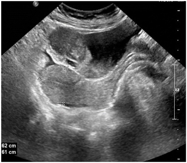
Inflammatory myofibroblastic tumors (IMTs) are rare mesenchymal neoplasms characterized by spindle-cell morphology with accompanying inflammatory infiltrates. Originally described in 1939, these tumors can arise in various anatomic locations, with the urinary bladder being a rare site of occurrence but the most common within the genitourinary tract. IMTs typically present as polypoid masses or firm submucosal nodules, often with painless hematuria in bladder cases. Histopathologically, IMTs are composed of myofibroblasts with myxoid stroma and mixed inflammatory cells, predominantly lymphocytes and plasma cells. Immunohistochemically, these tumors commonly express anaplastic lymphoma kinase1 (ALK1), vimentin, smooth muscle actin (SMA), and cytokeratin, with ALK1 serving as a crucial marker for diagnosis. This report details the case of a 31-year-old female presenting with hematuria, found to have a soft tissue mass in the urinary bladder (5.0 × 3.0 cm). Imaging revealed a well-defined lesion with vascularity. Histopathological examination confirmed an IMT, with immunohistochemistry showing diffuse ALK1 positivity, patchy SMA staining, and variable desmin expression, consistent with the diagnosis. IMTs are generally considered neoplasms of intermediate malignant potential. While metastasis is exceedingly rare in bladder IMTs, local recurrence has been reported, particularly in cases of incomplete surgical resection. Recent advances highlight the role of ALK inhibitors in managing unresectable cases, enabling partial cystectomy in select patients. This article underscores the importance of achieving complete surgical excision and highlights the role of ALK expression in diagnosis and differentiation from other spindle-cell neoplasms. Further studies are needed to elucidate the molecular and clinical factors influencing prognosis and to refine treatment strategies for IMTs.
Keywords: ALK; ALK1; Inflammatory myofibroblastic tumor (IMT); urinary bladder.
© The Author(s) 2024.
Conflict of interest statement
The author(s) declared no potential conflicts of interest with respect to the research, authorship, and/or publication of this article.
Figures


References
-
- Abu Asbeh Y, Naroditsky I, Katz A, et al. Unusual presentation of an inflammatory myofibroblastic tumor. CTSNet, Inc. 10.25373/ctsnet.5233969 (2017, accessed 21 July 2017). - DOI
-
- Golbert ZV, Pletnev SD. On pulmonary “pseudotumours”. Neoplasma 1967; 14(2): 189–198. - PubMed
-
- Patel A. Pathology of inflammatory myofibroblastic tumour. Glob J Med Microbiol Sci 2022; 10(1): 166.
-
- Fletcher CDM, Bridge JA, Hogendoorn PCW, et al. , eds. WHO classification of tumours of soft tissue and bone. pathology and genetics of tumours of soft tissue and bone. 4th ed. Lyon: IARC Press, 2013.
Publication types
LinkOut - more resources
Full Text Sources
