. 2024 Dec 3;9(50):49986-49999.
doi: 10.1021/acsomega.4c09411. eCollection 2024 Dec 17.

Systematic Analysis of Disulfidptosis-Related lncRNAs in Hepatocellular Carcinoma with Vascular Invasion Revealed That AC131009.1 Can Promote HCC Invasion and Metastasis through Epithelial-Mesenchymal Transition

Affiliations

Systematic Analysis of Disulfidptosis-Related lncRNAs in Hepatocellular Carcinoma with Vascular Invasion Revealed That AC131009.1 Can Promote HCC Invasion and Metastasis through Epithelial-Mesenchymal Transition

Xuefeng Gu et al. ACS Omega. .

Abstract

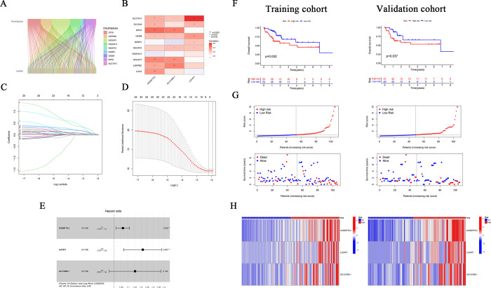
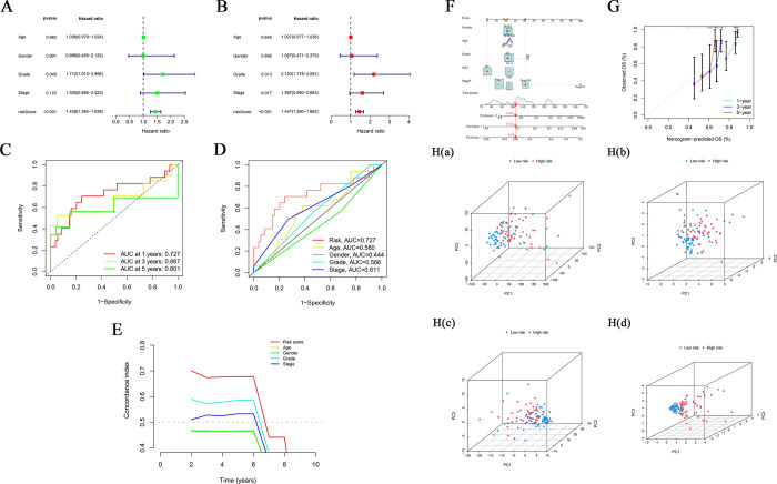
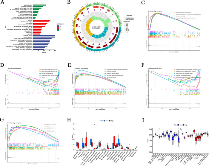
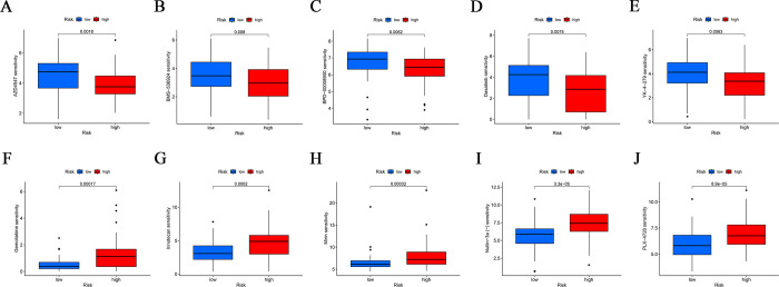
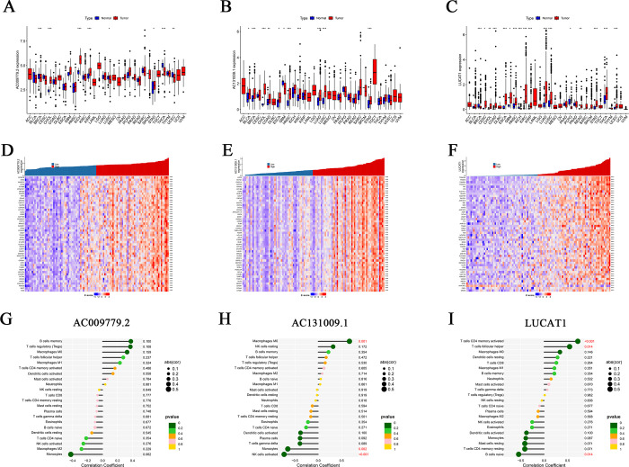
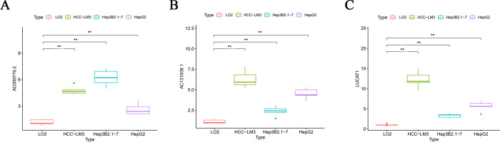
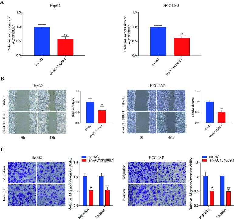
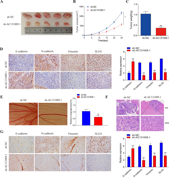
Disulfidptosis, a recently identified pathway of cellular demise, served as the focal point of this research, aiming to pinpoint relevant lncRNAs that differentiate between hepatocellular carcinoma (HCC) with and without vascular invasion while also forecasting survival rates and responses to immunotherapy in patients with vascular invasion (VI+). First, we identified 300 DRLRs in the TCGA database. Subsequently, utilizing univariate analysis, LASSO-Cox proportional hazards modeling, and multivariate analytical approaches, we selected three DRLRs (AC009779.2, AC131009.1, and LUCAT1) with the highest prognostic value to construct a prognostic risk model for VI+ HCC patients. Multivariate Cox regression analysis revealed that this model is an independent prognostic factor for predicting overall survival that outperforms traditional clinicopathological factors. Pathway analysis demonstrated the enrichment of tumor and immune-related pathways in the high-risk group. Immune landscape analysis revealed that immune cell infiltration degrees and immune functions had significant differences. Additionally, we identified valuable chemical drugs (AZD4547, BMS-536924, BPD-00008900, dasatinib, and YK-4-279) for high-risk VI+ HCC patients. In-depth bioinformatics analysis was subsequently conducted to assess immune characteristics, drug susceptibility, and potential biological pathways involving the three hub DRLRs. Furthermore, the abnormally elevated transcriptional levels of the three DRLRs in HCC cell lines were validated through qRT-PCR. Functional cell assays revealed that silencing the expression of lncRNA AC131009.1 can inhibit the migratory and invasive capabilities of HCC cells, a finding further corroborated by the chorioallantoic membrane (CAM) assay. Immunohistochemical analysis and hematoxylin-eosin staining (HE) staining provided preliminary evidence that AC131009.1 may promote the invasion and metastasis of HCC cells by inducing epithelial-mesenchymal transition (EMT) in both subcutaneous xenograft models and orthotopic HCC models within nude mice. To summarize, we developed a risk assessment model founded on DRLRs and explored the potential mechanisms by which hub DRLRs promote HCC invasion and metastasis.


Conflict of interest statement

The authors declare no competing financial interest.

Figures

Figure 1
Figure 1
Construction of the prognostic disulfidptosis-related lncRNA (DRLR) risk model for hepatocellular carcinoma (HCC).} (A) Sankey diagram showing the coexpression patterns of disulfidptosis-associated genes with DRLRs in VI HCC. (B) coexpression between 10 disulfidptosis-related genes and three prognostic lncRNAs independent of disulfidptosis. (C, D) Prognostic prediction model using the least absolute shrinkage and selection operator (LASSO)-Cox regression analysis. (E) Multivariable Cox regression analysis determined 3 hub DRLRs. (F) Kaplan–Meier survival curves for risk cohorts in both validation and training data sets. (G) distribution of risk scores and survival status within HCC cohorts. (H) Heatmap representing the expression of the three prognostic DRLRs in the examined cohorts.
Figure 2
Figure 2
Independent prognostic factor analysis based on VI+ HCC patient overall survival (OS).} (A) In the univariate Cox analysis, notable differences were observed in pathological characteristics and risk scores, which were statistically significant. (B) Multivariate Cox analysis also revealed significant statistical disparities in pathological characteristics and risk computations. (C) The time receiver operating characteristic (timeROC) curve predicts the 1-, 3-, and 5-year OS among patients with VI+ HCC. (D) The MultiROC assessment demonstrated that the predictive accuracy of the proposed risk model surpasses other clinical parameters. (E) According to the C-index, the prognostic precision of the risk model outperforms other clinical measures. (F) A nomogram constructed from the 3-DRLR signature score provides a prognostic prediction. (G) The calibration curve assists in forecasting overall survival (OS) at 1-year, 3-year, and 5-years. (H) Principal component analysis (PCA) was conducted to differentiate between high-risk and low-risk groupings across (Ia) all genes, (Ib) disulfidoptosis-related genes, (Ic) DRLRs, and (Id) prognostic markers associated with 3-DRLRs.
Figure 3
Figure 3
Elucidates the outcomes of Gene Ontology (GO) and Gene Set Enrichment Analysis (GSEA). (A) Displaying a bar chart representing the ten GO terms with the highest significance in enrichment. (B) Showing a circle diagram derived from GO enrichment analysis results. GSEA showed substantial discrepancies in the enrichment of the TCGA VI+ HCC cohort for the c2.all.v2022.1.Hs.symbols.gmt gene set between high-risk (C) and low-risk cohorts (D), as determined by the 3-DRLR signature. Similarly, the c5.all.v2022.1.Hs.symbols.gmt gene set showed differences between the high-risk (E) and low-risk cohorts (F). (G) Highlights significant enrichment of the h.all.v2022.1.Hs.symbols.gmt gene set in the high-risk cohort. (H) presents the percentages of 22 immune cell types in both the high- and low-risk groups as computed by the CIBERSORT methodology. (I) Comparison of immune function scores between patients categorized into low-risk and high-risk cohorts.
Figure 4
Figure 4
Drug sensitivity profiles for VI+ HCC patients in high-risk and low-risk groups based on the 3-DRLR signature. Analysis of the distribution of the half maximal inhibitory concentration (IC50) revealed significant differences between patients in the high-risk group and low-risk group for (A) AZD4547, (B) BMS-536924, (C) BPD-00008900, (D) dasatinib, (E) YK-4–279, (F) gemcitabine, (G) irinotecan, (H) mirin, (I) Nutlin-3a (−), and (J) PLX-4720.
Figure 5
Figure 5
Risk hub prognostic lncRNAs analysis in the 3-DRLR signature. Pancancer analysis showed differential expression of AC009779.2 (A), AC131009.1 (B), and LUCAT1 (C) in tumor and paracancerous tissues. Coexpression of the top 50 genes and 3 disulfidptosis-independent prognostic hub lncRNAs. The correlation heatmap shows the top 50 gene significantly coexpressed with AC009779.2 (D), AC131009.1 (E), and LUCAT1 (F). The relationship between the relative quantities of 22 immune cell types and the expression levels of lncRNA AC009779.2 (G), AC131009.1 (H), and LUCAT1 (I).
Figure 6
Figure 6
Validation of expression levels for prognostic hub lncRNAs within the context of the 3-DRLR model across different HCC cell lines. Expression levels of AC009779.2 (A), AC131009.1 (B), and LUCAT1 (C) were determined in HCC-LM3, Hep3B2.1–7, and HepG2 cell lines and contrasted against the normal liver cell line, L02. Data are presented as the mean ± standard deviation (SD) with n = 5. Statistically significant differences are indicated by **P < 0.01.
Figure 7
Figure 7
Impacts of lncRNA AC131009.1 on HCC cell migration and invasion. (A) The knockdown efficiency of sh-AC131009.1 compared with the negative control (NC) in HCC-LM3 and HepG2 cells was measured by qRT-PCR. (B) Wound healing assays of HepG2 and HCC-LM3 cells transfected with sh-AC131009.1 and sh-NC. (C) Transwell assays of HepG2 and HCC-LM3 cells transfected with sh-NC and sh-AC131009.1. Data are presented as mean ± SD (n = 3). Significant differences are denoted by **P < 0.01.
Figure 8
Figure 8
Impact of lncRNA AC131009.1 on HCC Cell Proliferation and Metastasis. Tumour dimensions and masses for the sh-AC131009.1 and sh-NC cohorts are depicted in panels A, B, and C. (D) The protein expression levels of E-cadherin, N-cadherin, vimentin and slug were detected in the tumor tissues in each group by IHC. (E) Chicken embryos were exposed to conditioned media (CM) for 4 days, photographed, and their vascular growth was quantified. (F) H&E staining is used to assess the tumor burden in the livers of mice in each group (magnified 40 times and 400 times, respectively). (G) IHC staining is used to measure the expression of E-cadherin, N-cadherin, vimentin, and slug in liver tumors of mice in each group. Data represented as mean ± SD for n = 5. Significant difference is indicated by *P < 0.05 and **P < 0.01.

Similar articles

Cited by

References

    1. Sung H.; Ferlay J.; Siegel R. L.; Laversanne M.; Soerjomataram I.; Jemal A.; Bray F. Global cancer statistics 2020: GLOBOCAN estimates of incidence and mortality worldwide for 36 cancers in 185 countries. CA Cancer J. Clin. 2021, 71, 209–49. 10.3322/caac.21660. - DOI - PubMed
    1. Llovet J. M.; Kelley R. K.; Villanueva A.; Singal A. G.; Pikarsky E.; Roayaie S.; Lencioni R.; Koike K.; Zucman-Rossi J.; Finn R. S. Hepatocellular carcinoma. Nat. Rev. Dis Primers. 2021, 7, 6.10.1038/s41572-020-00240-3. - DOI - PubMed
    1. Yang Y. Q.; Wen Z. Y.; Liu X. Y.; Ma Z. H.; Liu Y. E.; Cao X. Y.; Hou L.; Xie H. Current status and prospect of treatments for recurrent hepatocellular carcinoma. World J. Hepatol. 2023, 15, 129–150. 10.4254/wjh.v15.i2.129. - DOI - PMC - PubMed
    1. Sangro B.; Sarobe P.; Hervás-Stubbs S.; Melero I. Advances in immunotherapy for hepatocellular carcinoma. Nat. Rev. Gastroenterol. Hepatol. 2021, 18, 525–543. 10.1038/s41575-021-00438-0. - DOI - PMC - PubMed
    1. Zongyi Y.; Xiaowu L. Immunotherapy for hepatocellular carcinoma. Cancer Lett. 2020, 470, 8–17. 10.1016/j.canlet.2019.12.002. - DOI - PubMed

LinkOut - more resources