. 2024 Dec 4;9(50):49899-49912.
doi: 10.1021/acsomega.4c08830. eCollection 2024 Dec 17.

Stachys byzantina K. Koch in the Treatment of Skin Inflammation: A Comprehensive Evaluation of Its Therapeutic Properties

Affiliations

Stachys byzantina K. Koch in the Treatment of Skin Inflammation: A Comprehensive Evaluation of Its Therapeutic Properties

José Alisson da Silva Lima et al. ACS Omega. .

Abstract

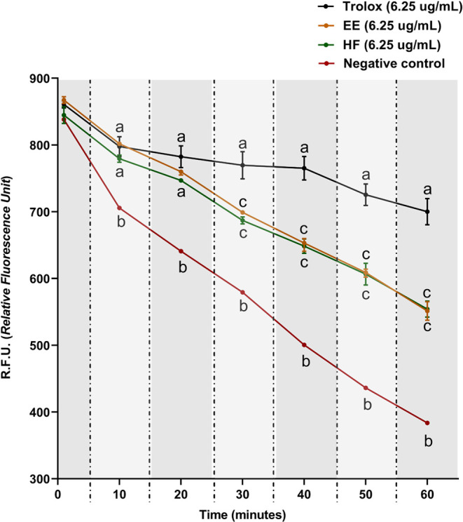
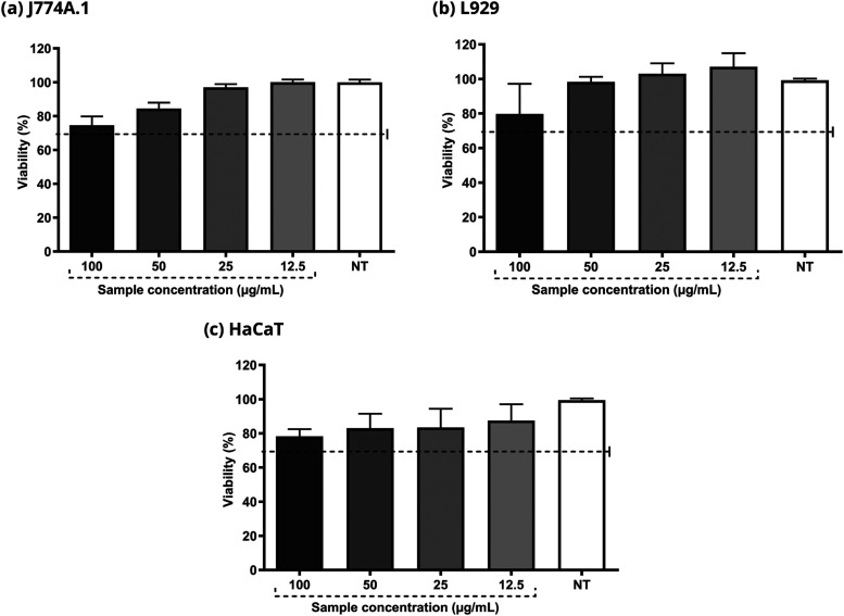
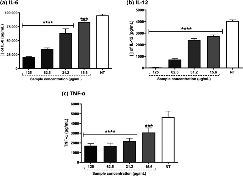
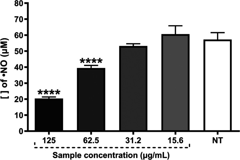
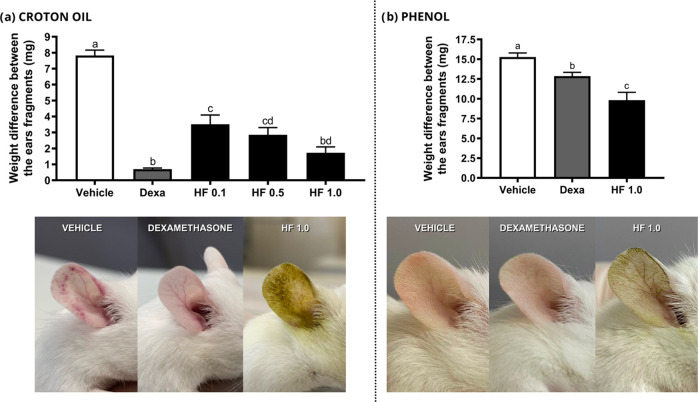
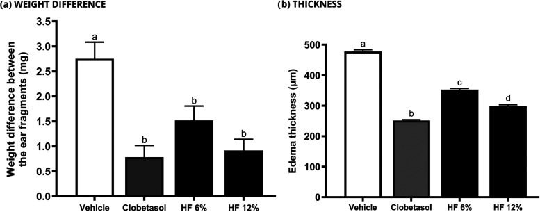
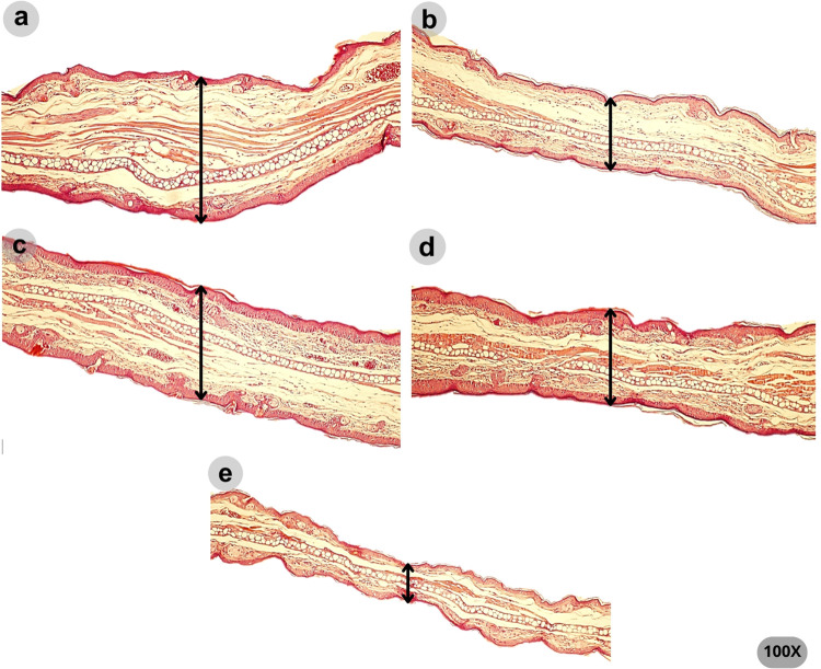
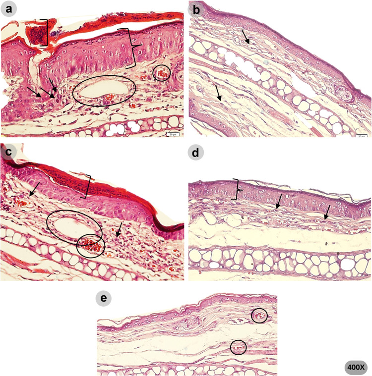
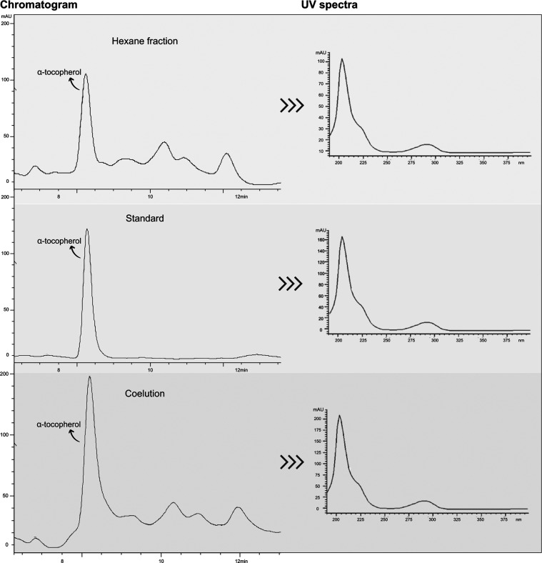
Stachys byzantina is a plant widely cultivated for food and medicinal purposes. Stachys species have been reported as anti-inflammatory, antibacterial, anxiolytic, and antinephritic agents. This study aimed to evaluate the anti-inflammatory potential of the ethanolic extract (EE) from the aerial parts of S. byzantina and its most promising fraction in models of acute and chronic inflammation, including a psoriasis-like mouse model. The EE was fractionated into hexane (HF), dichloromethane (DF), ethyl acetate (AF), and hydroalcoholic (HD) fractions. Screening for anti-inflammatory activity based on nitric oxide inhibition (IC50 μg/mL: HF 24.29 ± 5.87, EE 176.45 ± 18.65), hydroxyl radical scavenging (HF 3.89 ± 0.61, EE 6.38 ± 2.25), β-carotene/linoleic acid assay (HF 10.13 ± 3.81, EE 25.64 ± 2.12), and ORAC identified HF as the most active fraction. Topical application of HF effectively reduced croton oil- and phenol-induced ear edema in mice, with no statistical difference to the reference drugs. A formulation containing HF showed significant activity in the imiquimod-induced psoriasis model, reducing pro-inflammatory cytokines and nitric oxide production in macrophages, with no cytotoxicity to skin cells. Phytochemical analysis of HF revealed the presence of terpenes, steroids (491.68 ± 4.75 mg/g), phenols (34.30 ± 4.96 mg/g), flavonoids (151.77 ± 6.66 mg/g), and α-tocopherol, which was identified and quantified by HPLC-UV analysis (10.56 ± 0.97 mg/g of HF). These findings highlight the therapeutic potential of S. byzantina for skin inflammation, particularly contact dermatitis and psoriasis, encouraging further studies, including in human volunteers.


Conflict of interest statement

The authors declare no competing financial interest.

Figures

Figure 1
Figure 1
HF and EE kinetics in ORAC assay. Two-way ANOVA followed by Bonferroni test. The values obtained represent the mean ± s.e.m. of R.F.U. (Relative Fluorescence Unit). The data were obtained by six readings, each one being carried out within 10 min. Equal letters indicate no statistical difference (p < 0.05).
Figure 2
Figure 2
Evaluation of HF cytotoxicity in J774A.1, L929 and HaCaT cells. The concentrations used varied between 12.5 and 100 μg/mL. DMEM culture medium was used as the negative control. Viability above 70% characterizes a nontoxic sample.
Figure 3
Figure 3
Determination of IL-6, IL12, and TNF-α cytokines in J774A.1 cell line stimulated with E. coli LPS. HF concentrations range from 125 to 15.6 μg/mL. ANOVA followed by the Dunnet test. Significant values: ****p < 0.0001 and ***p < 0.001.
Figure 4
Figure 4
Determination of NO radical concentration in J774A.1 cell line stimulated with LPS 3 from E. coli and IFN-γ. HF concentrations ranged from 125 to 15.6 μg/mL. ANOVA 4 followed by the Dunnet test. Significant values: ****p < 0.0001.
Figure 5
Figure 5
Effect of HF on the inflammatory stimulus induced by croton oil and phenol, in addition to representative photographs of the clinical appearance of mice ears (n = 7 and 8 animals, respectively). Negative control (vehicle - acetone), dexamethasone (Dexa) 0.1 mg/20 μL and HF at 0.1; 0.5 and 1.0 mg/20 μL were topically administered immediately after topical application of 2.5% croton oil and phenol 10% (v/v). Values in each column represent the mean ± s.e.m. of the weight difference between ear fragments (mg). ANOVA, followed by the Newman-Keuls test. Equal letters indicate no statistical difference (p < 0.05).
Figure 6
Figure 6
Effect of HF on the weight difference between ear fragments in the imiquimod-induced ear edema test (n = 8 animals). Negative control (vehicle - formulation), clobetasol 0.5 mg/g, HF 6 and 12% were topically applied for 10 days to investigate the antipsoriatic activity. Values in each column represent the mean ± s.e.m. of weight (mg) and thickness (μm) of the right ear edema fragments for each group. ANOVA, followed by the Newman-Keuls test. Means represented by equal letters indicate no statistical difference (p < 0.05).
Figure 7
Figure 7
Representative photomicrographs (100× magnification) of the ear tissues on the last day of the imiquimod-induced ear edema test. The variation in edema thickness between groups is shown by the arrow. Means for each group were as follows: (a) vehicle (477.68 μm), (b) clobetasol (251.45 μm), (c) HF 6% (352.84 μm), (d) HF 12% (299.03 μm), and (e) left ear (186.17 μm).
Figure 8
Figure 8
Representative photomicrographs (400× magnification) of the ear tissue on the last day of the imiquimod-induced ear edema test. The presence of nuclei in the corneal layer indicates parakeratosis (in brackets). Leukocyte infiltrate is indicated by arrows. Vasodilation was highlighted by a circle. Increased epidermal thickness (hyperkeratosis), typical of psoriasis lesions is shown in braces. (a) vehicle, (b) clobetasol, (c) HF 6%, (d) HF 12%, and (e) left ear.
Figure 9
Figure 9
Chromatogram and UV spectra obtained by HPLC-UV analysis. The identification of α-tocopherol was carried out by comparing the UV spectra and retention time, and by coelution. Chromatographic conditions: Methanol and UHQ water (98:2, v/v) as mobile phase, at a flow rate of 1.3 mL/min. α-tocopherol standard at 25 μg/mL and PHEX at 1000 μg/mL were diluted in the mobile phase. The Injection volume was 20 μL, and the temperature was 25 °C. Agilent Eclipse Plus C18 column was used. UV detection was performed at 205 nm.

Similar articles

References

    1. Gallegos-Alcalá P.; Jiménez M.; Cervantes-garcía D.; Córdova-dávalos L. E.; Gonzalez-curiel I.; Salinas A. Glycomacropeptide Protects against Inflammation and Oxidative Stress and Promotes Wound Healing in an Atopic Dermatitis Model of Human Keratinocytes. Foods 2023, 12, 193210.3390/foods12101932. - DOI - PMC - PubMed
    1. Kabashima K.; Honda T.; Ginhoux F.; Egawa G. The immunological anatomy of the skin. Nat. Rev. Imunol. 2019, 19, 19–30. 10.1038/s41577-018-0084-5. - DOI - PubMed
    1. Wagener F.A.D.T.G.; Carels C. E.; Lundvig D. M. S. Targeting the redox balance in inflammatory skin conditions. Int. J. Mol. Sci. 2013, 14, 9126–9167. 10.3390/ijms14059126. - DOI - PMC - PubMed
    1. Khan A. Q.; Agha M. V.; Sultan M. K.; Sheikhan A.; Younis S. M.; Tamimi M. A.; Alam M.; Ahma A.; Uddin S.; Buddenkotte J.; Steinhoff M. Targeting deregulated oxidative stress in skin inflammatory diseases: An update on clinical importance. Biomed. Pharmacother. 2022, 154, 11360110.1016/j.biopha.2022.113601. - DOI - PubMed
    1. Poetker D. M.; Reh D. D. A comprehensive review of the adverse effects of systemic corticosteroids. Otolaryngol. Clin. North Am. 2010, 43, 753–768. 10.1016/j.otc.2010.04.003. - DOI - PubMed

LinkOut - more resources