. 2025 Jan 14;97(1):427-435.
doi: 10.1021/acs.analchem.4c04542. Epub 2024 Dec 23.

Millisecond Label-Free Single Peptide Detection and Identification Using Nanoscale Electrochromatography and Resistive Pulse Sensing

Affiliations

Millisecond Label-Free Single Peptide Detection and Identification Using Nanoscale Electrochromatography and Resistive Pulse Sensing

Maximillian Chibuike et al. Anal Chem. .

Abstract

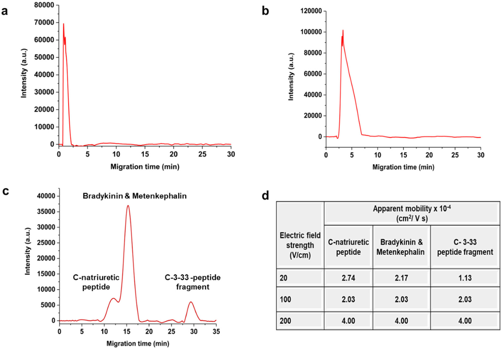
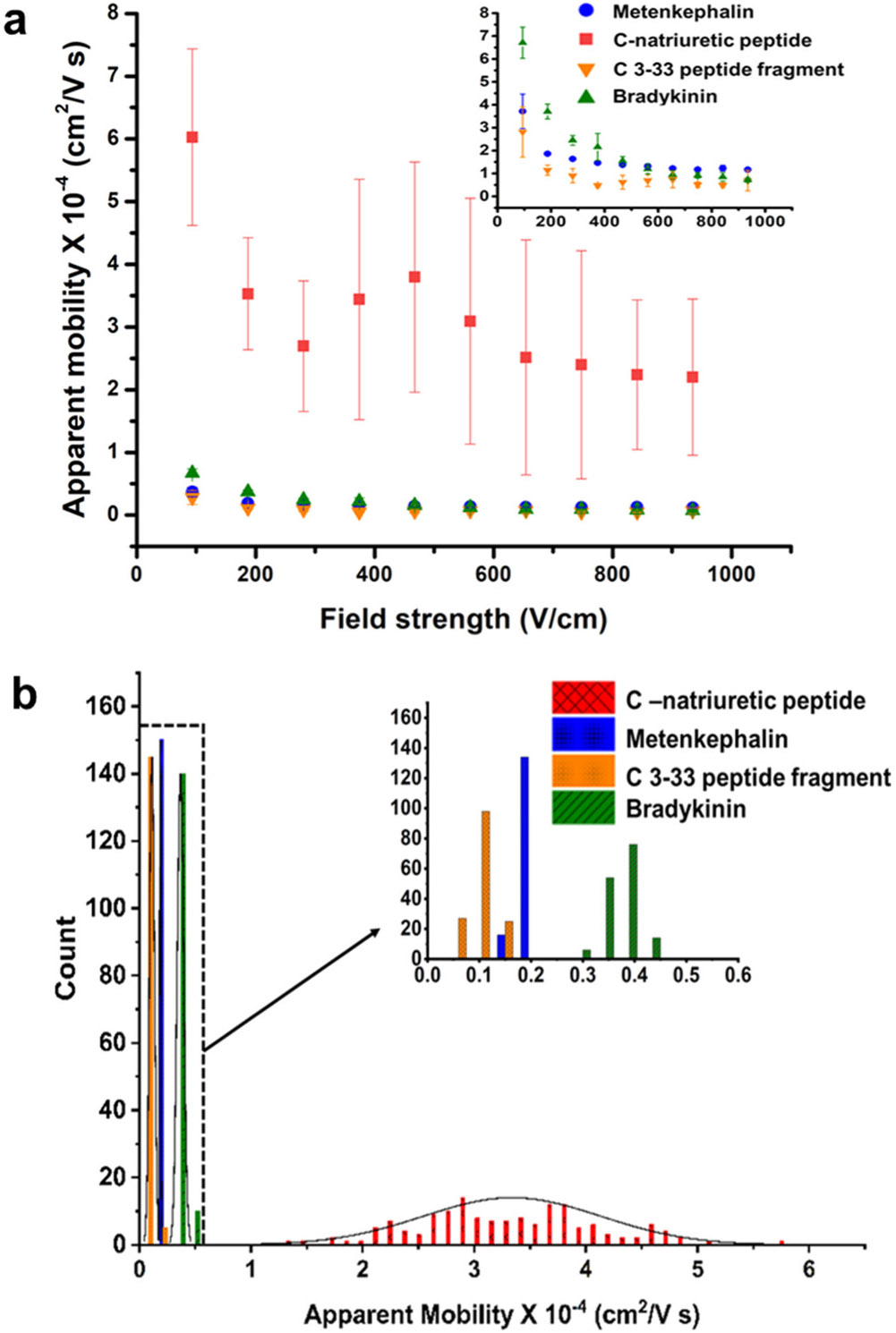
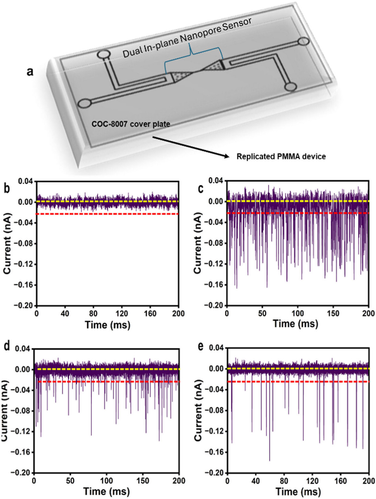
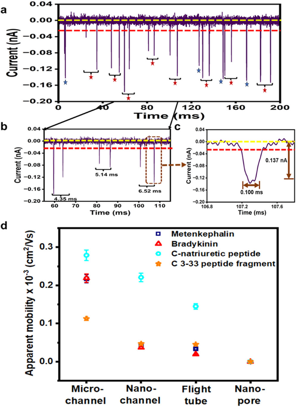
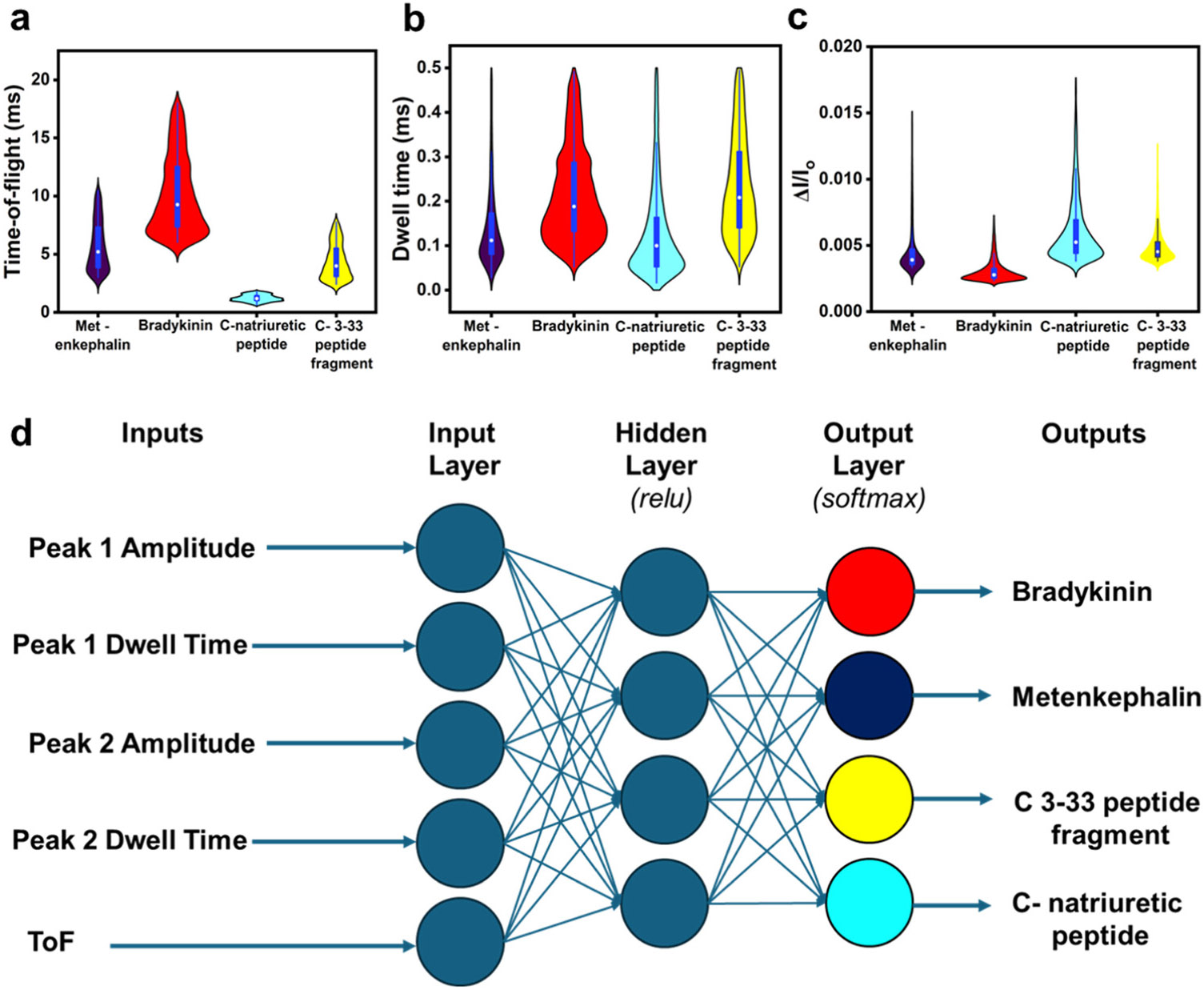
We are developing a unique protein identification method that consists of generating peptides proteolytically from a single protein molecule (i.e., peptide fingerprints) with peptide detection and identification carried out using nanoscale electrochromatography and label-free resistive pulse sensing (RPS). As a step in realizing this technology, we report herein the nanoscale electrochromatography of model peptides using thermoplastic columns with surfaces engineered to identify peptides via their molecularly dependent mobility (i.e., time-of-flight, ToF). ToFs were elucidated using a dual in-plane nanopore sensor, which consisted of two in-plane nanopores placed on either end of the nanoelectrochromatography column. The surface of the nanocolumn, which consisted of poly(methyl methacrylate) (PMMA), was activated with an O2 plasma, creating surface carboxylic acid groups (-COOH) inducing a surface charge on the column wall as well as affecting its hydrophilicity. To understand scaling effects, we carried out microchip and nanochannel electrochromatography of the peptides labeled with an ATTO 532 reporter to allow for single-molecule tracking. Our results indicated that the apparent mobilities of the model peptides did not allow for their separation in a microchannel, but when performed in a nanocolumn, clear differences in their apparent mobilities could be observed especially when operated at high electric field strengths. We next performed label-free detection of peptides using the dual in-plane nanopore sensor with the two pores separated by a 5 μm (length) column with a 50 nm width and depth. When a single peptide molecule passed through an in-plane nanopore, the sensor read a pair of resistive pulses with a time difference equivalent to ToF. We identified the peptides by evaluating their ToF, normalized RPS current transient amplitude (ΔI/I0), and RPS peak dwell time (td). We could identify the model peptides with nearly 100% classification accuracy at the single-molecule level using machine learning with a single molecule measurement requiring <10 ms.


Figures

Figure 1.
Figure 1.
Microchip electropherograms of model peptides in PMMA/COC microchips having dimensions of 50 μm × 100 μm (depth × width, respectively) with a 5 cm total column length (effective length = 4 cm). The electrochromatography was performed using 10 μM peptides labeled with ATTO 532. The peptides were detected using a laser-induced fluorescence system equipped with a 20 mW, 532 nm excitation laser and an electric field strength of (a) 200 V/cm, (b) 100 V/cm, and (c) 20 V/cm. (d) Listing of the apparent mobilities of the peptides at different electric field strengths. The cover plate used to enclose the fluid network was a COC 8007. Prior to thermal fusion bonding, both the cover plate and the substrate were activated with UV/O3 at 20 mW/cm2 for 17 min
Figure 2.
Figure 2.
(a) Apparent mobility (cm2/(V s)) vs electric field strength of the model peptides in PMMA/COC (substrate/cover plate) nanochannel devices. The inset shows an expanded view of apparent mobilities vs electric field strength of bradykinin, metenkephalin, and C-peptide 3–33. The electrophoresis was performed on 100 nM peptides labeled with ATTO 532. (b) Histograms of the apparent mobilities of peptides at 180 V/cm in PMMA/COC nanochannel devices. The histograms were fit to Gaussian functions and each bin width represented 1.5 × 106 cm2/(V s). The inset shows the histograms of apparent mobilities of peptides between (0–0.65) × 10−4 cm2/(V s). The devices represent hybrid devices in which a low Tg cover plate (COC 8007) was thermally fusion bonded to the higher Tg substrate (PMMA). Prior to assembly, the device was activated with an O2 plasma for 1 min
Figure 3.
Figure 3.
Electrokinetic transport of metenkephalin through PMMA/COC dual in-plane nanopore sensors assembled at 75 °C and 175 psi bonding pressure. (a) A schematic of the PMMA/COC 8007 dual in-plane nanopore sensor. The sensors were hybrid devices in which a low Tg cover plate (COC 8007) was thermally fusion bonded at 175 psi and 75 °C to a higher Tg substrate (PMMA). Before assembly, both the cover plate and the substrate were activated for 1 min with an O2 plasma. A 200 ms current transient trace of signal amplitudes was obtained for (b) blank, (c) 100, (d) 10, and (e) 1 nM solutions of metenkephalin in 0.5× PBS, 0.5 M KCl at pH 7.4 with an applied voltage of 2.5 V using the PMMA/COC dual in-plane sensor device. The event rate increased with an increase in concentration. All RPS measurements on the peptides were performed without a fluorescent label (i.e., label-free). The dashed red line shows the amplitude threshold used to reduce the possibility of scoring false events.
Figure 4.
Figure 4.
Using the PMMA/COC dual in-plane nanopore sensor to measure RPS parameters and ToF. (a) A 200 ms trace of the current–time signal acquired under a bias voltage of 2.5 V with a bandwidth of 10 kHz and a sampling frequency of 250 kHz. The yellow and red lines represent the baseline and detection threshold, respectively. The red stars indicate paired peaks, which correspond to a single metenkephalin peptide as the molecule electrokinetically moves through the pores and flight tube. Peak pairs were selected based on peak pair selection criteria (see the main text). The blue stars are peaks that did not meet the selection criteria. (b) Example of paired peaks with ToFs calculated. (c) An expanded current transient peak showing peak amplitude and td. (d) Plot of peptides’ apparent mobility in different channel dimensions: microchip electrophoresis, nanochip electrophoresis (110 nm × 110 nm; no in-plane pores), flight tube (50 nm × 50 nm), and nanopore (~10 nm apparent width). Microchannel, nanochannel, and dual in-plane nanopore sensor devices were fabricated in PMMA/COC devices.
Figure 5.
Figure 5.
Violin plots of ToFs, td, and ΔI/I0 for the label-free identification of single peptide molecules. 1 nM of each peptide was translocated through the dual in-plane nanopore sensor fabricated in PMMA/COC with a 5 μm length nanoflight tube. The nanoelectrochromatography was carried out using 0.5× PBS, 0.5 M KCl (pH 7.4), and an applied voltage of 2.5 V. The plots for each peptide show the medians (white circle) of (a) ToF, (b) td, and (c) ΔI/I0 for metenkephalin, bradykinin, C-natriuretic peptides, and C 3–33 peptide. The bars represent the range of data for each peptide within the 1.5 interquartile range (IQR). The μapp was calculated from ToFs. Mood’s median test at 0.05 confidence level evaluated using originPro 2023 data analysis software showed that TOFs, dwell times, and ΔI/I0 populations across the peptides were statistically different (p < 0.05). (d) Classification of peptides with machine learning via a neural network. Accuracy was 95.6 and 100% for the training and validation set, respectively

Similar articles

References

    1. Davis L. Basic Methods in Molecular Biology; Elsevier, 2012.
    1. Zhao Q; Jayawardhana DA; Wang D; Guan XJ Phys. Chem. B 2009, 113, 3572–3578. - PubMed
    1. Restrepo-Pérez L; Joo C; Dekker C Nat. Nanotechnol 2018, 13, 786–796. - PubMed
    1. Solntceva V; Kostrzewa M; Larrouy-Maumus G Front. Cell. Infect. Microbiol 2021, 10, No. 621452. - PMC - PubMed
    1. Kasicka V. Electrophoresis 1999, 20, 3084–3105. - PubMed

MeSH terms

LinkOut - more resources