. 2025 Feb;14(4):e2403000.
doi: 10.1002/adhm.202403000. Epub 2024 Dec 23.

Structured Polymers Enable the Sustained Delivery of Glucocorticoids within the Intra-Articular Space

Affiliations

Structured Polymers Enable the Sustained Delivery of Glucocorticoids within the Intra-Articular Space

Ana Crastin et al. Adv Healthc Mater. 2025 Feb.

Abstract

Intra-articular glucocorticoid injections are effective in controlling inflammation and pain in arthritides but restricted by short duration of action and risk of joint degeneration. Controlled drug release using biocompatible hydrogels offers a unique solution, but limitations of in situ gelation restrict their application. Gellan sheared hydrogels (GSHs) retain the advantages of hydrogels, however their unique microstructures lend themselves to intra-articular application - capable of shear thinning under force but restructuring at rest to enhance residence. This study examined GSHs for extended intra-articular glucocorticoid delivery of prednisolone (10 mg mL-1); demonstrating links between material mechanics, steroid release, and preclinical assessment of efficacy in synoviocyte culture and transgenic(TNF)197Gkl (TNFtg) murine model of arthritis. GSHs demonstrated sustained release, with typical Fickian profiles over 18 days. Moreover, systems showed good stability under extended culture, with inherent cell-compatibility and suppression of inflammatory synoviocyte activation. In TNFtg animals, GSHs suppressed synovitis (70.08%, p < 0.05), pannus formation (45.01%, p < 0.05), and increased articular cartilage (82.23%, p < 0.05) relative to vehicle controls. The extended profile of steroid release from injectable GSH formulations holds promise in the treatment and management of inflammatory arthritides such as rheumatoid and osteoarthritis, representing a step-change in intra-articular drug delivery to suppress long-term joint inflammation.

Keywords: arthritis; gellan; glucocorticoid; intra‐articular; sheared hydrogel.


Conflict of interest statement

Both L.M. Grover and R.J.A. Moakes are inventors of a patent describing the production of sheared gels and are founders in a company seeking to commercialize the technology (Healome Therapeutics Ltd).

Figures

Figure 1
Figure 1
A) Schematic of known gellan sheared hydrogel structure supported by representative microscopy image of polymer strands after separation in polyethylene glycol (scale bar 50 µm). B) Representative images of blue dye labeled gellan gum sheared hydrogel (GSH) (1.5% (w/v), 10 mM NaCl; Left) and red dye labeled saline (right) following administration using a 30‐gauge needle to a plastic model of the femur‐tibia articular space and over ten 90 ° mechanical articulations captured using a high‐speed camera. Black arrows denote saline egress from joint. ci‐iii) Viscosity test, Amplitude sweep test, and Frequency sweep test of GSH formulations (1,5, and 2% gellan at 10 mM NaCl, of 1,5 and 2% gellan at 10 mM NaCl) determined by linear rheology. D) Injection pressure (Pa) required to eject GSH formulations (1,5% gellan with 10 mM and 20 mM NaCl, 2% gellan with 10 mM and 20 mM NaCl and 3% gellan with 10 mM and 20 mM NaCl) through a 30‐gauge needle relative to distilled water. Data presented as mean ± SEM of at least 3 replicates per gel formulation.
Figure 2
Figure 2
A) Gel weights (1.5‐3% gellan (w/v), 10–20 mM NaCl, 50 µg mL−1 cortisol) after being maintained in RPMI culture media at 0 and 21 days. B) Accumulation of cortisol into media determined by ELISA after incubation with gellan sheared hydrogel (GSH) formulations (1.5‐3% gellan (w/v), 10 mM NaCl, 50 µg mL−1 cortisol) over 30 days. C) Accumulation of cortisol into media from gels determined by ELISA after incubation with GSH formulations (1.5% gellan (w/v), 10–20 mM NaCl, 5 µg/ml cortisol) over 24 days. D) H3‐labelled cortisol detection in culture media and within GHS (1.5% (w/v) gellan, 10 mM NaCl) at day 5 relative to matched GSH at day 0. E) mRNA expression (AU) of Gilz in human primary macrophage cultures treated for 24 h with conditioned media collected from cortisol loaded GSH (1.5% (w/v) gellan, 10 mM NaCl, cortisol 5 µM) at days 3, 5, 8, 10, and 12, determined by quantitative RT‐PCR. Data are presented as mean ± SEM of at least 3 replicates per gel formulation. Statistical significance was determined using one‐way ANOVA with Tukey's multiple comparisons test (* p ≤ 0.05, **p ≤ 0.01, *** p ≤ 0.001).
Figure 3
Figure 3
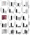
All groups consisted of treatments with either 25 µL of gellan sheared hydrogel (GSH) (1.5% gellan (w/v), 10 mM NaCl,) containing either vehicle (Gel), 5 µM cortisol (Cort Gel) relative to untreated control for 48 h for functional analysis or 24 h for mRNA analysis. A) Representative image by microscopy (scale 50 µl), B) cell viability determined by MTT, and C) phagocytosis determined by Vybrant™ E. coli phagocytosis assay in primary human macrophages. D‐F) mRNA expression (AU) of GILZ, TNFA, and CD163 determined by quantitative RT‐PCR and G‐H) cytokine release of TNFα and IL‐12 determined by ELISA in primary macrophages. I) Representative image of TRAP‐stained primary osteoclasts by microscopy (scale 50 µl). J) mRNA expression (AU) of CTSK determined by quantitative RT‐PCR, K) Primary human osteoclast perimeter (µM), and L) Average Bone erosion scores (AU) in primary human osteoclasts. M) Cell viability determined by MTT and N‐P) mRNA expression (AU) of GILZ, TNFA, and CD163 determined by quantitative RT‐PCR in primary human fibroblast‐like synoviocyte culture. Q) Representative image by microscopy (scale 50 µL) of primary human myotube culture. R‐T) mRNA expression (AU) of GILZ, IL6, and FOXO1 determined by quantitative RT‐PCR in primary human myotubes. Data are presented as mean ± SEM of at least three primary cultures from three independent patient donors. Statistical significance was determined using one‐way ANOVA with Tukey's multiple comparisons test (* p ≤ 0.05, **p ≤ 0.01, *** p ≤ 0.001).
Figure 4
Figure 4
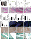
All groups consisted of intra‐articular injection with 10 µL of either saline control (CON), blank GSH (GEL: 488 1.5% gellan (w/v), 10 mM NaCl), methylprednisolone sodium succinate (PRED: 10 mg mL−1) or GSH loaded with methylprednisolone sodium succinate (PRED GEL: Gel: 1.5% gellan (w/v), 10 mM NaCl, Pred 10 mg mL−1). A) Representative image of Coomassie blue dye loaded gellan sheared hydrogel (GSH) administered into the intra‐articular space in the TNFtg murine model of polyarthritis. B) Body weight, C) disease score, and D) Inflammatory paw score D) measured over 21 days in the TNFtg model of polyarthritis following intra‐articular injection. E) Representative sagittal images of formalin‐fixed paraffin embedded knee joints, stained with hematoxylin and eosin, and F) measurement of synovitis, G) sub‐chondral pannus formation, and H) surface cartilage area in TNFtg animals at day 21 following treatment. I‐K) Fluorescence intensity/um2 of synovial tissue as a measure of expression of TNFα and podoplanin, and representative images of fluorescent staining for RANKL, TNFa, podoplanin, and DAPI, determined by confocal immunofluorescence area in TNFtg animals at day 21 following treatment. L) Representative GILZ staining within synoviocytes was determined in the same groups within articular synovium by immunohistochemistry. M) Representative alcian blue staining within the articular space was performed in TNFtg animals after 21 days following injection with either saline control (Con) or GSH loaded with methylprednisolone sodium succinate at 20 and 60X magnification. Black arrows denote positively stained GSHs. Data are presented as mean ± SEM of at least three six animals per group. Statistical significance was determined using one‐way ANOVA with Tukey's multiple comparisons test (* p ≤ 0.05, **p ≤ 0.01, *** p ≤ 0.001). (scale bars, 50 µm).
Figure 5
Figure 5
Schematic demonstrating the in vivo study design. TNF‐tg animals were maintained for 36 days from birth up to onset of early joint inflammation before receiving one of three therapeutic intervention wings by intra‐articular injection into the right leg, consisting of either gellan sheared hydrogel loaded with saline, gellan sheared hydrogel loaded with methylprednisolone sodium succinate (10 mg mL−1), or methylprednisolone sodium succinate made up in saline. All animals received a saline control injection into the left leg for the purpose of providing a matched control. Animals were scored for systemic and local disease activity up to day 58 when animal were euthanized and tissues harvested for analysis. (ii), schematic showing the known polymer structure of gellan sheared hydrogel and the predicted location of methylprednisolone both at rest in the resting joint and under mechanical articulation. (ii), schematic detailing the known changes in local inflammatory signaling mediated by glucocorticoid loaded hydrogels in primary cell cultures of joint cells, including osteoclasts, synovial macrophages, and synovial fibroblasts.

References

    1. Guo Q., Wang Y., Xu D., Nossent J., Pavlos N. J., Xu J., Bone Res. 2018, 6, 15. - PMC - PubMed
    1. Tong L., Yu H., Huang X., Shen J., Xiao G., Chen L., Wang H., Xing L., Chen D., Bone Res. 2022, 10, 60. - PMC - PubMed
    1. Deyle G. D., Allen C. S., Allison S. C., Gill N. W., Hando B. R., Petersen E. J., Dusenberry D. I., Rhon D. I., N. Engl. J. Med. 2020, 382, 1420. - PubMed
    1. Armstrong R. D., English J., Gibson T., Chakraborty J., Marks V., Ann. Rheum. Dis. 1981, 40, 571. - PMC - PubMed
    1. Hajialilo M., Ghorbanihaghjo A., Valaee L., Kolahi S., Rashtchizadeh N., Amirkhiz M. B., Malekmahdavi I., Khabbazi A., Clin. Rheumatol. 2016, 35, 2887. - PubMed

MeSH terms