Review
. 2025 Jan 30;45(1):1-18.
doi: 10.1042/BSR20240576.

Electron transfer in multicentre redox proteins: from fundamentals to extracellular electron transfer

Affiliations
Review

Electron transfer in multicentre redox proteins: from fundamentals to extracellular electron transfer

Büşra Bayar et al. Biosci Rep. .

Abstract

Multicentre redox proteins participate in diverse metabolic processes, such as redox shuttling, multielectron catalysis, or long-distance electron conduction. The detail in which these processes can be analysed depends on the capacity of experimental methods to discriminate the multiple microstates that can be populated while the protein changes from the fully reduced to the fully oxidized state. The population of each state depends on the redox potential of the individual centres and on the magnitude of the interactions between the individual redox centres and their neighbours. It also depends on the interactions with binding sites for other ligands, such as protons, giving origin to the redox-Bohr effect. Modelling strategies that match the capacity of experimental methods to discriminate the contributions of individual centres are presented. These models provide thermodynamic and kinetic characterization of multicentre redox proteins. The current state of the art in the characterization of multicentre redox proteins is illustrated using the case of multiheme cytochromes involved in the process of extracellular electron transfer. In this new frontier of biological electron transfer, which can extend over distances that exceed the size of the individual multicentre redox proteins by orders of magnitude, current experimental data are still unable, in most cases, to provide discrimination between incoherent conduction by heme orbitals and coherent band conduction.


Conflict of interest statement

The authors declare no conflict of interests.

Figures

Figure 1
Figure 1. Illustration of three scenarios for proteins with the same apparent mid-point potential (200 mV), but where the distribution of the potentials of the individual independent n = 1 redox centres is different.
The curves were calculated considering the sum of contributions from eqn 1. The curve in red was calculated considering that all centres have a potential E0 of 200 mV; the curve in blue was calculated considering that all centres have different potentials E0 of 100, 200, and 300 mV; and the curve in green was calculated considering that two centres have a potential E0 of 227 mV and one has a potential E0 of 83 mV.
Figure 2
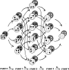
Figure 2. Diagram illustrating the total microstates to be considered for a protein with one redox centre
(A), two redox centres (B), four redox centres and an acid–base centre (C). Black and white dots represent reduced and oxidized redox centres, respectively. White and grey circles represent deprotonated and protonated microstates, respectively. Crossed and striped circles represent the fully reduced and the semi-reduced protein.
Figure 3
Figure 3. Titration curves calculated using eqn 1 for a mid-point potential of 200 mV, illustrating the effects of cooperativity on the redox behavior.
The red curve was calculated considering n = 1 and represents a Nernst curve with no cooperativity. The blue curve was calculated considering n = 2 and demonstrates the effect of positive cooperativity, characterized by a steeper slope. The green curve was calculated considering n = 0.5 and represents the effect of negative cooperativity, where the slope is flatter than the Nernstian reference. All curves were calculated considering the same midpoint potential.
Figure 4
Figure 4. Schematic representation of the 32 microstates of a protein with four redox centres coupled to one acid–base centre, which upon reaction with an electron donor can be interconverted through 64 possible electron transfer microsteps.
Black and white dots represent reduced and oxidized redox centres, respectively, while white and grey rounded squares represent deprotonated and protonated microstates, respectively.
Figure 5
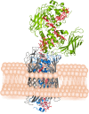
Figure 5. Representation of the MtrCAB structure from S. baltica (PDB ID: 6R2Q).
The transmembrane porin MtrB (gray) accommodates the MHC MtrA (blue) and associates with extracellular cytochrome MtrC (green). Hemes are represented in red sticks.
Figure 6
Figure 6. Three-dimensional structures of the porin–cytochromes predicted by AlphaFold 2: MtrD: AF-Q8e.g.30; DmsE: AF-Q8E9C4; PioA: AF-A1EBT2; MtoA: AF-D5CMQ0; and CwcA: AF-A0A0L6W663.
The hemes were inserted into the structures as described in the literature [76]. For MtrA, the experimentally reported structure was used (PDB code: 6R2Q_A).
Figure 7
Figure 7. Three-dimensional structures of the polymerising MHC nanowires.
Hemes are numbered according to the order of attachment to the polypeptide chain. Portions of heme arrangement similarity are present in all except OmcZ, and dashed lines connect hemes with similar arrangements. PDB codes used: 7TFS_A (OmcE), 6EF8_A (OmcS), 7LQ5_A (OmcZ), 8E5G_A (AvECN), and 8E5F (PcECN). The scale bar is represented at the bottom left corner.

Similar articles

Cited by

References

    1. Logan B.E., Rabaey K. Conversion of wastes into bioelectricity and chemicals by using microbial electrochemical technologies. Science. 2012;337:686–690. doi: 10.1126/science.1217412. - DOI - PubMed
    1. Kim Y., Logan B.E. Microbial desalination cells for energy production and desalination. Desal. 2013;308:122–130. doi: 10.1016/j.desal.2012.07.022. - DOI
    1. Cui Y., Lai B., Tang X. Microbial fuel cell-based biosensors. Biosensors. 2019;9:92. doi: 10.3390/bios9030092. - DOI - PMC - PubMed
    1. Al-Mamun A., Ahmed W., Jafary T., Nayak J.K., Al-Nuaimi A., Sana A. Recent advances in microbial electrosynthesis system: metabolic investigation and process optimization. Biochem. Eng. J. 2023;196:108928. doi: 10.1016/j.bej.2023.108928. - DOI
    1. Rehnlund D., Lim G., Philipp L.A., Gescher J. Nanowired electrodes as outer membrane cytochrome-independent electronic conduit in Shewanella oneidensis. i. Sci. 2022;25:103853. doi: 10.1016/j.isci.2022.103853. - DOI - PMC - PubMed

Publication types

LinkOut - more resources