

The evolutionary modifications of a GoLoco motif in the AGS protein facilitate micromere formation in the sea urchin embryo
- PMID: 39714020
- PMCID: PMC11666239
- DOI: 10.7554/eLife.100086
The evolutionary modifications of a GoLoco motif in the AGS protein facilitate micromere formation in the sea urchin embryo
Abstract
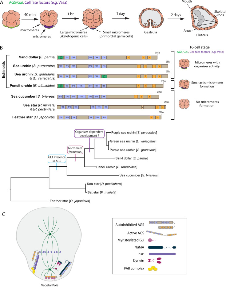
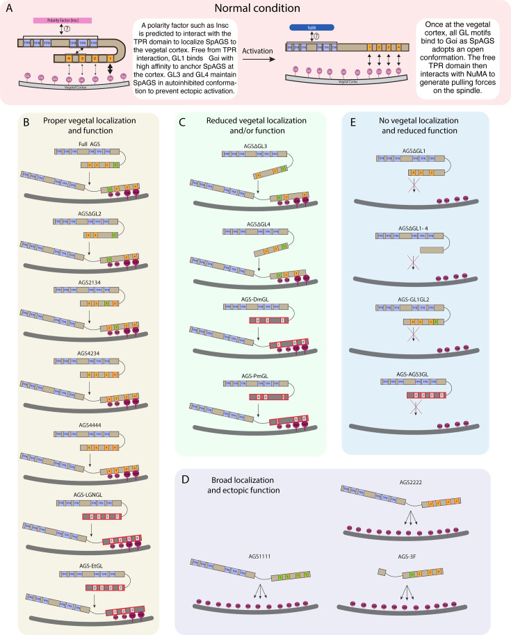
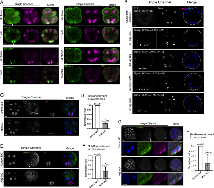
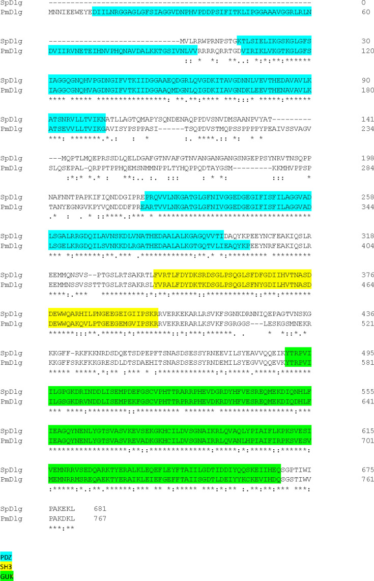
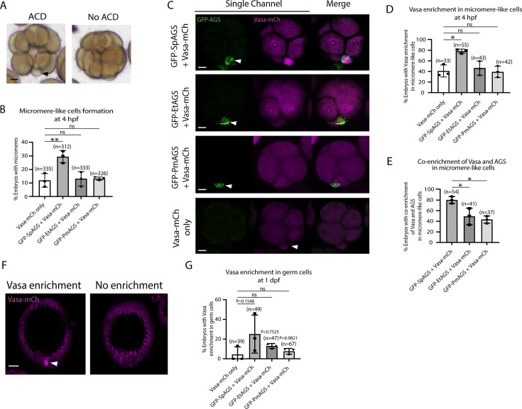
The evolutionary introduction of asymmetric cell division (ACD) into the developmental program facilitates the formation of a new cell type, contributing to developmental diversity and, eventually, species diversification. The micromere of the sea urchin embryo may serve as one of those examples: an ACD at the 16-cell stage forms micromeres unique to echinoids among echinoderms. We previously reported that a polarity factor, activator of G-protein signaling (AGS), plays a crucial role in micromere formation. However, AGS and its associated ACD factors are present in all echinoderms and across most metazoans. This raises the question of what evolutionary modifications of AGS protein or its surrounding molecular environment contributed to the evolutionary acquisition of micromeres only in echinoids. In this study, we learned that the GoLoco motifs at the AGS C-terminus play critical roles in regulating micromere formation in sea urchin embryos. Further, other echinoderms' AGS or chimeric AGS that contain the C-terminus of AGS orthologs from various organisms showed varied localization and function in micromere formation. In contrast, the sea star or the pencil urchin orthologs of other ACD factors were consistently localized at the vegetal cortex in the sea urchin embryo, suggesting that AGS may be a unique variable factor that facilitates ACD diversity among echinoderms. Consistently, sea urchin AGS appears to facilitate micromere-like cell formation and accelerate the enrichment timing of the germline factor Vasa during early embryogenesis of the pencil urchin, an ancestral type of sea urchin. Based on these observations, we propose that the molecular evolution of a single polarity factor facilitates ACD diversity while preserving the core ACD machinery among echinoderms and beyond during evolution.
Keywords: AGS; asymmetric cell division; developmental biology; evolutionary modification; sea urchin embryo; sea urchins.
© 2024, Emura, Wavreil et al.
Conflict of interest statement
NE, FW, AF, MY No competing interests declared
Figures


Update of
-
The evolutionary modifications of a GoLoco motif in the AGS protein facilitate micromere formation in the sea urchin embryo.bioRxiv [Preprint]. 2024 Oct 15:2024.06.30.601440. doi: 10.1101/2024.06.30.601440. bioRxiv. 2024. Update in: Elife. 2024 Dec 23;13:RP100086. doi: 10.7554/eLife.100086. PMID: 39005292 Free PMC article. Updated. Preprint.
Similar articles
-
The evolutionary modifications of a GoLoco motif in the AGS protein facilitate micromere formation in the sea urchin embryo.bioRxiv [Preprint]. 2024 Oct 15:2024.06.30.601440. doi: 10.1101/2024.06.30.601440. bioRxiv. 2024. Update in: Elife. 2024 Dec 23;13:RP100086. doi: 10.7554/eLife.100086. PMID: 39005292 Free PMC article. Updated. Preprint.
-
Evolutionary modification of AGS protein contributes to formation of micromeres in sea urchins.Nat Commun. 2019 Aug 22;10(1):3779. doi: 10.1038/s41467-019-11560-8. Nat Commun. 2019. PMID: 31439829 Free PMC article.
-
Micromere formation and its evolutionary implications in the sea urchin.Curr Top Dev Biol. 2022;146:211-238. doi: 10.1016/bs.ctdb.2021.10.008. Epub 2021 Dec 3. Curr Top Dev Biol. 2022. PMID: 35152984 Free PMC article.
-
Maternal factors regulating symmetry breaking and dorsal-ventral axis formation in the sea urchin embryo.Curr Top Dev Biol. 2020;140:283-316. doi: 10.1016/bs.ctdb.2019.10.007. Epub 2019 Nov 22. Curr Top Dev Biol. 2020. PMID: 32591077 Review.
-
A conserved role for the nodal signaling pathway in the establishment of dorso-ventral and left-right axes in deuterostomes.J Exp Zool B Mol Dev Evol. 2008 Jan 15;310(1):41-53. doi: 10.1002/jez.b.21121. J Exp Zool B Mol Dev Evol. 2008. PMID: 16838294 Review.
Cited by
-
Unique metabolic regulation of micromeres contributes to gastrulation in the sea urchin embryo.Nat Commun. 2025 Aug 11;16(1):7410. doi: 10.1038/s41467-025-62697-8. Nat Commun. 2025. PMID: 40790037 Free PMC article.
References
-
- Bate CM, Carr V, Graziadei PPC, Hirsch HVB, Hughes A, Ingle D, Leventhal AG, Monti Graziadei GA, Rubel EW, Saxod R, Scheibel AB, Scheibel ME, Silver J. In: Handbook of Sensory Physiology. Jacobson M, editor. Berlin, Heidelberg: Springer-Verlag; 1978. Development of sensory systems; pp. 1–53. - DOI
MeSH terms
Grants and funding
LinkOut - more resources
Full Text Sources
