. 2025 Feb 13;69(2):e0117424.
doi: 10.1128/aac.01174-24. Epub 2024 Dec 23.

Durlobactam in combination with β-lactams to combat Mycobacterium abscessus

Affiliations

Durlobactam in combination with β-lactams to combat Mycobacterium abscessus

Eunjeong Shin et al. Antimicrob Agents Chemother. .

Abstract

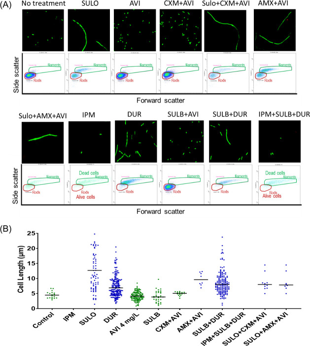
Mycobacterium abscessus (Mab) presents significant clinical challenges. This study evaluated the synergistic effects of a β-lactam and β-lactamase inhibitor combination against Mab and explored the underlying mechanisms. Synergy was assessed through MIC tests and time-kill studies, and binding affinities of nine β-lactams and BLIs to eight target receptors (L,D-transpeptidases [LDT] 1-5, D,D-carboxypeptidase, penicillin-binding protein [PBP] B, and PBP-lipo) were assessed using mass spectrometry and kinetic studies. Thermal stability and morphological changes were determined. Imipenem demonstrated high binding affinity to LDTs and PBPs, with extremely low inhibition constants (Ki,app; ≤0.002 mg/L for LDT1-2, ≤0.6 mg/L for PBPs), while cephalosporins, sulopenem, tebipenem, and amoxicillin exhibited moderate to low binding affinity. Durlobactam inactivated BlaMab and LDT/PBPs more potently than avibactam. The Ki,apps of durlobactam for PBP B, PBP-lipo, and LDT2 were below clinically achievable unbound concentrations, while avibactam's Ki,app for LDT/PBPs exceeded the clinical concentrations. Single β-lactam treatments resulted in minimal killing (~1 log10 reduction). Although avibactam yielded no effect, combinations with avibactam showed a significant reduction (~4 log10 CFU/mL). Durlobactam alone showed ~2 log10 reduction, and when combined with imipenem or two β-lactams, durlobactam achieved near-eradication of Mab, surpassing the current therapy (amikacin + clarithromycin + imipenem/cefoxitin). Inactivation of PBP-lipo by sulopenem, imipenem, durlobactam, and amoxicillin (with avibactam) led to morphological changes, showing filaments. This study demonstrates the mechanistic basis of combinations therapy, particularly imipenem + durlobactam, in overcoming β-lactam resistance in Mab.

Keywords: Mycobacterium abscessus; beta-lactamase inhibitors; beta-lactams; synergistic bacterial killing.


Conflict of interest statement

The authors declare no conflict of interest.

Figures

Fig 1
Fig 1
Time-kill curves were generated for single β-lactams: imipenem (IPM) (A), sulopenem (SULO) (D), and tebipenem (TBP) (G), as well as their combinations with either ceftaroline (CFT), cefuroxime (CXM), or amoxicillin (AMX) in the absence of β-lactamase inhibitors. Panels B, E, and H show a single or combination of two β-lactams in the presence of 4 mg/L avibactam (AVI). Panels C, F, and I represent single or dual β-lactams in the presence of 12 mg/L durlobactam (DUR) and 7 mg/L sulbactam (SUL). The SOC combinations of amikacin (AMK) + clarithromycin (CLM) + IPM (shown in pink band) or cefoxitin (FOX) (shown in purple band) were included for comparison of the killing effects. The concentrations of each drug used in this assay were clinically achievable: IPM at 12 mg/L, CFT at 8 mg/L, CXM at 8 mg/L, AMX at 2 mg/L, SULO at 2 mg/L, TBP at 1.5 mg/L, AMK at 12 mg/L, CLM at 0.3 mg/L, and FOX at 7 mg/L. To counteract thermal degradation, the following supplementation was performed every 48 h: 75% for IPM, 33% for SULO, 54% for TBP, 50% for CXM, and 55% for FOX.
Fig 2
Fig 2
A classification tree was constructed based on bacterial burden reduction in log10 CFU/mL to determine the relative importance of predictor variables in the CART analysis. This CART analysis was performed with time-kill data collected on day 5 after dosing. Each split in the tree represents different regimens, such as β-lactamase inhibitors (BLIs), dual β-lactams (DBL), or BLI types. Data are average ± SD on the log10 scale, along with the number of treatment (or control) arms for various drug combinations in each category.
Fig 3
Fig 3
Redundancy and interaction between β-lactams or β-lactamase inhibitors (imipenem, avibactam, and durlobactam) and LDTs 1–4, D,D-carboxypeptidase, PBP B, PBP-lipo, and BlaMab.
Fig 4
Fig 4
Chemical structures of β-lactams and β-lactamase inhibitors (A) ceftaroline (CFT); (B) sulopenem (SULO); (C) tebipenem (TBP); (D) imipenem (IPM); (E) cefuroxime (CXM); (F) avibactam (AVI); (G) durlobactam (DUR); (H) amoxicillin (AMX); (I) sulbactam (SUL); (J) cefoxitin (FOX) as well as the chemical structures of (K) amikacin and (L) clarithromycin. When ceftaroline, sulopenem, tebipenem, imipenem, and cefuroxime bind to LDTs or PBPs, they bind either as whole compounds or as fragments (Table 2). The portions expected to be removed upon binding to proteins are highlighted in green.
Fig 5
Fig 5
Binding of avibactam (AVI), amoxicillin (AMX), cefoxitin (FOX), durlobactam (DUR), imipenem (IPM), sulopenem (Sulo), ceftaroline (CFT), and cefuroxime (CXM) with PBP-lipo. The intensities of the PBP-lipo complex bands were quantified using GelAnalyzer software and normalized to the intensity of PBP-lipo without β-lactams (PBP-lipo Apo), which was set to 1. The low intensity of the PBP-lipo and Bocillin complex indicates that β-lactams are strongly bound to PBP-lipo.
Fig 6
Fig 6
Confocal microscopy and flow cytometry assay, characterizing morphology changes in the Mycobacterium abscessus ATCC 19977 strain. Microscope image (×60) and forward scatter vs side scatter plot (A) exhibited that inactivation of PBP-lipo by sulopenem (SULO), amoxicillin (AMX) with avibactam (AVI), imipenem (IPM), or durlobactam (DUR) led to filamentous cells with significant bacterial killing at 24 h. (B) Cell length was determined through confocal microscopy image analysis. The lengths of all cells in the microscope sample at 24 h were measured, and the line represents the median value.

References

    1. Nessar R, Cambau E, Reyrat JM, Murray A, Gicquel B. 2012. Mycobacterium abscessus: a new antibiotic nightmare. J Antimicrob Chemother 67:810–818. doi:10.1093/jac/dkr578 - DOI - PubMed
    1. Jhun BW, Moon SM, Jeon K, Kwon OJ, Yoo H, Carriere KC, Huh HJ, Lee NY, Shin SJ, Daley CL, Koh WJ. 2020. Prognostic factors associated with long-term mortality in 1445 patients with nontuberculous mycobacterial pulmonary disease: a 15-year follow-up study. Eur Respir J 55:55. doi:10.1183/13993003.00798-2019 - DOI - PubMed
    1. Benwill JL, Wallace RJ Jr. 2014. Mycobacterium abscessus: challenges in diagnosis and treatment. Curr Opin Infect Dis 27:506–510. doi:10.1097/QCO.0000000000000104 - DOI - PubMed
    1. Esther CR, Esserman DA, Gilligan P, Kerr A, Noone PG. 2010. Chronic Mycobacterium abscessus infection and lung function decline in cystic fibrosis. J Cyst Fibros 9:117–123. doi:10.1016/j.jcf.2009.12.001 - DOI - PMC - PubMed
    1. Maurer FP, Rüegger V, Ritter C, Bloemberg GV, Böttger EC. 2012. Acquisition of clarithromycin resistance mutations in the 23S rRNA gene of Mycobacterium abscessus in the presence of inducible erm(41). J Antimicrob Chemother 67:2606–2611. doi:10.1093/jac/dks279 - DOI - PubMed

Publication types

MeSH terms

LinkOut - more resources