. 2025 Jan 31;99(1):e0149724.
doi: 10.1128/jvi.01497-24. Epub 2024 Dec 23.

Single-chain antibody gene therapy strategy based on high-throughput screening triggers sustained antiviral activity in the body

Affiliations

Single-chain antibody gene therapy strategy based on high-throughput screening triggers sustained antiviral activity in the body

Liang Zhao et al. J Virol. .

Abstract

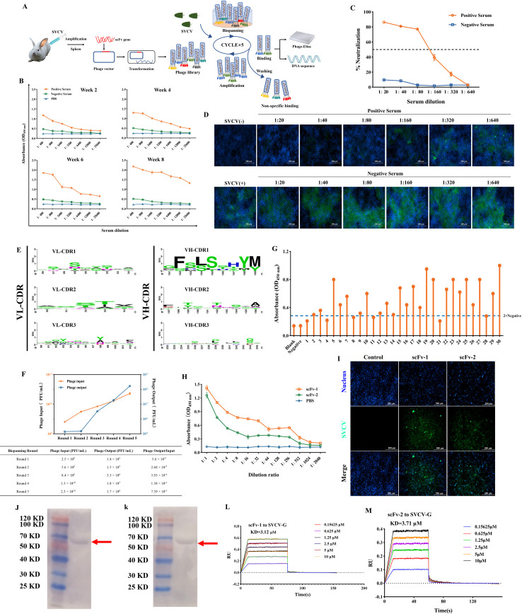
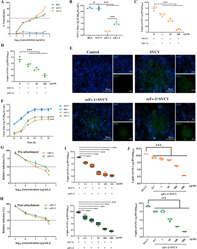
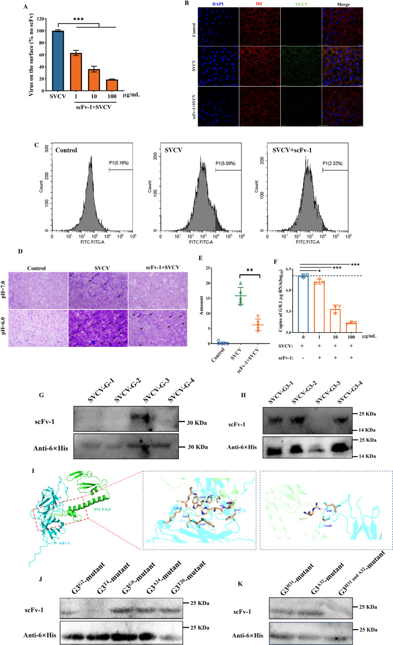
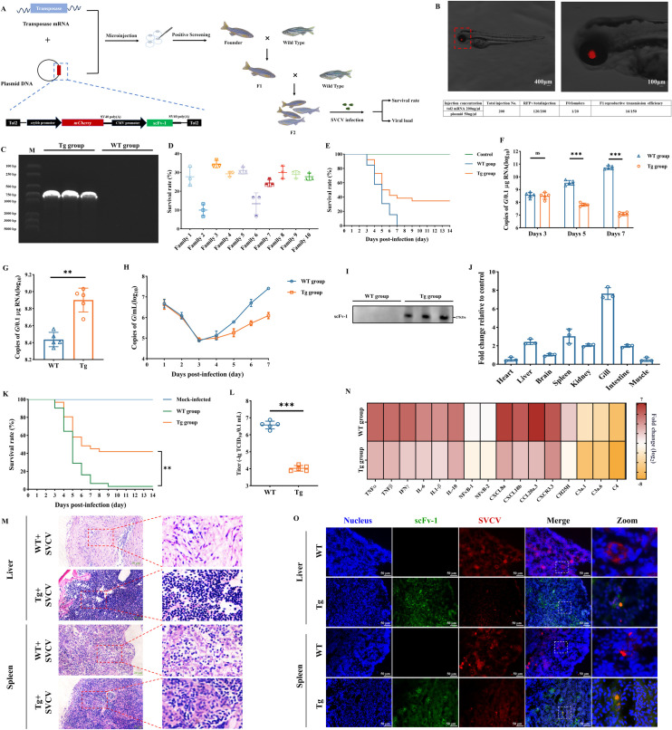
The occurrence of viral diseases poses a huge threat and impact on human public health safety and the development of the animal and fishery industry. Here, a strain of single-chain antibody fragment, scFv-1, was isolated from the phage antibody display library construct by immunizing New Zealand white rabbits with rhabdovirus. In vitro analysis showed that the single-chain antibody could inhibit the infection of the virus in multiple pathways, including adsorption, fusion, and release. In vivo analysis revealed scFv-1 had a preventive and protective effect against the infection of virus. In addition, we describe that transposon-based transport of neutralizing genes allows for long-term, continuous expression, avoiding the need for lifelong, repeated passive immunization for treatment. In sum, high-throughput screening of neutralization genes based on phage display technology and transposon vector-based gene transfer provides effective methods for treating and preventing diseases and avoiding repetitive passive immunotherapy. This study also provides a reference for the prevention and treatment of unknown pathogens.IMPORTANCELivestock and fisheries play an important role in economic development and food security. The frequent outbreaks of viral diseases have caused great losses to the livestock industry, while the increase in drug resistance caused by the use of antibiotics as well as the potential risks to human health have raised serious concerns. Here, we constructed a phage display antibody library by immunizing New Zealand white rabbits with purified rhabdovirus and selected a single-chain antibody, scFv-1, with good neutralizing activity, which was validated and found to be able to block multiple phases of the virus and thus play a neutralizing role. In addition, we describe that transposon-based transport of neutralizing genes allows for long-term, continuous expression, reducing the need for lifelong, repeated passive immunization for treatment. Our work not only provides methods for the prevention and treatment of viral diseases but also provides the body with long-lasting and even permanent protection against repeated passive immunization.

Keywords: antibody gene transfer; neutralization gene therapy; single-chain antibodies; viral diseases.


Conflict of interest statement

The authors declare no conflict of interest.

Figures

Fig 1
Fig 1
Construction of anti-SVCV phage library and the biopanning and identification of single-chain antibody. (A) Schematic diagram of the biopanning strategy for anti-SVCV-specific scFv’s. The purified SVCV was used as the target, and the immunopositive phage clones were further identified through sequencing and enzyme-linked immunosorbent assay (ELISA) after five rounds of biopanning. (B) The titers of polyclonal antibodies in serum at different time points after immunization were analyzed using ELISA. (C) The neutralization effect of serum samples on SVCV at the eighth week after immunization was evaluated by the MTT assay. The serum was diluted from 1:20 to 1:640 in a twofold ratio, and the serum samples were re-tested three times. Results are presented as mean ± SD. (D) Indirect immunofluorescence assay was used to detect the neutralizing effect of serum on SVCV, which increased with the increase in serum concentration. Blue indicates the nucleus; green denotes SVCV. Scale: 200 µm. (E) Diversity analysis of the constructed SVCV immune rabbit phage display single-chain antibody library was performed by randomly selecting multiple single colonies for sequencing analysis. (F) Monitoring of the output:input ratio of phages in five consecutive rounds of biopanning. (G) Randomly select 30 positive phage plaques from the phage library after five rounds of biological selection for ELISA detection. (H) Affinity of purified scFv’s to SVCV detected by indirect ELISA. Results are presented as mean ± SD. (I) Indirect immunofluorescence assay was used to detect the specific reaction between scFv’s and SVCV in infected cells. Blue indicates the nucleus; green denotes SVCV. Scale: 200 µm. (J and K) The ability of the scFv’s to bind to each of the SVCV proteins by Western blot assay; samples were prepared using purified virus particles. (L and M) The affinity of the scFv’s for SVCV-G protein was detected by biolayer interferometry assay.
Fig 2
Fig 2
Neutralization characteristics of anti-SVCV-specific scFv’s in vitro. (A) The neutralization effect of scFv’s on SVCV was evaluated by the MTT method. Results are presented as mean ± SD. (B) Titer analysis of virus after scFv treatment. The viral load was evaluated by real-time quantitative PCR and expressed as copies of the SVCV G gene. Values are presented as mean ± SD. The viral load of cells infected with SVCV was detected after scFv-1 (C) and scFv-2 (D) treatments, respectively. ns, no significance, ***P < 0.001. (E) The neutralization effect of scFv on SVCV was detected using indirect immunofluorescence assay. The nucleus was stained with DAPI (4',6-diamidino-2-phenylindole, blue fluorescence), and the SVCV showed green fluorescence. Scale: 200 µm. (F) The purified scFv protein with a final concentration of 100 µg/mL was pre-incubated with SVCV at 25°C for 1 h, and then co-incubated with cells. The supernatant was collected at 24, 36, 48, 60, 72, and 96 h to determine TCID50. The values at different time points were used to reflect the growth curve of SVCV. (G) The scFv’s were pre-incubated with SVCV at 4°C for 1 h, followed by incubation of the antibody-virus mixture with EPC cells at 4°C for 1 h. After removal of non-adsorbed virus, the culture was maintained for 48 h, and the neutralization ability was assessed by MTT assay. (H) The EPC cells were co-incubated with SVCV at 4°C for 1 h, followed by addition of the scFv’s for 1 h. Subsequently, the medium was replaced with M199 containing 2% FBS, and the culture was continued for 48 h; the neutralization ability was assessed by MTT assay. The viral load was evaluated by RT-qPCR to reflect the ability of the scFv-1 (I) and scFv-2 (J) to neutralize SVCV at pre-attachment or post-attachment stage. Values are presented as mean ± SD. ***P < 0.001.
Fig 3
Fig 3
Evaluation of neutralization mechanism of the scFv-1. (A) The scFv-1 was pre-incubated with SVCV at 4°C for 1 h, and then the antibody-virus mixture was incubated with EPC cells at 4°C for 2 h. Subsequently, the cells were washed to remove the non-adsorbed virus, and the viral load on the cell surface was detected using RT-qPCR. Results are presented as mean ± SD. *P < 0.05, ***P < 0.001. (B) Virus on the cell surface was detected by indirect immunofluorescence using confocal microscopy. Red indicates the cell membrane; blue indicates the nucleus; green indicates the SVCV. Oil mirror, ×63. (C) Detection of fluorescence intensity of viral particles on the cell surface by flow cytometry. (D) The fusion of virus with cell membrane was inhibited by scFv-1 under acidic conditions. (E) Quantitative analysis of the number of syncytia. (F) The EPC cells were co-incubated with SVCV at 25°C for 2 h, and then scFv-1 was added to the co-incubation for 18 h. Subsequently, the supernatant was collected, and the viral load was detected by RT-qPCR. Results are presented as mean ± SD. *P < 0.05, ***P < 0.001. (G) The SVCV-G protein was divided into four segments using the truncated segmentation method, and the binding ability of the scFv-1 to them was detected using the WB assay after induced expression. (H) The SVCV-G3 protein was divided into four segments using the truncated segmentation method, and the binding ability of scFv-1 to these segments was detected using the WB assay after induced expression. (I) Details of interaction between scFv-1 and SVCV-G3. The right panel shows the hydrogen bound at the binding interface between scFv-1 (blue) and SVCV-G3 (green). (J and K) The amino acid sites at which the scFv-1 binds to the SVCV-G3 were sequentially targeted for mutation, and the ability of the scFv-1 to bind to the mutant was detected using the WB assay.
Fig 4
Fig 4
Prophylactic and therapeutic effect of scFv-1 against SVCV infection in vivo. (A) Schematic diagram of challenge and therapeutic strategy. Zebrafish were treated with the scFv-1 (concentration of 10 mg/kg) after intraperitoneal injection of SVCV virus for 6 h. The viral load was evaluated by RT-qPCR and expressed as copies of the SVCV G gene. Values are presented as mean ± SD. (B–D) The viral load in the liver, spleen, and kidney of SVCV-infected zebrafish on day 7 after scFv-1 treatment. ns, no sigficance, ***P < 0.001 (n = 5). (E) Survival rate of SVCV-infected fish within 14 days after scfv-1 treatment; the survival curve was calculated by GraphPad Prism software. (F) The virus titer analysis of zebrafish after scFv-1 treatment (n = 5). Data are presented as mean ± SD. ***P < 0.001. (G) After scFv-1 treatment, hematoxylin-eosin staining was used to observe pathological changes in liver and spleen tissues. (H) Schematic diagram of challenge and prophylactic strategy. Zebrafish were challenged with SVCV after intraperitoneal injection of scFv-1 (concentration of 12.5 mg/kg) for 6 h. (I–K) The viral load in the liver, spleen, and kidney of SVCV-infected zebrafish on day 7 after scFv-1 pre-treatment (n = 5). ns, no significance, ***P < 0.001. (L) Kaplan-Meier survival curve of SVCV-infected fish after scFv-1 pre-treatment within 14 days. (M) The virus titer analysis of zebrafish after scFv-1 treatment (n = 5). Data are presented as mean ± SD. ***P < 0.001. (N and O) Distribution and metabolic analysis at different time points after intraperitoneal injection of fluorescein isothiocyanate (FITC)-labeled scFv-1 in zebrafish (n = 3).
Fig 5
Fig 5
Construction of zebrafish expressing scFv-1 and evaluation of therapeutic effects. (A) Schematic diagram of the construction strategy for zebrafish with constitutively expressing neutralizing antibody. (B) Positive individuals were screened through fluorescence microscopy observation, and the eyes of the positive individuals were red due to the expression of mCherry protein. The enlarged details of the eyes are shown in the image on the right. Scale: 200 µm. (C) Zebrafish were randomly selected from F2 generation-positive individuals for PCR amplification of the scFv-1 gene, and the amplification products were verified by agarose electrophoresis. Randomly select wild zebrafish were used as a control. (D) Survival rate of F2 generation-positive zebrafish individuals from different families within 14 days after infection with SVCV; the survival curve was calculated by GraphPad Prism software. (E) The survival rate of zebrafish in the Tg and WT groups after SVCV immersion infection within 14 days; the survival curve was calculated by GraphPad Prism software. The viral load was evaluated by RT-qPCR and expressed as copies of the SVCV G gene. Results are presented as mean ± SD. (F) The viral load in the zebrafish of the Tg group and the WT group after infection with SVCV on days 3, 5, and 7.ns, no significance, ***P < 0.001. (G) The viral load of dying zebrafish in the Tg group and the WT group after infection with SVCV. **P < 0.01. (H) Aquaculture water samples were collected for seven consecutive days; virus load was detected using the RT-qPCR method. Results are presented as mean ± SD. (I) Zebrafish were randomly selected from F2 generation-positive individuals, and the total protein was extracted after removing the head. The expression of scFv-1 protein was detected using the WB method. (J) The expression of the scFv-1 gene in zebrafish (Tg group, 2 months old) heart, liver, brain, spleen, kidney, gill, intestine, and muscle tissues (n = 3). (K) The survival rate of zebrafish (2 months old) after SVCV infection within 14 days; the survival curve was calculated by GraphPad Prism software. (L) The virus titer analysis of zebrafish infected with SVCV (n = 5). Data are presented as mean ± SD. ***P < 0.001. (M) Analysis of pathological sections of the liver and spleen of zebrafish infected with SVCV. (N) Heat map display of cytokine and chemokine analysis after zebrafish infection with SVCV. (O) Neutralization of SVCV by scFv-1 protein expressed in vivo using immunohistochemical analysis.

Similar articles

References

    1. Burton DR. 2023. Antiviral neutralizing antibodies: from in vitro to in vivo activity. Nat Rev Immunol 23:720–734. doi:10.1038/s41577-023-00858-w - DOI - PMC - PubMed
    1. Newman J, Thakur N, Peacock TP, Bialy D, Elrefaey AME, Bogaardt C, Horton DL, Ho S, Kankeyan T, Carr C, Hoschler K, Barclay WS, Amirthalingam G, Brown KE, Charleston B, Bailey D. 2022. Neutralizing antibody activity against 21 SARS-CoV-2 variants in older adults vaccinated with BNT162b2. Nat Microbiol 7:1180–1188. doi:10.1038/s41564-022-01163-3 - DOI - PMC - PubMed
    1. Guo Y, Zhang G, Yang Q, Xie X, Lu Y, Cheng X, Wang H, Liang J, Tang J, Gao Y, et al. . 2023. Discovery and characterization of potent pan-variant SARS-CoV-2 neutralizing antibodies from individuals with Omicron breakthrough infection. Nat Commun 14:3537. doi:10.1038/s41467-023-39267-x - DOI - PMC - PubMed
    1. Suryadevara N, Shrihari S, Gilchuk P, VanBlargan LA, Binshtein E, Zost SJ, Nargi RS, Sutton RE, Winkler ES, Chen EC, Fouch ME, Davidson E, Doranz BJ, Chen RE, Shi P-Y, Carnahan RH, Thackray LB, Diamond MS, Crowe JE Jr. 2021. Neutralizing and protective human monoclonal antibodies recognizing the N-terminal domain of the SARS-CoV-2 spike protein. Cell 184:2316–2331. doi:10.1016/j.cell.2021.03.029 - DOI - PMC - PubMed
    1. Pegu A, Borate B, Huang Y, Pauthner MG, Hessell AJ, Julg B, Doria-Rose NA, Schmidt SD, Carpp LN, Cully MD, Chen X, Shaw GM, Barouch DH, Haigwood NL, Corey L, Burton DR, Roederer M, Gilbert PB, Mascola JR, Huang Y. 2019. A meta-analysis of passive immunization studies shows that serum-neutralizing antibody titer associates with protection against SHIV challenge. Cell Host Microbe 26:336–346. doi:10.1016/j.chom.2019.08.014 - DOI - PMC - PubMed

Publication types

LinkOut - more resources