

About bacteriophage tail terminator and tail completion proteins: structure of the proximal extremity of siphophage T5 tail
- PMID: 39714170
- PMCID: PMC11784142
- DOI: 10.1128/jvi.01376-24
About bacteriophage tail terminator and tail completion proteins: structure of the proximal extremity of siphophage T5 tail
Abstract
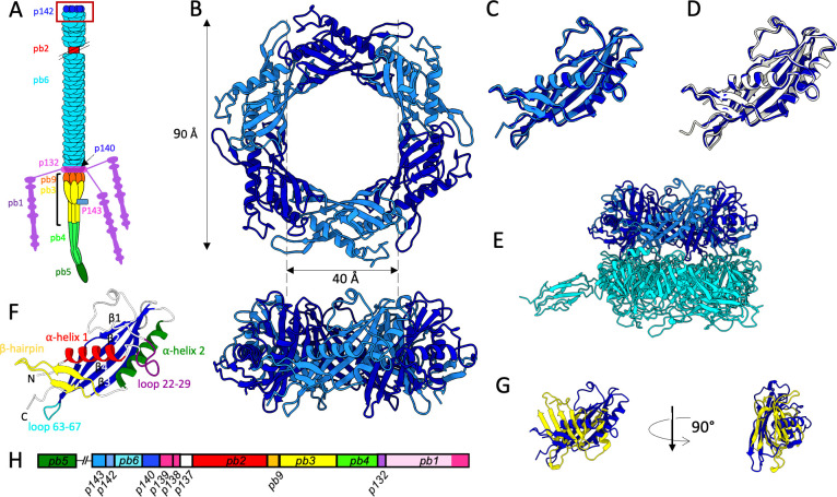
Bacteriophages are viruses infecting bacteria. The vast majority of them bear a tail, allowing host recognition, cell wall perforation, and DNA injection into the host cytoplasm. Using electron cryo-microscopy (cryo-EM) and single particle analysis, we determined the organization of the tail proximal extremity of siphophage T5 that possesses a long flexible tail and solved the structure of its tail terminator protein p142 (TrP142). It allowed us to confirm the common evolutionary origin between T5 TrPp142 and other known or putative TrPs from siphophages, myophages, and bacterial tail-like machines, despite very poor sequence conservation. By also determining the structure of the T5 tail proximal extremity after interaction with T5 bacterial receptor FhuA, we showed that no conformational changes occur in TrPp142 and confirmed that the infection signal transduction is not carried by the tube itself. We also investigated the location of T5 Neck1 or tail completion protein p143 (TCPp143) and showed, thanks to a combination of cryo-EM and structure prediction using Alphafold2, that it is not located at the capsid-to-tail interface as suggested by its position in the genome, but instead, very unexpectedly, on the side of T5 tail tip, and that it appears to be monomeric. Based on structure comparison with other putative TCPs predicted structures, this feature could not be shared by other TCPs and questions the affiliation of p143 to this family of protein.IMPORTANCEBacteriophages, viruses infecting bacteria, are the most abundant living entities on Earth. They are present in all ecosystems where bacteria develop and are instrumental in the regulation, diversity, evolution, and pathogeny of microbial populations. Moreover, with the increasing number of pathogenic strains resistant to antibiotics, virulent phages are considered a serious alternative or complement to classical treatments. 96% of all phages present a tail that allows host recognition and safe channeling of the DNA to the host cytoplasm. We present the atomic model of the proximal extremity of the siphophage T5 tail, confirming structural similarities with other phages. This structure, combined with results previously published and further explored, also allowed a review and a discussion on the role and localization of a mysterious tail protein, the tail completion protein, which is known to be present in the phage tails, but that was never identified in a phage structure.
Keywords: bacteriophage T5; cryo-electron microscopy; tail completion protein; tail terminator protein.
Conflict of interest statement
The authors declare no conflict of interest.
Figures


Similar articles
-
The In Situ Structure of T-Series T1 Reveals a Conserved Lambda-Like Tail Tip.Viruses. 2025 Feb 28;17(3):351. doi: 10.3390/v17030351. Viruses. 2025. PMID: 40143278 Free PMC article.
-
Comparative anatomy of siphophage tails before and after interaction with their receptor.Curr Opin Struct Biol. 2025 Jun;92:103045. doi: 10.1016/j.sbi.2025.103045. Epub 2025 Apr 24. Curr Opin Struct Biol. 2025. PMID: 40279683 Review.
-
Drugs for preventing postoperative nausea and vomiting in adults after general anaesthesia: a network meta-analysis.Cochrane Database Syst Rev. 2020 Oct 19;10(10):CD012859. doi: 10.1002/14651858.CD012859.pub2. Cochrane Database Syst Rev. 2020. PMID: 33075160 Free PMC article.
-
Antidepressants for pain management in adults with chronic pain: a network meta-analysis.Health Technol Assess. 2024 Oct;28(62):1-155. doi: 10.3310/MKRT2948. Health Technol Assess. 2024. PMID: 39367772 Free PMC article.
-
Systemic pharmacological treatments for chronic plaque psoriasis: a network meta-analysis.Cochrane Database Syst Rev. 2017 Dec 22;12(12):CD011535. doi: 10.1002/14651858.CD011535.pub2. Cochrane Database Syst Rev. 2017. Update in: Cochrane Database Syst Rev. 2020 Jan 9;1:CD011535. doi: 10.1002/14651858.CD011535.pub3. PMID: 29271481 Free PMC article. Updated.
Cited by
-
Structure of the Staphylococcus aureus bacteriophage 80α neck shows details of the DNA, tail completion protein, and tape measure protein.Structure. 2025 Jun 5;33(6):1063-1073.e2. doi: 10.1016/j.str.2025.03.007. Epub 2025 Apr 1. Structure. 2025. PMID: 40174589
References
-
- Pell LG, Liu A, Edmonds L, Donaldson LW, Howell PL, Davidson AR. 2009. The X-ray crystal structure of the phage lambda tail terminator protein reveals the biologically relevant hexameric ring structure and demonstrates a conserved mechanism of tail termination among diverse long-tailed phages. J Mol Biol 389:938–951. doi:10.1016/j.jmb.2009.04.072 - DOI - PubMed
Publication types
MeSH terms
Substances
Grants and funding
LinkOut - more resources
Full Text Sources
