. 2025 Feb 4;13(2):e0259524.
doi: 10.1128/spectrum.02595-24. Epub 2024 Dec 23.

Listeria monocytogenes use multiple mechanisms to disseminate from the intestinal lamina propria to the mesenteric lymph nodes

Affiliations

Listeria monocytogenes use multiple mechanisms to disseminate from the intestinal lamina propria to the mesenteric lymph nodes

Joshua S Nowacki et al. Microbiol Spectr. .

Abstract

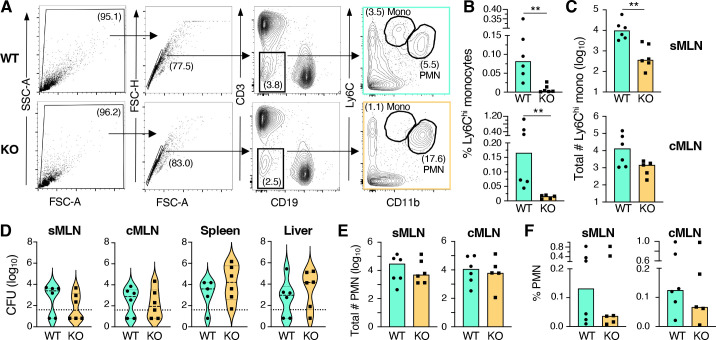
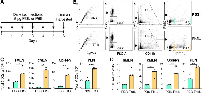
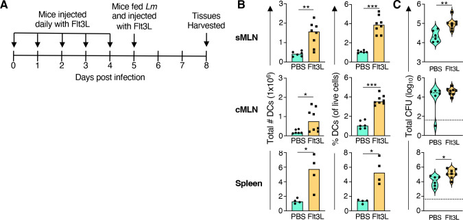
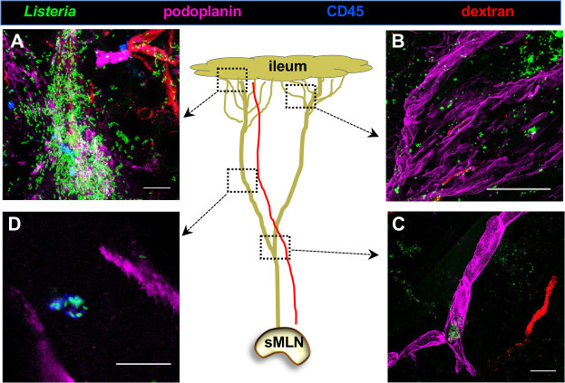
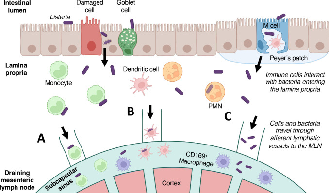
Listeria monocytogenes are facultative intracellular bacterial pathogens that cause foodborne disease in humans. The bacteria can use the surface protein InlA to invade intestinal epithelial cells or transcytose across M cells in the gut, but it is not well understood how the bacteria traffic from the underlying lamina propria to the draining mesenteric lymph nodes (MLN). Previous studies indicated that L. monocytogenes associated with both monocytes and dendritic cells in the intestinal lamina propria. We show here that CCR2-/- mice had a significant reduction in Ly6Chi monocytes in the MLN but no change in bacterial burden following foodborne infection; thus, dissemination of L. monocytogenes associated with monocytes is not required for colonization of the MLN. To block CCR7-mediated trafficking of dendritic cells from the lamina propria, we treated mice with anti-VEGFR3 antibody (clone AFL4) prior to and during infection but did not see a change in dendritic numbers in the MLN as had been previously reported with other anti-VEGFR3-specific antibodies. However, increasing the number of circulating dendritic cells by treating mice with rFlt3L resulted in a significant increase in L. monocytogenes in the lymph nodes that drain the small intestine and the spleen. Whole-mount fluorescent microscopy of lymphatic vessels following ligated loop infection revealed both free-floating L. monocytogenes and cell-associated bacteria within lymphatic vessels. Together, these results suggest that L. monocytogenes can use multiple, redundant mechanisms to disseminate from the gut tissue to the MLN.

Importance: Consumption of the foodborne bacterial pathogen Listeria monocytogenes results in a wide spectrum of human disease from mild self-limiting gastroenteritis to life-threatening infections of the bloodstream, brain, and placenta. It is not well understood how the bacteria migrate from the intestines to the draining mesenteric lymph nodes, which are thought to serve as the last barrier to prevent systemic infections. Results presented here reveal multiple redundant mechanisms L. monocytogenes can use to disseminate from the ileum or colon to the mesenteric lymph nodes.

Keywords: facutatively intracellular pathogens; foodborne; gastrointestinal infection; lymph nodes.


Conflict of interest statement

The authors declare no conflict of interest.

Figures

Fig 1
Fig 1
Inhibition of monocyte egress had no impact on Listeria burdens in the mesenteric lymph nodes. C57BL/6 (WT) and CCR2−/− (KO) mice were fed 1.7–3.6 × 10⁸ CFU of Lm SD2001, and tissues were collected 3 days post infection. Pooled data for n = 6 mice analyzed in three independent experiments are shown. (A) Gating strategy used to identify Ly6Chi monocytes (Mono) and neutrophils (PMN). Median percentage of live cells (B) and absolute number of monocytes (C) in MLN draining the small intestine (sMLN) and colon (cMLN). (D) Violin plots of the Lm SD2001 burden in the tissues 3 dpi; the dashed lines represent median, the dotted lines represent the limits of detection, and the vertical segments represent the interquartile range. Median number of PMN (E) and percentage of live cells (F) in both subsets of MLN as determined by flow cytometry. Significance was calculated for all panels using Mann-Whitney test for unpaired data (**P < 0.01).
Fig 2
Fig 2
Treatment with anti-VEGFR3 clone AFL4 did not significantly alter CCR7+ cell recruitment to the MLN. Pooled data for n = 4 mice analyzed in four different experiments are shown. (A) BALB/cByJ mice were injected i.p. with 250 mg either anti-VEGFR3 (VEG) or isotype control (ISO) antibody and then fed 1–5 × 10⁸ CFU of Lm SD2000. Three additional i.p. injections of the same antibody occurred every 12 h after that and tissues were harvested at 72 hpi post infection for flow cytometry and CFU quantification. (B) Gating strategy used to define CD11c+ DC in MLN (pre-gated on live cells using FSC-A and SSC-A). (C) Median percentage of CD11c+ DCs in both subsets of MLN. (D) Gating strategy used to define CD3+ T cells in MLN (pre-gated on live cells). (E) Median percentage of T cells in both subsets of MLN. (F) Violin plots of the Lm SD2000 burden in the tissues 3 dpi; the dashed lines represent median, and the dotted lines represent limits of detection. Significance was assessed using a Mann-Whitney test for unpaired data.
Fig 3
Fig 3
Six days of FLT3L injections were sufficient to increase the number and percentage of CD11c+ DC in the MLN fivefold. Pooled data for n = 3 mice per group analyzed in three independent experiments are shown. (A) Mice were injected (i.p.) with either phosphate-buffered saline (PBS) or 5 mg FMS-like tyrosine kinase 3 ligand (Flt3L) every day for 6 days and tissues were 24 h after the last injection. (B) Representative gating strategy for cells harvested from sMLN. Median number (C) and percentage (D) of CD11c+ DC as in lymphoid tissues including peripheral lymph nodes (PLN). Data from three independent experiments are shown. Significance calculated using a student’s t test for unpaired data (*P < 0.05; **P < 0.01; ***P < 0.001).
Fig 4
Fig 4
Increasing the number of dendritic cells resulted in increased bacterial burdens in the sMLN and spleen. (A) BALBc/ByJ mice were injected with either murine Flt3L or PBS for 6 days to expand DC differentiation and then infected with 2–8 × 10⁸ CFU of Lm SD2000. Tissues were collected 3 days post infection. Four independent experiments with n = 2 mice per group were performed. (B) Median number and percentage of live cells for CD11c+ DC in sMLN, cMLN, and spleen; CD11c+ DC were identified using the gating strategy shown in Fig. 3 only for experiments #3 and #4. (C) Violin plots of the Lm SD2000 burden in tissues; dashed lines represent medians, and the vertical segments represent the interquartile range. Dotted lines across the graphs represent limits of detection. Significance was calculated using Mann-Whitney test for unpaired data (*P < 0.05; **P < 0.01; ***P < 0.001).
Fig 5
Fig 5
Both cell-associated and free-floating L. monocytogenes were observed in mesentery lymphatic vessels during ligated loop infection. BALBc/ByJ mouse ileal loop was injected with 109 CFU GFP+ Listeria, and tissues were harvested 45 min post infection. Just prior to euthanasia, fluorescently conjugated dextran was injected intravenously. The center cartoon depicts a single plexus leading from the ileum to the draining sMLN; the dotted lines indicate the areas analyzed in the surrounding representative images. (A) Hundreds of L. monocytogenes are found proximal to the intestinal lamina propria. (B) A network of lymphatic vessels near the gut tissue. (C) Large collecting vessel more distal to the gut with a cluster of L. monocytogenes at the branch point. A blood vessel containing dextran is seen on the right. (D) A CD45+ cell with several attached Lm migrates through the vessel. Representative images from one of two independent experiments are shown; all images are Z stacks except (D) which shows a single z plane. Scale bars, 20 mM.
Fig 6
Fig 6
Proposed model of dissemination from intestinal lamina propria to the MLN. Ingested L. monocytogenes (purple) in the gut lumen can cross the mucosal barrier by either InlA-mediated uptake into epithelial cells or transcytosis via M cells. Once in the underlying lamina propria, Lm can interact with a variety of immune cells, including activated PMN and macrophages which promote clearance of extracellular bacteria. (A) Ly6Chi inflammatory monocytes do not readily phagocytose Listeria but may transport surface-associated bacteria to the MLN. (B) Migratory CD11chi CD103+ cDC can internalize L. monocytogenes and then migrate to draining lymph nodes to present antigen to T cells. Previous data suggest that Listeria survive for several hours within DC. (C) Extracellular bacteria that are not cleared by phagocytes can disseminated free floating through the afferent lymphatic vessels to enter the subcapsular sinus of the lymph node. Together, these three mechanisms make up our proposed routes of dissemination for Lm to travel from the LP to the MLN.

Similar articles

References

    1. Weis J, Seeliger HP. 1975. Incidence of Listeria monocytogenes in nature. Appl Microbiol 30:29–32. doi:10.1128/am.30.1.29-32.1975 - DOI - PMC - PubMed
    1. Schlech WF. 2019. Epidemiology and clinical manifestations of Listeria monocytogenes infection. Microbiol Spectr 7. doi:10.1128/microbiolspec.gpp3-0014-2018 - DOI - PMC - PubMed
    1. Mohapatra RK, Mishra S, Tuglo LS, Sarangi AK, Kandi V, Al Ibrahim AA, Alsaif HA, Rabaan AA, Zahan MK-E. 2024. Recurring food source-based Listeria outbreaks in the United States: an unsolved puzzle of concern? Health Sci Rep 7:e1863. doi:10.1002/hsr2.1863 - DOI - PMC - PubMed
    1. D’Orazio SEF. 2014. Animal models for oral transmission of Listeria monocytogenes. Front Cell Infect Microbiol 4:15. doi:10.3389/fcimb.2014.00015 - DOI - PMC - PubMed
    1. Melton-Witt JA, Rafelski SM, Portnoy DA, Bakardjiev AI. 2012. Oral infection with signature-tagged Listeria monocytogenes reveals organ-specific growth and dissemination routes in guinea pigs. Infect Immun 80:720–732. doi:10.1128/IAI.05958-11 - DOI - PMC - PubMed

LinkOut - more resources