. 2025 Jan 31;91(1):e0165424.
doi: 10.1128/aem.01654-24. Epub 2024 Dec 23.

Positive regulation of a LuxR family protein, MilO, in mildiomycin biosynthesis

Affiliations

Positive regulation of a LuxR family protein, MilO, in mildiomycin biosynthesis

Zhiyu Li et al. Appl Environ Microbiol. .

Abstract

Mildiomycin is a representative peptidyl nucleoside antibiotic and was first isolated from Streptoverticillium rimofaciens, which has been used as an important biological agent to control powdery mildew in plants. Despite its importance, the biosynthetic pathways and regulatory mechanisms remain to be fully elucidated. In this study, we identified MilO as a positive pathway-specific regulator of mildiomycin biosynthesis in the heterologous host Streptomyces avermitilis. Gene disruption of milO resulted in almost loss of mildiomycin production, and it was restored to the level comparable to that in the wild-type strain in complemented strain. Overexpression of milO using host native promoter rpsJp, engineered promotor SP44, and kasOp* led to a 50%, 6.5-fold, and 9.2-fold increase in mildiomycin production compared with the wild-type strain, respectively. Quantitative real-time PCR and electrophoretic mobility shift assay (EMSA) experiments revealed that MilO directly enhances the transcription of the milA gene by 20 folds after 48 h fermentation and indirectly regulates the transcription levels of other genes from milB to milM. Using DNase I footprinting assays, milO was revealed to bind to a 44 bp DNA sequence of the milA promoter region. The binding region consists of three imperfect direct repeats of TGTC(N)3CGGT separated by two-nucleotide spacers and each repeat is important to efficient binding to MilO. In addition, we identified two related compounds by overexpressing milO in a structural gene milN-deficient mutant. Taken together, this study indicates that pathway-specific regulator MilO is essential for mildiomycin biosynthesis and provides an effective strategy to improve the production of mildiomycin and its intermediates.IMPORTANCEAs an important biological agent to control powdery mildew on plants, mildiomycin has been commercialized and used in various plants. However, its regulatory mechanisms and biosynthetic pathways remain unknown. This study provides new insights into the regulation of mildiomycin biosynthesis through MilO, a LuxR family protein that modulates mildiomycin production by directly enhancing the transcription of milA. The yield of mildiomycin was significantly improved by overexpressing milO in a heterologous host. In addition, the positive regulatory effect of milO helped to discover two related compounds, which provide important clues for the timing of uploading of two amino acid side chains during mildiomycin biosynthesis for the first time. In brief, our findings on transcriptional regulation of mildiomycin biosynthesis by milO will be valuable to further increase the yield of mildiomycin and explore its biosynthetic pathways.

Keywords: LuxR regulator; MilO; Streptomyces avermitilis; gene overexpression; mildiomycin; shunt product; yield improvement.


Conflict of interest statement

The authors declare no conflict of interest.

Figures

Fig 1
Fig 1
Proposed biosynthetic pathway of mildiomycin. CMP, cytidine 5′-monophosphate. HM-CMP, 5-hydroxymethyl CMP. HMC, 5-hydroxymethylcytosine. HM-CGA, 5-hydroxymethyl cytosylglucuronic acid. MilA, CMP 5-hydroxymethylase. MilB, nucleotide hydrolase. MilC, cytosylglucuronic acid/hydroxymethyl cytosylglucuronic acid synthase. MilD, putative degT/dnrJ/eryC1/strS aminotransferase. MilG, putative cytosylglucuronate decarboxylase. MilM, putative pyridoxal phosphate–dependent arginine oxidases. MilN, putative dihydrodipicolinate synthetase. MilI, putative acyl carrier protein. MilH, putative tRNA-dependent aminoacyltransferase. The solid arrows represent reactions that have been elucidated, and the dashed arrows indicate proposed reactions. The changing groups are indicated in different colors. The shunt product compound 1 is highlighted in a yellow rectangle.
Fig 2
Fig 2
MilO is a LuxR family transcription regulator and is indispensable to mildiomycin biosynthesis. (A) Sequence alignment of MilO with other homologous proteins, the HTH domain was marked with the horizontal line. (B) Phylogenetic analysis of MilO and other LuxR-type regulators and the accession number of all proteins are listed in Table S4. REC, receiver domain. LAL, larger ATP-binding regulators of the LuxR. PAS, Per (period circadian protein), Arnt (Ah receptor nuclear translocator protein), Sim (single-minded protein). FHA, fork head-associated domain. (C) HPLC analysis of mildiomycin in WT (S. avermitilis::14A6), milO deletion mutant (ΔmilO), and complementary strain (ΔmilO::milO). The complementary milO is expressed under the promoter of kasOp*.
Fig 3
Fig 3
Exploring the effect of milO overexpression on mildiomycin titer. (A) HPLC analysis of mildiomycin production in WT-CK (WT::pPM927), WrO (WT::pPM927-rpsJp-milO), WsO (WT::pPM927-SP44-milO), and WkO (WT::pPM927-kasOp*-milO) (left); titers of mildiomycin from three replicative fermentations for each of strains were quantified (right). All strains were cultured in a fermentation medium for 7 days. (B) Time-course production of mildiomycin for three producer strains. (C) Time-course biomass of three mildiomycin producers. The OD595 values determined by the diphenylamine colorimetric method were proportional to the biomass.
Fig 4
Fig 4
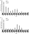
qPCR-based comparison of expression of mil genes in strains with varied expression of MilO. The cells were harvested from the fermentation broth after 48 h (A) and 96 h (B). Error bars were calculated by measuring the standard deviations of the data from three replicates of each sample. *, **, and *** indicate statistically significant results (P-value < 0.05, P-value < 0.01 and P-value < 0.001, respectively).
Fig 5
Fig 5
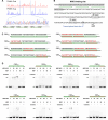
EMSAs to examine the binding of MilO to six promotors of transcriptional units. (A) mil gene organization and seven putative transcriptional units that are determined by RT-PCR analysis in panel B. The arrows show their orientation; the promoters for each of the transcriptional unit were indicated with P-I, P-II, P-III, P-IV, P-V, and P-VI (black triangles); the circled numbers denote the PCR fragment across two neighboring orfs. They are the same as those in panel B, which shows the RT-PCR analysis of the transcriptional units. The cDNA was generated by reverse transcription of total RNA samples, genomic DNA, and total RNA of WT were used as control. (C) 3 pmol FAM-labeled DNA probes for each promoter was incubated with 10 pmol or 20 pmol recombinant MilO at room temperature for 10 min. The unlabeled probe of 60 pmol (the last lane for each gel) was used as the control to demonstrate specific and competitive binding with MilO. 0.2 µg salmon sperm DNA was included in each reaction. The arrow indicates the shifted bands of the protein-DNA complexes. MBP, MBP-His8; MBP-MilO, MBP-His8-MilO.
Fig 6
Fig 6
DNA-binding properties of MilO targeting the MilA promoter region. (A) DNase I footprinting analysis of MilO in the promotor of MilA (probe P-I). The sequence of the protected region is indicated below the electropherograms, and the three 11 bp direct repeats sequence was shown in green. (B) Nucleotide sequence of the MilA promoter region. The predicted transcription start sites (TSS) are bolded and enlarged, the putative −10 and −35 regions are boxed out. (C) Schematic diagram to show the sequence of eight probes with various mutations at three direct repeats. The mutated nucleotide sequences are shown in red. The three direct repeat sequences are shown in gray and the spacers in white. The variants are shown in pink. Sizes are not proportional. (D) EMSAs of MilO binding to OBSWT and its variants (OBS, MilO binding site). The 44 bp FAM-labeled DNA oligos (0.5 pmol) were incubated with increasing concentrations of recombinant MilO (lane 2, 25 pmol; lane 3, 50 pmol). Lane 1, negative control without MilO; lane 4, MBP alone with 0.5 pmol labeled probe; lane 5, 50 pmol unlabeled probe was added to the reaction system for lane 2. 0.2 µg Salmon sperm DNA was included in each reaction mixture. MBP MBP-His8; MBP-MilO, MBP-His8-MilO.
Fig 7
Fig 7
Overexpression of MilO in milN mutant produces two cryptic shunt products. (A) HPLC analysis of the products of Streptomyces avermitilis-derived mutants. WT is Streptomyces avermitilis expressing the complete mildiomycin biosynthetic gene cluster on integrative cosmid 14A6. The sample after resin purification was shown in a solid line, and the flowthrough during purification was indicated by a dashed line. (B, C) The LC-Q-TOF-MS analysis (positive model) of compound 1 and compound 2. Both compounds were collected from the extract of ΔmilN::milO. Compound 1: [M + H]+ Cal. 242.1135, Obs. 242.1139, 1.65 ppm Error; Compound 2: [M + H]+ Cal. 212.1030, Obs. 212.1033, 1.41 ppm Error. The inferred structural formulas are shown above.

Similar articles

References

    1. Niu G, Tan H. 2015. Nucleoside antibiotics: biosynthesis, regulation, and biotechnology. Trends Microbiol 23:110–119. doi:10.1016/j.tim.2014.10.007 - DOI - PubMed
    1. Winn M, Goss RJM, Kimura K, Bugg TDH. 2010. Antimicrobial nucleoside antibiotics targeting cell wall assembly: recent advances in structure-function studies and nucleoside biosynthesis. Nat Prod Rep 27:279–304. doi:10.1039/b816215h - DOI - PubMed
    1. Harada S, Kishi T. 1978. Isolation and characterization of mildiomycin, a new nucleoside antibiotic. J Antibiot 31:519–524. doi:10.7164/antibiotics.31.519 - DOI - PubMed
    1. Harada S, Mizuta E, Kishi T. 1981. Structure of mildiomycin, a new antifungal nucleoside antibiotic. Tetrahedron 37:1317–1327. doi:10.1016/S0040-4020(01)92447-0 - DOI
    1. Feduchi E, Cosin M, Carrasco L. 1985. Mildiomycin: a nucleoside antibiotic that inhibits protein synthesis. J Antibiot 38:415–419. doi:10.7164/antibiotics.38.415 - DOI - PubMed

MeSH terms

Supplementary concepts

LinkOut - more resources