

Age-dependent effects of Delaire facemask therapy for class III malocclusion : Impact on maxillary sutures and palatal morphology
- PMID: 39714628
- PMCID: PMC12181118
- DOI: 10.1007/s00056-024-00564-9
Age-dependent effects of Delaire facemask therapy for class III malocclusion : Impact on maxillary sutures and palatal morphology
Abstract
Background and aim: Treatment effects of a combined rapid maxillary expansion (RME) and Delaire facemask (DFM) therapy have so far only been scientifically investigated through cephalometric analyses. The combination of cephalometric, dental cast and cone-beam computed tomography (CBCT) scan analysis was not yet used for investigating morphologic changes of the tooth-bearing palate. The aim of the present study was to determine whether patient age at treatment begin has an influence upon palatal length changes after RME/DFM therapy, and to what extent transverse palatal sutures contribute to these.
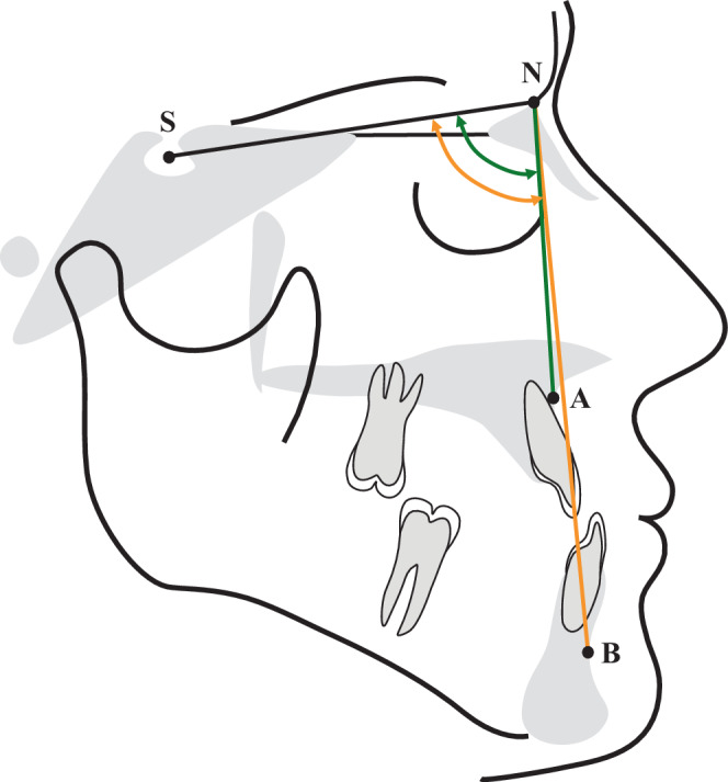
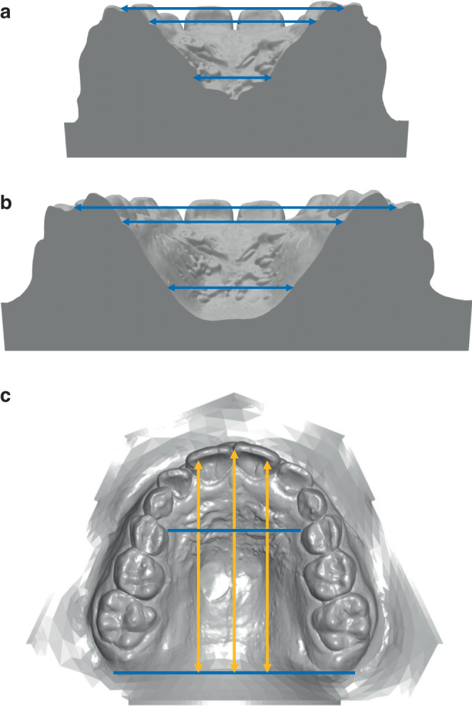
Patients and methods: In n = 6 patients (min 10.5 years, max 14.7 years) from a total group of n = 40, CBCT datasets showing all palatal sutures were visually assessed, and palatal morphology was compared with a dental cast analysis. In addition, lateral cephalograms and dental casts of n = 40 patients were divided into two groups (PG1: < 12 years, n = 20; PG2: ≥ 12 years, n = 20), and an analysis was performed to investigate changes in the tooth-bearing palate after RME/DFM treatment.
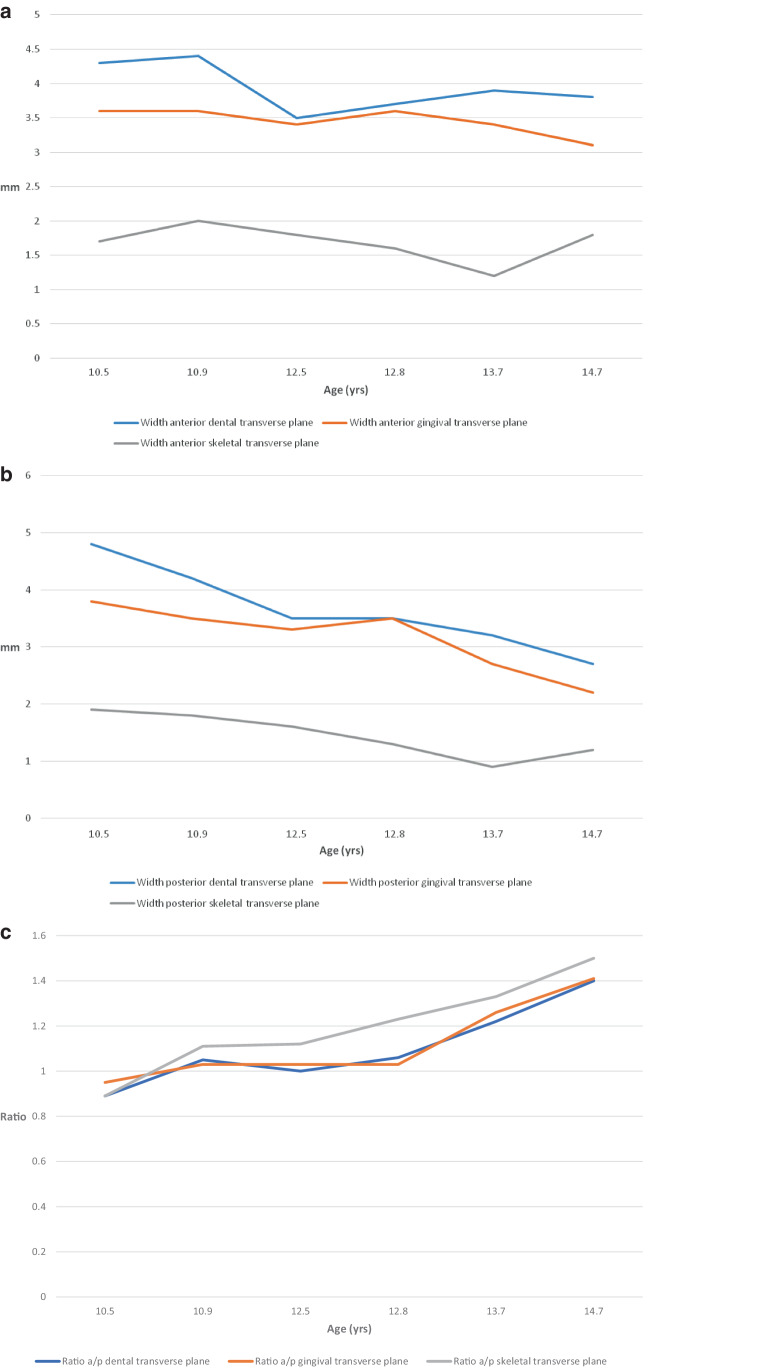
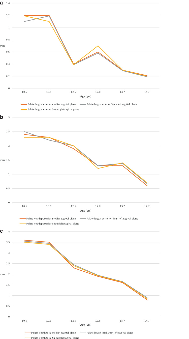
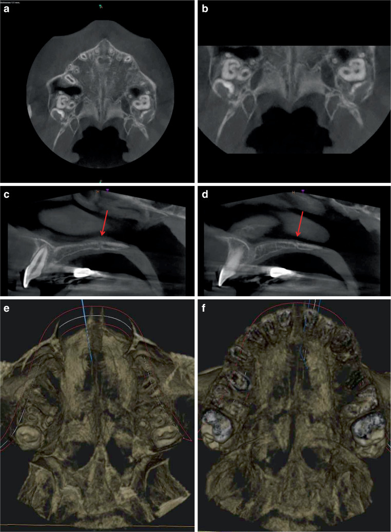
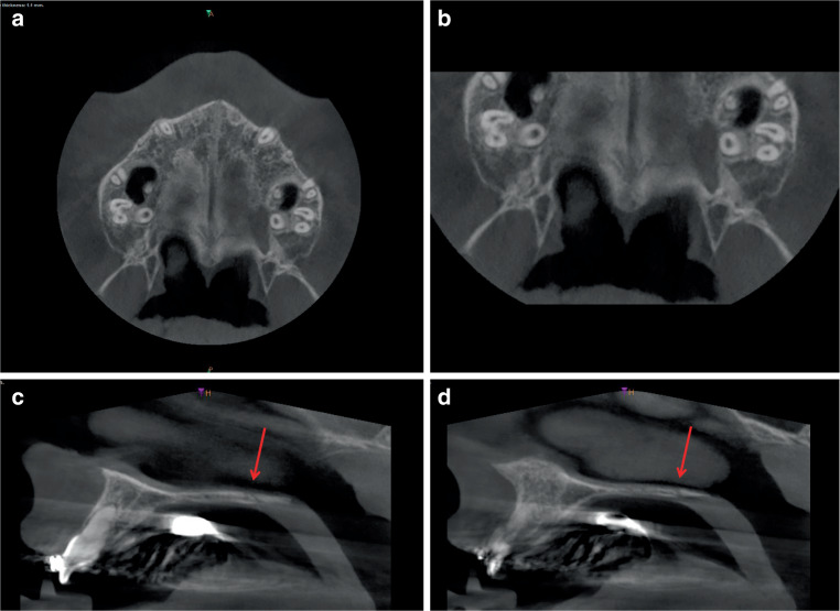
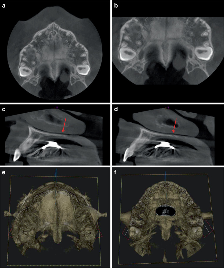
Results: The CBCT analysis showed that the median and transverse palatine sutures were always open. On the contrary, the pterygopalatomaxillary sutures were partially open only in the youngest patients. The transverse palatal suture showed age-dependent morphologic changes in the transverse and sagittal planes. The changes of the palatal width and length show clear differences between the two younger and the four older patients in the corresponding dental cast analysis. The cephalometric analysis showed that a significant improvement of the sagittal jaw relation due to ventral displacement of the maxilla during treatment occurred only in younger patients (< 12 years) despite similar initial findings in both patient groups. The dental cast analysis also revealed that changes are age-dependent: In PG1, the width increases posteriorly more than anteriorly; in PG2, this is reversed. The length increases are always significant in both patient groups, whereby the anterior, posterior, and total amounts are greater in PG1 than in PG2. In relative terms, the increases in both groups are greater posteriorly than anteriorly. There is a significant difference between the groups posteriorly and overall.
Conclusions: Morphological changes of the sutures appear to have a decisive influence on the success of RME/DFM therapy. The age-dependent reactions of pterygopalatomaxillary and transverse palatine sutures represent a further main therapeutic effect of DFM treatment in addition to those described by Delaire and explain the different changes in palate length before and after the age of 12. If the maximum effect of RME/DFM treatment is desired, it should be started before the age of 12. Treatment success is age-dependent.
Zusammenfassung: HINTERGRUND UND ZIEL: Die Behandlungseffekte einer kombinierten Therapie aus „rapid maxillary expansion“ (RME) und „facemask“ nach Delaire (DFM) wurden bislang nur durch kephalometrische Studien wissenschaftlich überprüft. Durch diese Therapie veranlasste morphologische Veränderungen des Gaumens sind dagegen bislang nicht kombiniert durch kephalometrische, Modell- und DVT(digitale Volumentomographie)-Analysen untersucht worden. Ziel der vorliegenden Studie war es festzustellen, ob das Alter der Patienten bei Behandlungsbeginn einen Einfluss auf die Längenveränderungen des Gaumens nach RME/DFM-Therapie hat und inwieweit die quer verlaufenden palatinalen Suturen dazu beitragen.
Patienten und methoden: Bei n = 6 Patienten (min. 10,5 Jahre, max. 14,7 Jahre) aus einer Gesamtgruppe von n = 40 wurden DVT-Datensätze im Bereich aller palatinalen Suturen visuell befundet und die Gaumenmorphologie mit einer Modell-Analyse abgeglichen. Zusätzlich wurden Fernröntgenseitenbilder und Modelle von n = 40 Patienten in zwei Gruppen unterteilt (PG1: < 12. Lebensjahr, n = 20; PG2: ≥ 12, n = 20), und es wurde eine Analyse durchgeführt, um Veränderungen des zahntragenden Gaumens nach der RME/DFM-Behandlung zu beurteilen.
Ergebnisse: Die DVT-Analyse zeigte, dass die Sutura palatina mediana und die Sutura palatina transversa immer geöffnet waren. Die pterygopalatomaxillären Suturen waren dagegen nur bei den jüngsten Patienten partiell geöffnet. Die Sutura palatina transversa zeigt in der Transversal- sowie in der Sagittalebene altersabhängige morphologische Veränderungen. Die Werte der Gaumenbreiten- und Gaumenlängenveränderungen zeigen bei der korrespondierenden Modellanalyse deutliche Unterschiede zwischen den beiden jüngeren und den 4 älteren Patienten. Die FRS-Analyse zeigt vergleichbare Anomalien in beiden Patientengruppen zu Behandlungsbeginn, eine signifikante Verbesserung der mesiobasalen Kieferbasenrelation durch sagittale Ventralverlagerung der Maxilla unter der Behandlung aber nur bei den jüngeren Patienten (< 12. Lebensjahr). In der Modellstudie sind die Veränderungen ebenfalls altersabhängig: Bei PG1 nimmt die Breite im posterioren Bereich mehr als anterior zu, bei PG2 ist dies umgekehrt. Die Längenzunahmen sind in beiden Patientengruppen immer signifikant, wobei sowohl anterior, posterior als auch gesamt die Beträge bei PG1 größer sind als bei PG2. Relativ betrachtet sind die Zunahmen in beiden Gruppen posterior größer als anterior. Ein signifikanter Unterschied zwischen den Gruppenwerten besteht posterior und gesamt.
Schlussfolgerungen: Morphologische Veränderungen der Suturen scheinen einen entscheidenden Einfluss auf den Erfolg der RME/DFM-Therapie zu haben. Die altersabhängigen Reaktionen der pterygopalatomaxillären Suturen und der Sutura palatina transversa stellen einen weiteren, zusätzlich zu den von Delaire beschriebenen therapeutischen Haupteffekt der DFM-Behandlung dar und erklären die unterschiedlichen Gaumenlängenveränderungen vor und nach dem 12. Lebensjahr. Will man den maximalen Effekt der RME/DFM-Behandlung erzielen, sollte sie vor dem 12. Lebensjahr begonnen werden. Der Behandlungserfolg ist altersabhängig.
Keywords: Cast analysis; Cone-beam computed tomography; Maxilla; Pterygopalatomaxillary sutures; Transverse palatine suture.
© 2024. The Author(s).
Conflict of interest statement
Declarations. Conflict of interest: G.S.M. Kinzinger, J. Hourfar, J.N. Sommer and J.A. Lisson declare that they have no competing interests. Ethical standards: This article does not contain any studies with human participants or animals performed by any of the authors. Ethical approval for this retrospective study was granted by the Ethical Committee of Saarland University, Germany (E 130/17 and E 170/12). For this type of study, formal consent is not required.
Figures
Similar articles
-
Age-dependent interactions of maxillary sutures during RME and their effects on palatal morphology : CBCT and dental cast analysis.J Orofac Orthop. 2022 Nov;83(6):412-431. doi: 10.1007/s00056-022-00429-z. Epub 2022 Oct 7. J Orofac Orthop. 2022. PMID: 36205766 Free PMC article.
-
The sutural and dentoskeletal effects of alternate expansion and constriction of deficient maxilla in young adults: a randomized controlled clinical trial.BMC Oral Health. 2025 Jul 13;25(1):1156. doi: 10.1186/s12903-025-06489-y. BMC Oral Health. 2025. PMID: 40653475 Free PMC article. Clinical Trial.
-
Long-term three-dimensional skeletal effects of hybrid hyrax with facemask versus mentoplate in growing Class III patients: a randomized controlled trial.Prog Orthod. 2025 Apr 21;26(1):14. doi: 10.1186/s40510-025-00561-7. Prog Orthod. 2025. PMID: 40254643 Free PMC article. Clinical Trial.
-
Systemic pharmacological treatments for chronic plaque psoriasis: a network meta-analysis.Cochrane Database Syst Rev. 2021 Apr 19;4(4):CD011535. doi: 10.1002/14651858.CD011535.pub4. Cochrane Database Syst Rev. 2021. Update in: Cochrane Database Syst Rev. 2022 May 23;5:CD011535. doi: 10.1002/14651858.CD011535.pub5. PMID: 33871055 Free PMC article. Updated.
-
Intravenous magnesium sulphate and sotalol for prevention of atrial fibrillation after coronary artery bypass surgery: a systematic review and economic evaluation.Health Technol Assess. 2008 Jun;12(28):iii-iv, ix-95. doi: 10.3310/hta12280. Health Technol Assess. 2008. PMID: 18547499
Cited by
-
Impact of malocclusion on oral health-related quality of life: insights from children with and without hypodontia.J Orofac Orthop. 2025 Mar 21. doi: 10.1007/s00056-025-00580-3. Online ahead of print. J Orofac Orthop. 2025. PMID: 40116911 English.
References
-
- Jäger A, Braumann B, Kim C, Wahner S (2001) Skeletal and dental effects of maxillary protraction in patients with angle class III malocclusion. A meta-analysis. J Orofac Orthop 62:275–284 - PubMed
-
- Graber TM, Chung DB, Aoba IT (1967) Dentofacial orthopedics versus orthodontics. J Am Dent Assoc 75:1145–1166 - PubMed
-
- Graber TM (ed) (1969) Current orthodontic concepts and techniques. WB Saunders, Philadelphia
-
- Saussouni V (1972) Dentofacial orthopedics: a critical review. Am J Orthod 61:255–269 - PubMed
-
- Sanborn RT (1955) Differences between the facial skeletal patterns of class III malocclusions and normal occlusion. Angle Orthod 25:208–222
MeSH terms
LinkOut - more resources
Full Text Sources
Miscellaneous
