. 2025 Apr;22(2):228-238.
doi: 10.1007/s10388-024-01102-7. Epub 2024 Dec 23.

p53 and Ki-67 combined with periodic acid-Schiff staining for the diagnosis of early stage esophageal squamous cell carcinoma lesions in biopsy specimens

Affiliations

p53 and Ki-67 combined with periodic acid-Schiff staining for the diagnosis of early stage esophageal squamous cell carcinoma lesions in biopsy specimens

Feifei Liu et al. Esophagus. 2025 Apr.

Abstract

Background: Esophageal cancer is highly prevalent in China, predominantly represented by squamous cell carcinoma. This retrospective study sought to evaluate the diagnostic efficacy of four staining protocols in identifying early stage esophageal squamous cell carcinoma (ESCC).

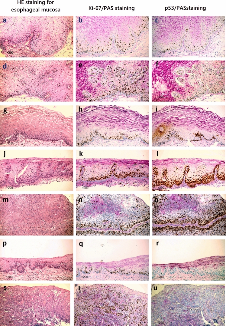
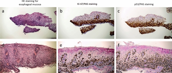
Methods: A consecutive series of ninety biopsy samples of esophageal mucosa, collected retrospectively from March 2016 to December 2019, were obtained at Beijing Chao-Yang Hospital, a tertiary care facility in Beijing, China. These samples were categorized into four groups: non-neoplastic squamous lesions (Non-NSL), low-grade dysplasia (LGD), high-grade dysplasia (HGD), and early stage ESCC. Baseline, molecular analyses (p53 by immunohistochemistry and Ki-67 by immunohistochemistry), and staining analyses (hematoxylin & eosin (HE) and periodic acid-Schiff (PAS) were conducted across the categories. The staining protocols included HE, HE + p53 + Ki-67, HE + p53 + Ki-67 + PAS, and HE + p53/PAS + Ki-67/PAS.

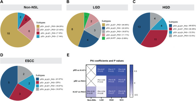
Results: Patients with HGD and ESCC were significantly older and had larger lesions. Elevated p53 and Ki-67 mutation rates were observed in HGD and ESCC, while increased PAS positivity was noted in RE and LGD. The p53, Ki-67, and PAS staining results showed mostly no correlation among the four groups. Abnormal Ki-67 basal layer distribution pattern correlated with histological grades, with higher proportions in HGD and ESCC. HE + p53 + Ki-67 + PAS and HE + p53/PAS + Ki-67/PAS demonstrated complete consistency with the reference standard, with weighted κ values of 1. HE + p53 + Ki-67 + PAS and HE + p53/PAS + Ki-67/PAS protocols exhibited 100% accuracy, sensitivity, and specificity for diagnosing ESCC or ESCC combined with HGD, outperforming the other protocols.

Conclusions: Incorporating specific staining protocols, particularly HE + p53 + Ki-67 + PAS and HE + p53/PAS + Ki-67/PAS, enhances the diagnostic accuracy for early stage ESCC, showing promise in advancing the pathology diagnostic pathway.

Keywords: Early diagnosis; Esophageal mucosa biopsy; Esophageal squamous cell carcinoma; Ki-67; P53; PAS staining; Retrospective study.


Conflict of interest statement

Declarations. Conflict of interest: The authors declare no competing interests. Ethical statement: The study was conducted in accordance with the Declaration of Helsinki and was approved by the ethics committee of Beijing Chao-Yang Hospital (No. 2023–598). Since this was a retrospective study and involved no direct patient intervention, informed consent was waived. However, all patient data were anonymized, and confidentiality was maintained throughout the study.

Figures

Fig. 1
Fig. 1
Pathological images obtained from esophageal mucosal biopsy samples, HE staining indicated non-neoplastic squamous lesions (Non-NSL) of esophageal mucosa, low-grade dysplasia (LGD) of esophageal mucosal tissue, high-grade dysplasia (HGD) of esophageal mucosal tissue, and esophageal squamous cell carcinoma (ESCC) of esophageal mucosal tissue. Non-NSL from esophageal mucosa: a HE staining illustrating reactive hyperplasia characterized by increased cellular growth. b Reactive hyperplastic lesions displaying scattered abnormal Ki-67 expression in basal layer cells, indicating proliferative activity. c Detection of p53 protein highlighting its presence in reactive hyperplasia. d HE staining showing proliferating papillary structures within the tissue. e Proliferating papillary structures displaying scattered abnormal Ki-67 expression in cells around the papillary formations, signifying active cell division. f Detection of p53 protein in proliferating papillary structures suggesting cellular abnormalities. LGD of esophageal mucosal tissue: g, j HE staining indicating dysplastic cells confined within the lower half of the epithelium. h, k Increased number of abnormal Ki-67 expression cells with a continuous distribution pattern, indicating heightened cellular proliferation. i, l Remarkable increase in the number of p53-positive cells, suggesting cellular abnormalities associated with dysplasia. HGD of esophageal mucosal tissue: m, p HE staining showing histological features characteristic of high-grade dysplasia. n, q Detection of Ki-67 protein indicating increased cellular proliferation. o, r p53 mutation, signifying abnormal cellular behavior associated with high-grade dysplasia. ESCC of esophageal mucosal tissue: s HE staining displaying atypical cells occupying the entire epithelial thickness, indicative of ESCC. t Marked increase in the number of abnormal Ki-67 expression cells, highlighting intense cellular proliferation. u Complete absence of p53 protein, suggesting abnormal cellular behavior associated with ESCC
Fig. 2
Fig. 2
Pairwise correlation and molecular typing distribution of p53, Ki-67, and PAS staining among various groups. A Pie charts stained with p53, Ki-67, and PAS in Non-NSL group. B Pie plots stained with p53, Ki-67, and PAS in LGD group. C Pie charts stained with p53, Ki-67, and PAS in HGD group. D Pie plots stained with p53, Ki-67, and PAS in early ESCC group. E Correlated heat maps stained with p53, Ki-67, and PAS between the four groups, with color bars representing the size of Phi coefficients. Note: p53-/ + represents the wild/mutant type of p53, Ki-67-/ + indicates the normal/abnormal expression of Ki-67, and PAS-/ + indicates that PAS + occupies the epithelial height < 1/2 and > 1/2
Fig. 3
Fig. 3
HE staining of epithelial tissue sections from two cases with intact and incomplete low-grade dysplasia (LGD) of esophageal mucosa. The upper part of the epithelial tissue of the esophageal mucosa was missing, and the atypical changes were initially diagnosed by HE staining of the remaining basal cells a HE staining showed slight atypia. b Ki-67/PAS staining showed abnormal expression of Ki-67 and expression of Ki-67- in basal cells in areas of cytopathic lesions where PAS staining was absent. c p53/PAS staining, PAS staining deletion of cytopathic areas, p53 is mutant expression. HE staining of a complete section of esophageal mucosal epithelial tissue was initially diagnosed as atypical cell changes: d HE staining showed slight atypia. e Ki-67/PAS staining showed abnormal expression of Ki-67 and expression of Ki-67 in basal cells in areas of cytopathic lesions where PAS staining was absent. f p53/PAS staining, PAS staining deletion of cytopathic areas, p53 is mutant expression

Similar articles

Cited by

References

    1. Sjoquist KM, Burmeister BH, Smithers BM, Zalcberg JR, Simes RJ, Barbour A, et al. Survival after neoadjuvant chemotherapy or chemoradiotherapy for resectable oesophageal carcinoma: an updated meta-analysis. Lancet Oncol. 2011;12:681–92. - PubMed
    1. Colvin H, Mizushima T, Eguchi H, Takiguchi S, Doki Y, Mori M. Gastroenterological surgery in Japan: The past, the present and the future. Ann Gastroenterol Surg. 2017;1:5–10. - PMC - PubMed
    1. Malik S, Sharma G, Sanaka MR, Thota PN. Role of endoscopic therapy in early esophageal cancer. World J Gastroenterol. 2018;24:3965–73. - PMC - PubMed
    1. Domper Arnal MJ, Ferrández Arenas Á, Lanas AÁ. Esophageal cancer: risk factors, screening and endoscopic treatment in western and eastern countries. World J Gastroenterol. 2015;21:7933–43. - PMC - PubMed
    1. Lao-Sirieix P, Fitzgerald RC. Screening for oesophageal cancer. Nat Rev Clin Oncol. 2012;9:278–87. - PubMed

MeSH terms

LinkOut - more resources