. 2024 Dec 23;20(12):e1012443.
doi: 10.1371/journal.ppat.1012443. eCollection 2024 Dec.

Immunodominant extracellular loops of Treponema pallidum FadL outer membrane proteins elicit antibodies with opsonic and growth-inhibitory activities

Affiliations

Immunodominant extracellular loops of Treponema pallidum FadL outer membrane proteins elicit antibodies with opsonic and growth-inhibitory activities

Kristina N Delgado et al. PLoS Pathog. .

Abstract

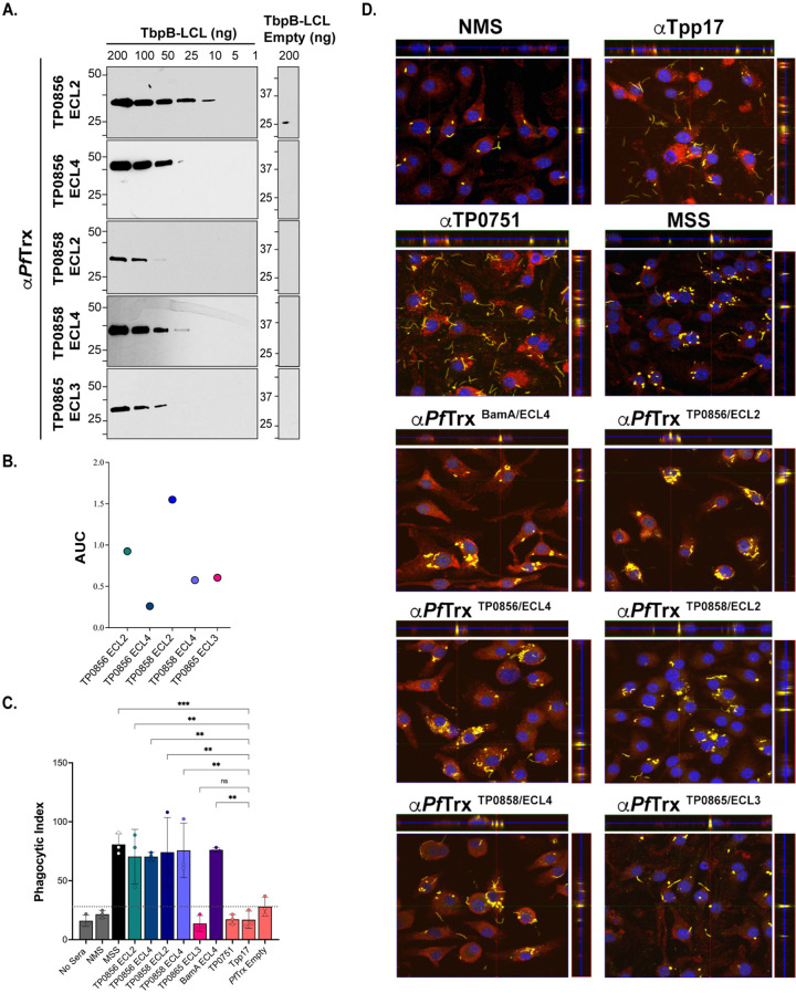
The global resurgence of syphilis has created a potent stimulus for vaccine development. To identify potentially protective antibodies against Treponema pallidum (TPA), we used Pyrococcus furiosus thioredoxin (PfTrx) to display extracellular loops (ECLs) from three TPA outer membrane protein families (outer membrane factors for efflux pumps, eight-stranded β-barrels, and FadLs) to assess their reactivity with immune rabbit serum (IRS). We identified five immunodominant loops from the FadL orthologs TP0856, TP0858 and TP0865 by immunoblotting and ELISA. Rabbits and mice immunized with these five PfTrx constructs produced loop-specific antibodies that promoted opsonophagocytosis of TPA by rabbit peritoneal and murine bone marrow-derived macrophages at levels comparable to IRS and mouse syphilitic serum. Heat-inactivated IRS and loop-specific rabbit and mouse antisera also impaired viability, motility, and cellular attachment of spirochetes during in vitro cultivation. The results support the use of ECL-based vaccines and suggest that loop-specific antibodies promote spirochete clearance via Fc receptor-independent as well as Fc receptor-dependent mechanisms.


Conflict of interest statement

The authors have declared that no competing interests exist.

Figures

Fig 1
Fig 1. Prediction of ECL boundaries.
trRosetta 3D models for outer membrane factors (OMFs), eight-stranded β-barrels (8SβBs), and FadLs (Panels A-C, respectively), depict ECL boundaries (ECL1-Salmon, ECL2-Blue, ECL3-Purple, ECL4-Green, ECL5-Yellow, ECL6-Cyan, ECL7-Dark Teal, and Hatch-Red) used to clone ECLs onto the PfTrx scaffold (see S1 Table).
Fig 2
Fig 2. Reactivity of scaffolded ECLs with Nichols IRS reveals immunodominant FadL ECLs.
Reactivity by immunoblot (left) and ELISA (right) of scaffolded FadL ECLs against sera from five Nichols immune rabbits. ELISA reactivity was measured as AUC corrected for PfTrx background (see Methods). n  =  3 wells per condition. Data are shown as mean ± SD. Significant differences (*p<0.05; ***p<0.001; or ****p<0.0001) between the means of the groups were determined by one-way ANOVA with Bonferroni’s correction for multiple comparisons. Color codes of ECLs are as follows: ECL1-Salmon, ECL2-Blue, ECL3-Purple, ECL4-Green, ECL5-Yellow, ECL6-Cyan, and ECL7-Dark Teal.
Fig 3
Fig 3. Comparative sequence analysis of the Nichols and SS14 FadLs and reactivity of Nichols FadL ECLs with SS14 IRS.
(A) Summary chart representing the number of variable residues within Nichols and SS14 FadLs. (B) Reactivity by immunoblot (left) and ELISA (right) of Nichols FadL ECLs with SS14 IRS. ELISA reactivity measured as AUC corrected for PfTrx background. n  =  3 wells per condition. Data are shown as mean ± SD. Significant differences (*p<0.05 or **p<0.01) between the means determined by one-way ANOVA with Bonferroni’s correction for multiple comparisons. Color codes of ECLs are as follows: ECL1-Salmon, ECL2-Blue, ECL3-Purple, ECL4-Green, ECL5-Yellow, ECL6-Cyan, and ECL7-Dark Teal.
Fig 4
Fig 4. Opsonic activity of rabbit antisera to PfTrx-scaffolded FadL ECLs.
(A) Immunoblot and (B) ELISA (AUC) reactivities of rabbit ECL antisera against the corresponding Tbpb-LCLECL and Tbpb-LCLEmpty. (C) TPA freshly harvested from rabbits was pre-incubated for 2 h with 10% heat-inactivated NRS, IRS, or rabbit antisera to PfTrx ECLs, Tpp17, or TP0751 followed by incubation with rabbit peritoneal macrophages for 4 h at an MOI 10:1. Phagocytic indices were determined from epifluorescence micrographs as described in Methods [18]. Significant differences (*p<0.05, **p<0.01, ***p<0.001 or ****p<0.0001) were determined by one-way ANOVA using Newman-Keuls correction for multiple comparisons. Bars represent mean ± SD, n = 3 wells per condition. (D) Representative confocal micrographs showing composites of 9–12 consecutive Z-stack planes with labeling of TPA, plasma membranes, and nuclei shown in green, red and blue, respectively.
Fig 5
Fig 5. Opsonic activity of mouse antisera to PfTrx-scaffolded FadL ECLs.
(A) Immunoblot and (B) ELISA (AUC) reactivities of sera from mice immunized with PfTrx scaffolded TP0856 ECL2 and ECL4, TP0858 ECL2 and ECL4, and TP0865 ECL3 against graded the corresponding Tbpb-LCLECL and Tbpb-LCLEmpty. (C) TPA freshly harvested from rabbits were pre-incubated for 2 h with 10% heat-inactivated NMS, MSS, and mouse antisera against PfTrx ECLs, Tpp17, and TP0751 followed by incubation with mouse BMDMs for 4 h at an MOI 10:1. Internalization of spirochetes was quantified from epifluorescence micrographs using the phagocytic index. Significant differences (*p<0.05 or **p<0.01) were determined by one-way ANOVA using Bonferroni’s correction for multiple comparisons. Bars represent mean ± SD, n = 3 wells per condition. (D) Representative confocal micrographs showing composites of 9–12 consecutive Z-stack planes with labeling of TPA, plasma membranes, and nuclei shown in yellow, red, and blue, respectively.
Fig 6
Fig 6. Rabbit and mouse antibodies impact growth of TPA Nichols and SS14 during in vitro cultivation.
(A) Enumeration by darkfield microscopy (DFM) at day 7 (solid circles) of spirochetes cultured with 10%, 5%, and 1% concentrations of the indicated rabbit sera, with initial seeding at 2.5 x 10⁶ per well (open circles). (B) Nichols and (C) SS14 TPA strains were seeded initially at 2.5 x 10⁶ per well (open shapes) and cultured with Nichols and SS14 IRS (black and cyan, respectively). On day 7 (solid shapes), spirochetes were harvested and enumerated. Homologous IRS and heterologous IRS are depicted as circles and diamonds, respectively. (D) Spirochetes harvested on day 7 (solid circles) were transferred to a fresh plate containing Sf1Ep cells and TpCM-2 without rabbit sera. On day 14 (solid squares), spirochetes were harvested and enumerated. (E) Enumeration by DFM of spirochetes (initial seeding 1.5 x 10⁶ per well) with 5% mouse antisera targeting FadL ECLs. On day 7, samples were harvested for analysis as described above. Each condition was performed with n = 3 replicates. Significant differences (****p<0.0001) were determined by two-way ANOVA with Tukey correction for multiple comparisons.
Fig 7
Fig 7. Transcriptional analysis of TPA OMP genes.
Pairwise normalized read counts for OMP genes in vivo (grey) and in vitro (black) were calculated using DESeq2 after processing of raw reads previously published by De Lay et al. [46]. A q-value < 0.05, indicated with *, was considered as a significant difference between normalized counts in vivo compared to in vitro. Only one gene, tp0856, was expressed at ≥2-fold higher/lower levels in TPA in vivo compared to in vitro (fold change indicated above the bars).

Update of

References

    1. Ghanem KG, Ram S, Rice PA. The modern epidemic of syphilis. N Engl J Med. 2020;382(9):845–54. doi: 10.1056/NEJMra1901593 . - DOI - PubMed
    1. Peeling RW, Mabey D, Chen XS, Garcia PJ. Syphilis. Lancet. 2023;402(10398):336–46. doi: 10.1016/S0140-6736(22)02348-0 . - DOI - PubMed
    1. Kojima N, Klausner JD. An update on the global epidemiology of syphilis. Curr Epidemiol Rep. 2018;5(1):24–38. Epub 2018/08/18. doi: 10.1007/s40471-018-0138-z ; PubMed Central PMCID: PMC6089383. - DOI - PMC - PubMed
    1. Gottlieb SL, Deal CD, Giersing B, Rees H, Bolan G, Johnston C, et al.. The global roadmap for advancing development of vaccines against sexually transmitted infections: Update and next steps. Vaccine. 2016;34(26):2939–47. doi: 10.1016/j.vaccine.2016.03.111 . - DOI - PMC - PubMed
    1. Kojima N, Konda KA, Klausner JD. Notes on syphilis vaccine development. Front Immunol. 2022;13:952284. Epub 20220728. doi: 10.3389/fimmu.2022.952284 ; PubMed Central PMCID: PMC9365935. - DOI - PMC - PubMed

Substances