. 2024 Dec 23;20(12):e1012817.
doi: 10.1371/journal.ppat.1012817. eCollection 2024 Dec.

Discovery of Nanosota-EB1 and -EB2 as Novel Nanobody Inhibitors Against Ebola Virus Infection

Affiliations

Discovery of Nanosota-EB1 and -EB2 as Novel Nanobody Inhibitors Against Ebola Virus Infection

Fan Bu et al. PLoS Pathog. .

Abstract

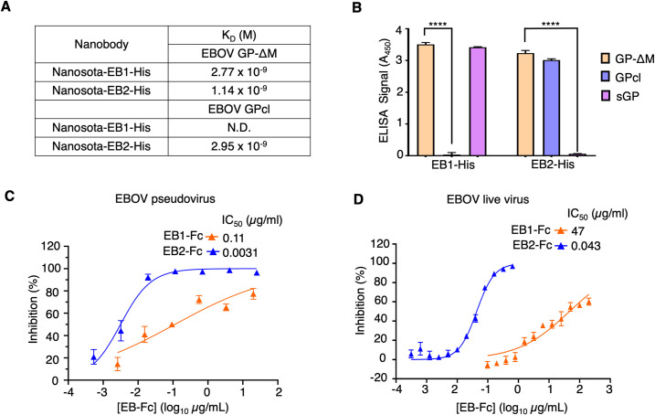
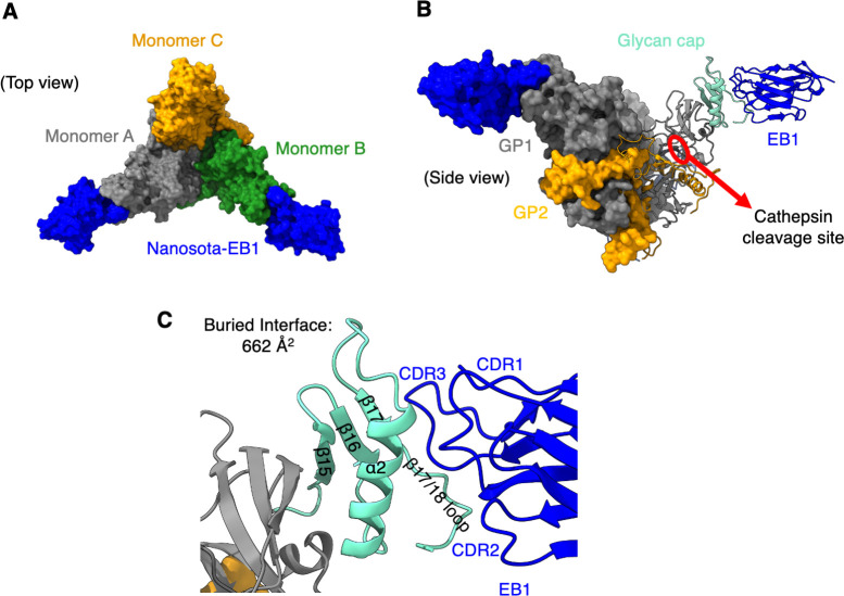
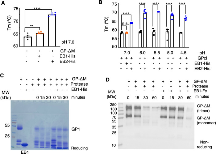
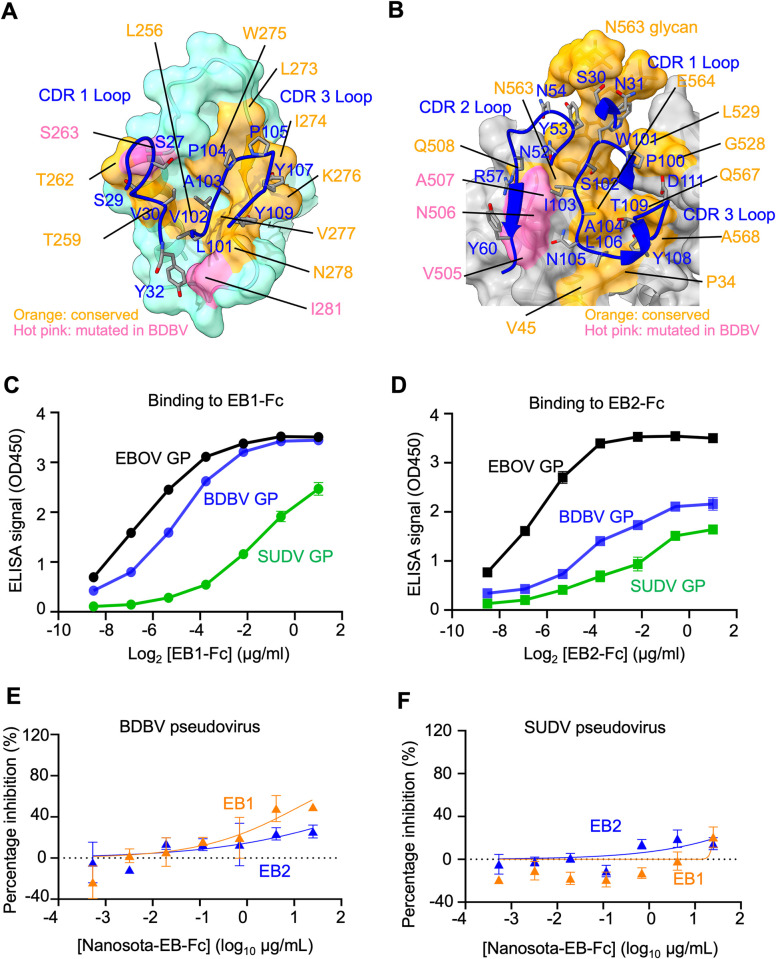
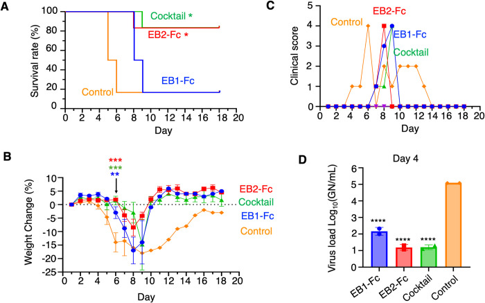
The Ebola filovirus (EBOV) poses a serious threat to global health and national security. Nanobodies, a type of single-domain antibody, have demonstrated promising therapeutic potential. We identified two anti-EBOV nanobodies, Nanosota-EB1 and Nanosota-EB2, which specifically target the EBOV glycoprotein (GP). Cryo-EM and biochemical data revealed that Nanosota-EB1 binds to the glycan cap of GP1, preventing its protease cleavage, while Nanosota-EB2 binds to critical membrane-fusion elements in GP2, stabilizing it in the pre-fusion state. Nanosota-EB2 is a potent neutralizer of EBOV infection in vitro and offers excellent protection in a mouse model of EBOV challenge, while Nanosota-EB1 provides moderate neutralization and protection. Nanosota-EB1 and Nanosota-EB2 are the first nanobodies shown to inhibit authentic EBOV. Combined with our newly developed structure-guided in vitro evolution approach, they lay the foundation for nanobody-based therapies against EBOV and other viruses within the ebolavirus genus.


Conflict of interest statement

B.E.W. and B.S. are co-founders of Turkey Creek Biotechnology. The University of Minnesota has filed patents on Nanosota-EB1 and Nanosota-EB2 with F.L, F.B., and G.Y. as inventors.

Figures

Fig 1
Fig 1. In Vitro Characterization of Two Novel Anti-EBOV Nanobodies, Nanosota-EB1 and -EB2.
(A) Binding affinities between His-tagged nanobodies and two versions of EBOV GP proteins (GP-ΔM and GPcl; see S1B Fig for their definitions) were measured by surface plasmon resonance (SPR). N.D., binding not detected. (B) Binding interactions between His-tagged nanobodies and three versions of the EBOV GP proteins (GP-ΔM, GPcl and sGP; see S1B Fig for their definitions) were evaluated by ELISA. A450: absorbances at 450 nm. Data are presented as mean ± SEM (n = 3). An unpaired two-tailed Student’s t-test was used to analyze the statistical differences between the indicated groups, with results indicated above each bar. ****P < 0.0001. (C) Efficacy of Fc-tagged nanobodies in neutralizing EBOV pseudoviruses. Retroviruses pseudotyped with full-length EBOV GP were used to infect Huh7 cells in the presence of Fc-tagged Nansota-EB1 or -EB2 at different concentrations. The efficacy of each nanobody against EBOV pseudoviruses was expressed as the concentration required to neutralize pseudovirus entry by 50% (IC50). Error bars represent SEM (n = 3). (D) Efficacy of Fc-tagged nanobodies in neutralizing authentic EBOV infection. Authentic EBOV was used to infect Huh7 cells in the presence of Fc-tagged Nanosota-EB1 or -EB2 at different concentrations. The efficacy of each nanobody against authentic EBOV infection was expressed as the concentration required to neutralize EBOV infection by 50% (IC50). Error bars represent SEM (n = 3). Since Nanosota-EB1-Fc could not fully block viral entry at any of the tested concentrations, the IC50 values for Nanosota-EB1-Fc are estimations.
Fig 2
Fig 2. Structural Basis for the Anti-EBOV Functions of Nanosota-EB1.
(A) Cryo-EM structure of EBOV GP-ΔM complexed with Nanosota-EB1 (top view; surface presentation). The three subunits of EBOV GP-ΔM are colored orange, gray, and green, respectively. Nanosota-EB1 is shown in blue. The trimeric GP-ΔM is bound by two Nanosota-EB1 molecules. (B) Cryo-EM structure of EBOV GP-ΔM complexed with Nanosota-EB1 (side view). The overall structure is shown in surface presentation, with one GP subunit and one Nanosota-EB1 molecule shown in cartoon presentation. Nanosota-EB1 binds to the glycan cap of EBOV GP. The glycan cap is colored cyan. The cathepsin cleavage site near the glycan cap is marked by a red circle. (C) The binding interface between Nanosota-EB1 and the glycan cap. Nanosota-EB1 binds to the β17 strand of the glycan cap, displacing the β18 strand and pushing it aside to form a loop.
Fig 3
Fig 3. Structural Basis for the Anti-EBOV Functions of Nanosota-EB2.
(A) Cryo-EM structure of EBOV GP-ΔM complexed with Nanosota-EB2 (top view; surface presentation). The three subunits of EBOV GP-ΔM are colored orange, gray, and green, respectively. Nanosota-EB2 is shown in blue. The N563 glycan involved in binding Nanosota-EB2 is shown in red. The trimeric GP-ΔM is bound by three Nanosota-EB2 molecules. (B) Cryo-EM structure of EBOV GP-ΔM complexed with Nanosota-EB2 (side view). The overall structure is shown in surface presentation, with one Nanosota-EB2 molecule and several membrane-fusion elements in GP shown in cartoon presentation, and the N563 glycan shown in sticks. (C) The binding interface between Nanosota-EB2 and GP. Nanosota-EB2 recognizes quaternary epitopes, including HR1, fusion loop, N-terminus of GP2, and β1/β2 strands of GP1. (D)-(H) Detailed interactions between Nanosota-EB2 and N563 glycan, HR1, fusion loop, N-terminus of GP2, and β1/β2 strands of GP1, respectively. Dotted lines indicate hydrogen bonds. Double arrows indicate hydrophobic interactions.
Fig 4
Fig 4. Biochemical Mechanisms for the Anti-EBOV Functions of Nanosota-EB1 and -EB2.
(A) Differential scanning fluorimetry (DSF) assay for assessing the impact of nanobodies on the thermal stability of EBOV GP-ΔM. His-tagged Nanosota-EB1 and -EB2 slightly and significantly increased the thermostability of EBOV GP-ΔM, respectively. Comparisons of the Tm values for GP-ΔM in the absence or presence of the nanobodies were performed using an unpaired two-tailed Student’s t-test. Error bars represent SEM (n = 6). **p<0.01, ****p<0.0001. (B) DSF assay for assessing the impact of His-tagged Nanosota-EB2 on the thermal stability of EBOV GPcl at lower pHs. Nanosota-EB2 significantly increased the thermostability of EBOV GPcl at low pH. Comparisons of the Tm values for GPcl in the absence or presence of Nanosota-EB2 were performed using an unpaired two-tailed Student’s t-test. Error bars represent SEM (n = 6). ****p<0.0001. n.s.: not significant. Note that this experiment could not be conducted for Nanosota-EB1 because Nanosota-EB1 does not bind to EBOV GPcl. (C) Glycan cap cleavage assay to evaluate the effect of Nanosota-EB1 on the protease sensitivity of the glycan cap, using SDS-PAGE under reducing conditions and Coomassie blue staining. Nanosota-EB1 presence slowed the thermolysin L cleavage of GP-ΔM. (D) Glycan cap cleavage assay to evaluate the effect of Nanosota-EB1 on the protease sensitivity of the glycan cap, using Western blot to detect the His tag on GP-ΔM under non-reducing conditions. Nanosota-EB1 presence again slowed thermolysin L cleavage of GP-ΔM. Each of the above experiments was performed three times, yielding consistent results.
Fig 5
Fig 5. Footprints of Nanosota-EB1 and Nanosota-EB2 on EBOV GP and Their Binding to the GP of EBOV-Related Ebolaviruses.
(A) Footprint of Nanosota-EB1 on the glycan cap of EBOV GP (surface representation). (B) Footprint of Nanosota-EB2 on the quaternary epitopes of EBOV GP (surface representation). Nanobody residues are labeled in blue. Nanobody-contacting residues on EBOV GP are categorized as follows: residues conserved between EBOV and BDBV or SUDV are marked in orange, while those differing between EBOV and BDBV or SUDV are highlighted in pink. (C) ELISA results showing the binding interactions between Nanosota-EB1-Fc and GP-ΔM from EBOV, BDBV, and SUDV. (D) ELISA results showing the binding interactions between Nanosota-EB2-Fc and GP-ΔM from EBOV, BDBV, and SUDV. (E) Neutralization efficacy of Fc-tagged nanobodies against BDBV pseudoviruses. (F) Neutralization efficacy of Fc-tagged nanobodies against SUDV pseudoviruses.
Fig 6
Fig 6. In Vivo Efficacy of Nanosota-EB1-Fc, -EB2-Fc, and Their Cocktail.
Groups of 10 mice were challenged with EBOV and then treated with the indicated nanobody, nanobody cocktail, or vehicle alone (control). Each nanobody was administered at a total dose of 50 mg/kg via intraperitoneal (IP) injection. For the cocktail, 25 mg/kg of each nanobody was given. Four animals were used for serum collection and subsequently removed from the study. The remaining 6 animals were monitored for 18 days. (A) Survival curves were generated using Kaplan-Meier analysis, and comparisons to the vehicle control were made using the Mantel-Cox test. *P<0.05. (B) Weight change for each animal was calculated from its starting weight. Statistical analysis was performed for the data collected on Day 6 using an Ordinary One-Way ANOVA. The average weight change of each experimental group was compared to that of the vehicle control. Error bars on Day 6 represent SEM (n = 3 for control; n = 6 for treatment groups). **P<0.01; ***P<0.001. (C) The highest clinical score for each group on each day is displayed. (D) Viral loads were measured in mice. Genome copy numbers (GN) were back-calculated from Cq values using a standard curve of synthetic RNA corresponding to the amplicon. Statistical analysis was performed using an Ordinary One-Way ANOVA with multiple comparisons, where each experimental group was compared to the vehicle control. Error bars represent SEM (n = 4). ****P<0.0001.

Similar articles

Cited by

References

    1. Jacob ST, Crozier I, Fischer WA, Hewlett A, Kraft CS, Vega M-AdL, et al.. Ebola virus disease. Nature reviews Disease primers. 2020;6(1):13. doi: 10.1038/s41572-020-0147-3 - DOI - PMC - PubMed
    1. Kuhn JH, Amarasinghe GK, Basler CF, Bavari S, Bukreyev A, Chandran K, et al.. ICTV virus taxonomy profile: Filoviridae. Journal of General Virology. 2019;100(6):911–2. doi: 10.1099/jgv.0.001252 - DOI - PMC - PubMed
    1. Leroy EM, Labouba I, Maganga G, Berthet N. Ebola in West Africa: The outbreak able to change many things. Clinical Microbiology and Infection. 2014;20(10):O597–O9. doi: 10.1111/1469-0691.12781 - DOI - PubMed
    1. Leroy EM, Epelboin A, Mondonge V, Pourrut X, Gonzalez J-P, Muyembe-Tamfum J-J, et al.. Human Ebola outbreak resulting from direct exposure to fruit bats in Luebo, Democratic Republic of Congo, 2007. Vector-borne and zoonotic diseases. 2009;9(6):723–8. doi: 10.1089/vbz.2008.0167 - DOI - PubMed
    1. Liu J, Trefry JC, Babka AM, Schellhase CW, Coffin KM, Williams JA, et al.. Ebola virus persistence and disease recrudescence in the brains of antibody-treated nonhuman primate survivors. Science translational medicine. 2022;14(631):eabi5229. doi: 10.1126/scitranslmed.abi5229 - DOI - PubMed

MeSH terms