

A novel placement method of a calibration-free pH capsule for continuous wireless measurement of intragastric pH in horses
- PMID: 39715411
- PMCID: PMC11665962
- DOI: 10.1111/jvim.17273
A novel placement method of a calibration-free pH capsule for continuous wireless measurement of intragastric pH in horses
Abstract
Background: Current methods to measure intragastric pH in horses have limitations. A wireless capsule has been designed for continuous esophageal pH monitoring in humans.
Objectives: To (1) determine the feasibility and describe the methodology of measuring intragastric pH wirelessly in horses; and (2) determine attachment duration of the capsules.
Animals: Eleven healthy adult horses.
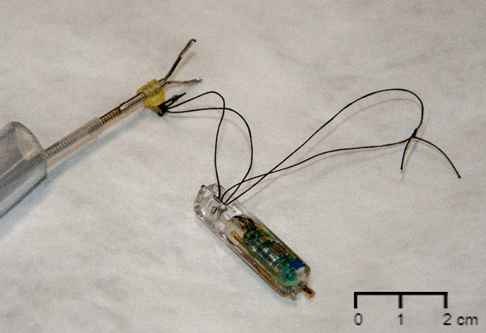
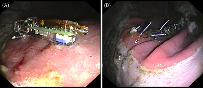
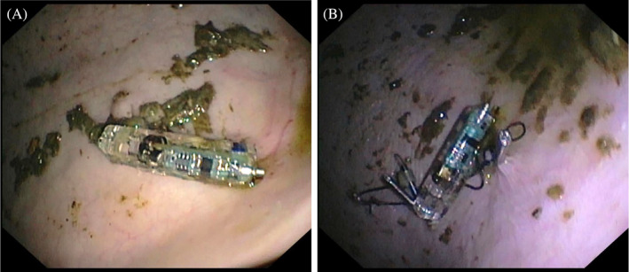
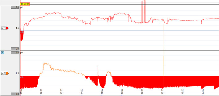
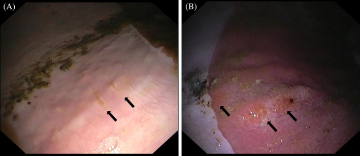
Methods: Capsules were attached to squamous and glandular gastric mucosa under gastroscopic guidance, using suture loops and 1 to 4 hemostasis clips. pH was continuously recorded using a wireless recorder in both fed and fasted states. Gastroscopy was performed daily to assess capsule attachment and any mucosal damage. Data were analyzed using commercially available software. Values are reported as median (interquartile range).
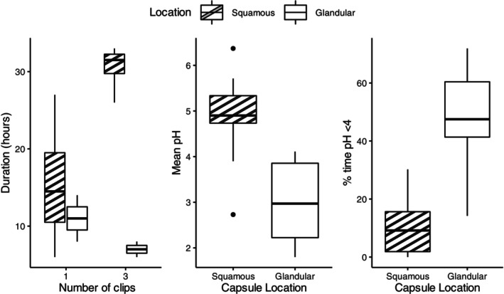
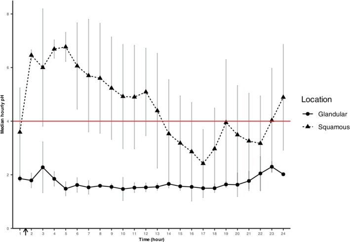
Results: Capsules were successfully placed and data obtained in squamous (n = 11) and glandular (n = 7) regions. The overall duration of squamous capsule attachment was 27 hours (15-32); 1 clip (n = 4) was 15 hours (11-20), 2 clips (n = 2) was 20 hours (16-23), 3 clips (n = 4) was 32 hours (30-32), and 4 clips (n = 1) was 33 hours. The overall duration of glandular capsule attachment was 10 hours (8-21); 1 clip (n = 2) was 11 hours (10-13), 2 clips (n = 2) was 19 hours (14-23), 3 clips (n = 2) was 7 hours (7-8), and 4 clips (n = 1) was 158 hours. There was no substantial damage to the gastric mucosa as a consequence of attachment.
Conclusions and clinical importance: This novel technique enables the wireless measurement of intragastric pH in horses at known locations under fed and fasted conditions, providing a viable alternative for continuous monitoring in both research and clinical scenarios.
Keywords: EGGD; EGUS; ESGD; equine; gastric; ulcers.
© 2024 The Author(s). Journal of Veterinary Internal Medicine published by Wiley Periodicals LLC on behalf of American College of Veterinary Internal Medicine.
Conflict of interest statement
Authors declare no conflict of interest.
Figures
Similar articles
-
The effect of a hay grid feeder on feed consumption and measurement of the gastric pH using an intragastric electrode device in horses: a preliminary report.Equine Vet J. 2014 Jul;46(4):484-7. doi: 10.1111/evj.12175. Epub 2013 Nov 18. Equine Vet J. 2014. PMID: 23991941 Clinical Trial.
-
Bravo capsule placement in the gastric cardia: a novel method for analysis of proximal stomach acid environment.Am J Gastroenterol. 2005 Aug;100(8):1721-7. doi: 10.1111/j.1572-0241.2005.41733.x. Am J Gastroenterol. 2005. PMID: 16086707
-
A novel placement method of the Bravo wireless pH monitoring capsule for measuring intragastric pH.Dig Dis Sci. 2009 Mar;54(3):578-85. doi: 10.1007/s10620-008-0399-3. Epub 2008 Jul 23. Dig Dis Sci. 2009. PMID: 18649136
-
Equine gastric ulcer syndrome in adult horses.Vet J. 2022 May-Jun;283-284:105830. doi: 10.1016/j.tvjl.2022.105830. Epub 2022 Apr 25. Vet J. 2022. PMID: 35472513 Review.
-
Assessment of gastric acidity in intensive care patients: intermittent pH registration cannot replace continuous pH monitoring.Intensive Care Med. 1996 Mar;22(3):220-5. doi: 10.1007/BF01712240. Intensive Care Med. 1996. PMID: 8727435 Review.
References
-
- Chameroy KA, Nadeau JA, Bushmich SL, Dinger JE, Hoagland TA, Saxton AM. Prevalence of non‐glandular gastric ulcers in horses involved in a university riding program. J Equine Vet Sci. 2006;26:207‐211.
-
- Argenzio RA. Comparative pathophysiology of nonglandular ulcer disease: a review of experimental studies. Equine Vet J. 1999;31:19‐23. - PubMed
-
- Merritt AM, Sanchez LC, Burrow JA, Church M, Ludzia S. Effect of GastroGard and three compounded oral omeprazole preparations on 24 h intragastric pH in gastrically cannulated mature horses. Equine Vet J. 2003;35:691‐695. - PubMed
MeSH terms
Grants and funding
LinkOut - more resources
Full Text Sources
