. 2024 Dec 23;15(1):10724.
doi: 10.1038/s41467-024-54901-y.

Development of mirror-image monobodies targeting the oncogenic BCR::ABL1 kinase

Affiliations

Development of mirror-image monobodies targeting the oncogenic BCR::ABL1 kinase

Nina Schmidt et al. Nat Commun. .

Abstract

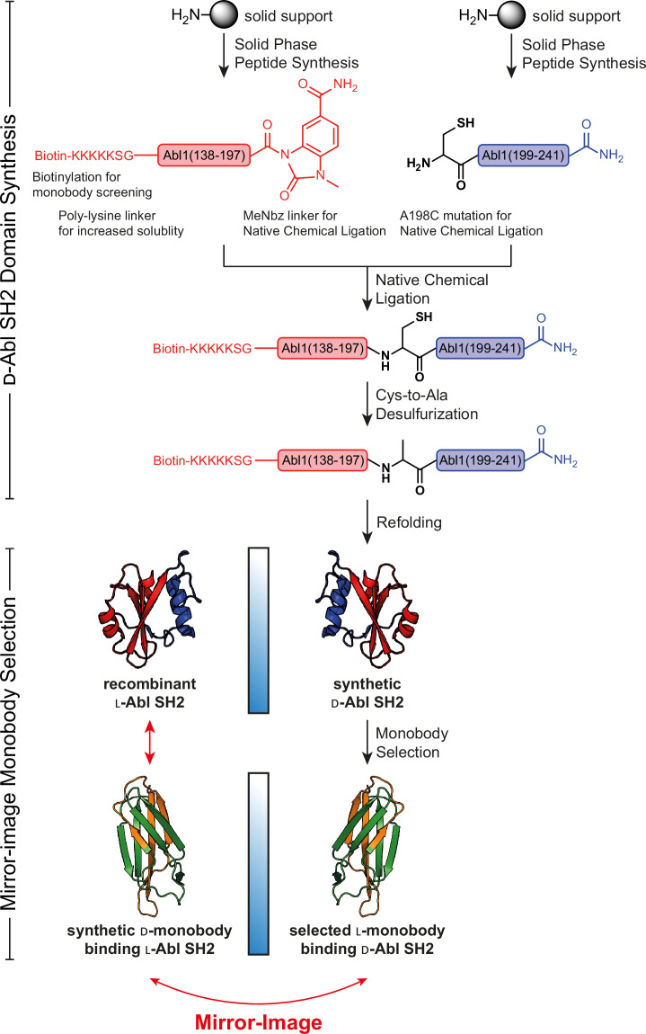
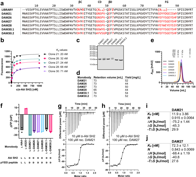
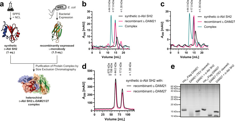
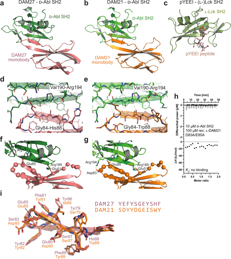
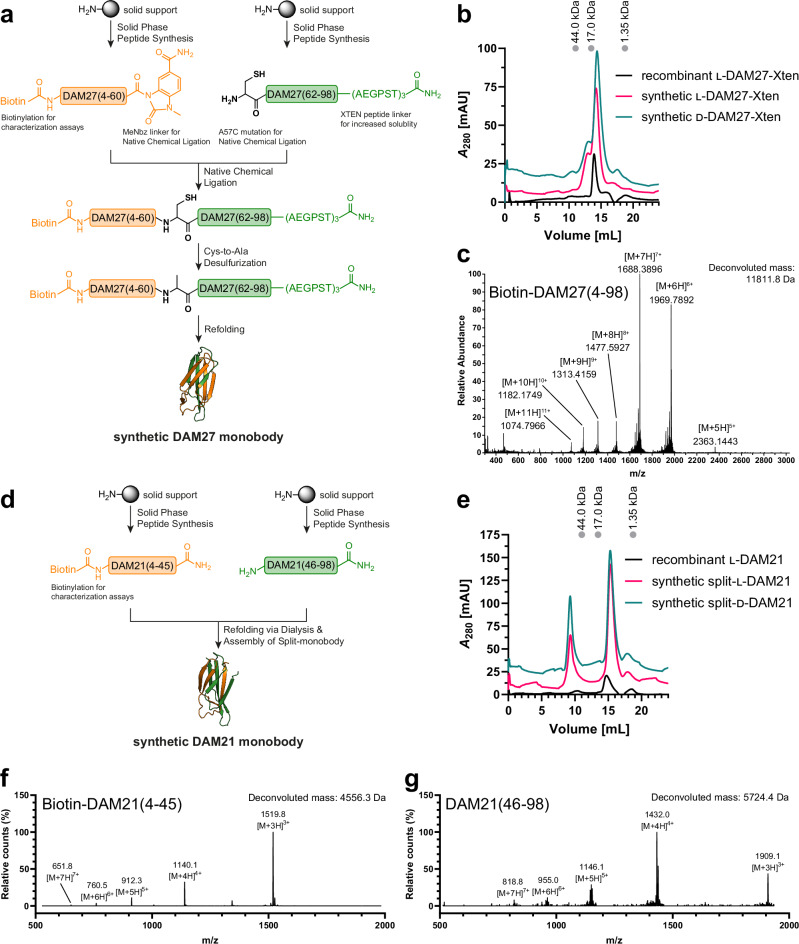
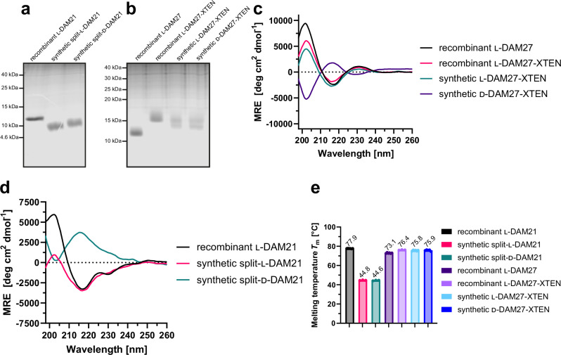
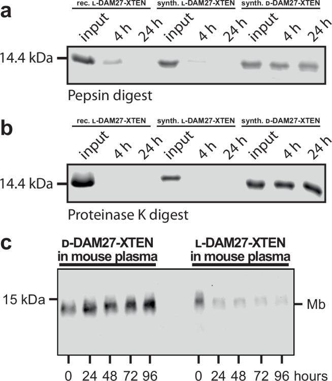
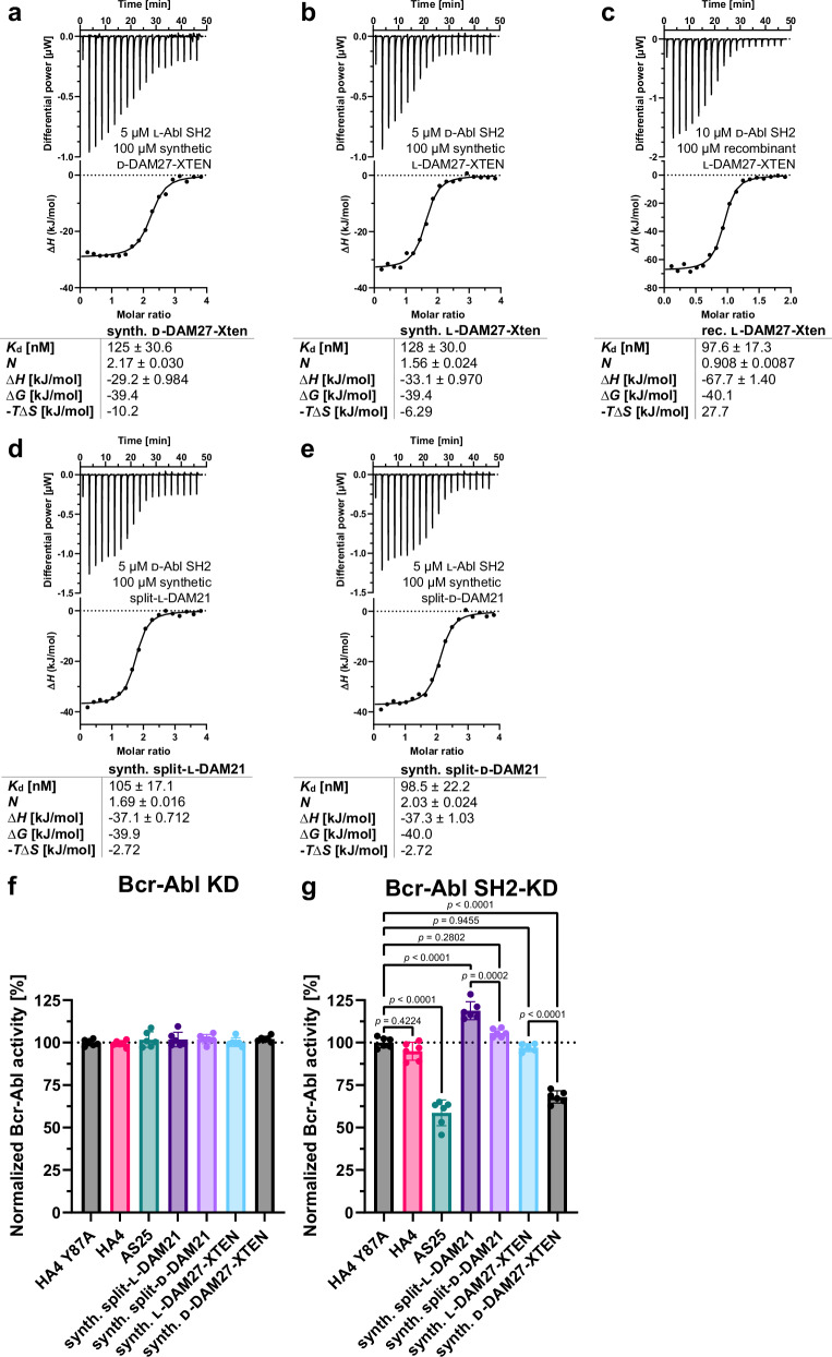
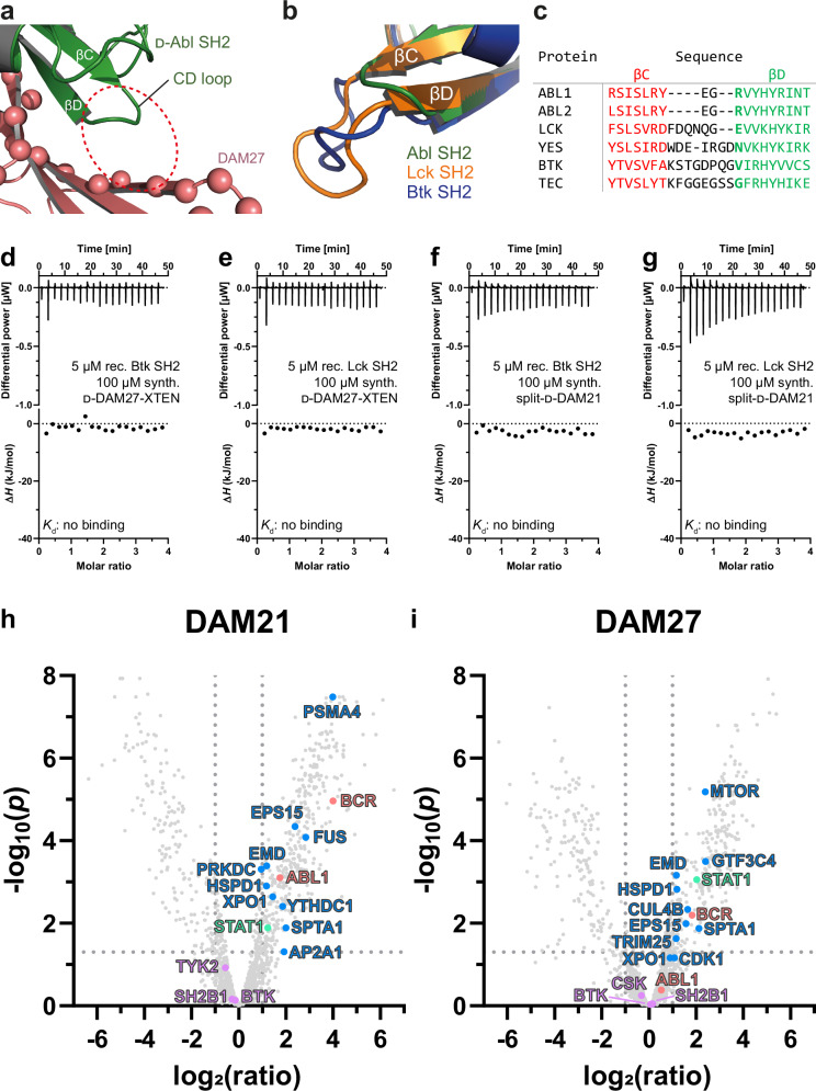
Mirror-image proteins, composed of D-amino acids, are an attractive therapeutic modality, as they exhibit high metabolic stability and lack immunogenicity. Development of mirror-image binding proteins is achieved through chemical synthesis of D-target proteins, phage display library selection of L-binders and chemical synthesis of (mirror-image) D-binders that consequently bind the physiological L-targets. Monobodies are well-established synthetic (L-)binding proteins and their small size (~90 residues) and lack of endogenous cysteine residues make them particularly accessible to chemical synthesis. Here, we develop monobodies with nanomolar binding affinities against the D-SH2 domain of the leukemic tyrosine kinase BCR::ABL1. Two crystal structures of heterochiral monobody-SH2 complexes reveal targeting of the pY binding pocket by an unconventional binding mode. We then prepare potent D-monobodies by either ligating two chemically synthesized D-peptides or by self-assembly without ligation. Their proper folding and stability are determined and high-affinity binding to the L-target is shown. D-monobodies are protease-resistant, show long-term plasma stability, inhibit BCR::ABL1 kinase activity and bind BCR::ABL1 in cell lysates and permeabilized cells. Hence, we demonstrate that functional D-monobodies can be developed readily. Our work represents an important step towards possible future therapeutic use of D-monobodies when combined with emerging methods to enable cytoplasmic delivery of monobodies.


Conflict of interest statement

Competing interests: A.Ko. and S.K. are listed as inventors on issued and pending patents on the monobody technology filed by the University of Chicago (US Patent 9512199 B2 and related pending applications). The other authors declare no competing interests.

Figures

Fig. 1
Fig. 1. Workflow of Bcr-Abl SH2 synthesis as d-target for mirror-image monobody screening.
The d-Bcr-Abl SH2 domain was prepared by native chemical ligation (NCL) and subsequent desulfurization from two fragments produced by solid-phase peptide synthesis (SPPS), as previously described. After refolding and structure validation, the d-target was subjected to monobody selection through phage and yeast display yielding l-monobody binders. Following binder characterization, most promising monobodies were synthesized in d-configuration resulting in d-monobodies targeting the natural l-Bcr-Abl SH2 domain.
Fig. 2
Fig. 2. l-monobody selection and characterization against d-Bcr-Abl SH2.
a Amino acid sequences of d-Abl SH2 l-monobody binders (DAM) generated by phage and yeast display selection based on the combinatorial ‘side-and-loop’ library. In the library designs, “X” denotes a mixture of 30% Tyr, 15% Ser, 10% Gly, 5% Phe, 5% Trp, and 2.5% each of all the other amino acids except for Cys; “O” denotes a mixture of Asn, Asp, His, Ile, Leu, Phe, Tyr, and Val; “U” denotes a mixture of His, Leu, Phe, and Tyr; and “Z” denotes a mixture of Ala, Glu, Lys, and Thr. A hyphen indicates a deletion. b Binding titrations in the yeast surface display format to estimate binding affinities of l-monobodies to d-Abl SH2. The mean fluorescence intensities of yeast cells displaying a monobody are plotted as a function of the concentration of the target and fitted to a 1:1 binding model. c Sodium dodecyl sulfate-polyacrylamide gel electrophoresis (SDS-PAGE) analysis (performed once), (d) retention volumes in size exclusion chromatography (SEC) purification and expression yields, and (e) SEC chromatograms of selected l-DAMs recombinantly expressed in E. coli. (f) Competitive fluorescence polarization (FP) assay of l-DAMs incubated with d-Abl SH2 and fluorescently labeled d-pYEEI peptide binders in comparison with previously published l-monobodies HA4 (pY peptide competitor) and AS25 (allosteric binder) against l-Abl SH2 incubated with the corresponding l-pYEEI peptide. Measured data from two independent experiments (depicted as dots) were averaged. gj Isothermal titration calorimetry (ITC) measurements of recombinant l-monobodies (g) DAM21 and (h) DAM27 titrated to the synthetic d-Abl SH2 domain. Each panel shows the raw heat signal of an ITC experiment (top) and the integrated calorimetric data of the area of each peak (bottom). The continuous line represents the best fit of the data based on a 1:1 binding model computed from the MicroCal software. Binding parameters including Kd value, stoichiometry (N), enthalpy (∆H), free enthalpy (∆G) and −TS calculated from the fit of each experiment are shown in (i) and (j). A representative measurement of at least two ITC experiments for each monobody is shown. Source data of (a, c and f) are provided as a Source Data file.
Fig. 3
Fig. 3. Complex formation and structure elucidation of the heterochiral d-Abl SH2:l-monobody interaction.
a Workflow for generation of the heterochiral d-Abl SH2:l-monobody complex. d-Abl SH2 was produced by solid-phase peptide synthesis (SPPS) and native chemical ligation (NCL) and the l-monobody by bacterial expression and subsequent purification. After mixing, the complex was purified by size exclusion chromatography (SEC). b, c Analytical SEC of complex formation between synthetic d-Abl SH2 and recombinantly expressed (b) l-DAM21 and (c) l-DAM27. d Preparative SEC of d-Abl SH2:l-monobody complexes generated for crystallization condition screening. All chromatograms include a calibration of the column with standard proteins on top. e Sodium dodecyl sulfate-polyacrylamide gel electrophoresis (SDS-PAGE) analysis of the complex formation (performed once). Source data of (e) are provided as a Source Data file.
Fig. 4
Fig. 4. Crystal structures of heterochiral monobody-Abl SH2 complexes.
a, b Ribbon representation of the d-Abl SH2 domain structure (green) bound to the (a) DAM27 or (b) DAM21 monobodies in l-configuration. Arg171 of the SH2 FLVRES motif, which is critical for pY binding, as well as Glu85 in the FG loop of the monobodies is shown in stick representation. c Structure of an (l-) phosphotyrosine peptide (pYEEI, stick representation) bound to the (l-)SH2 domain of the Src family kinase Lck (PDB: 1LKK). The critical Arg residue of the FLVRES motif in the pY binding pocket is shown in stick representation. d, e Close-up view of the d-SH2:(l-)monobody interface (DAM27, panel d; DAM21, panel e) highlighting the heterochiral “rippled” anti-parallel β-sheet between strand Gly84 to Trp88/His88 in DAM21/27 of the monobody FG loop with Val190 to Arg194 of the βD-strand of the d-Abl SH2 domain. f, g Close-up view of the d-SH2:(l-)monobody interface (DAM27, panel f; DAM21, panel g). The ionic interactions discussed in the “Results” section are shown as sticks and are labeled. The position of randomized residues in the monobody side-and-loop library is shown as balls. h Isothermal titration calorimetry (ITC) measurements of recombinant l-DAM21 D83A/E85A mutant titrated to the synthetic d-Abl SH2 domain. The panel shows the raw heat signal of an ITC experiment (top) and the integrated calorimetric data of the area of each peak (bottom). i Structural superposition and sequence alignment of the FG loops of the DAM27 and DAM21 monobodies.
Fig. 5
Fig. 5. DAM21 and DAM27 synthesis, refolding and purification.
a Strategy of DAM27 synthesis. The N- and C-terminal fragments are shown in orange and green, respectively, and synthesized via solid-phase peptide synthesis (SPPS). The N-terminal peptide corresponds to the biotinylated DAM27(4-60) fragment with C-terminal MeDbz linker, which is activated to MeNbz on resin, and the C-terminal peptide resembles DAM27(62-98) with an N-terminal cysteine (Cys61) residue. After cleavage from the resin, both peptides can undergo native chemical ligation (NCL) with subsequent desulfurization to yield full-length monobody DAM27(4-98). MeDbz: 3-amino-4-(methylamino)benzoic acid; MeNbz: N-acyl-N-methylacylurea. b Size exclusion chromatography (SEC) and (c) high-performance liquid chromatography-mass spectrometry (HPLC-MS) analysis of the final refolded l- and d-DAM27 proteins in comparison with the recombinantly expressed l-DAM27-XTEN reveals similar SEC retention volumes and expected masses of the synthetic proteins. d Strategy of DAM21 synthesis. The N- and C-terminal peptides in orange and green obtained by SPPS represent the biotinylated DAM21(4-45) and DAM21(46-98) fragments, respectively. After mixing of the peptides and refolding by dialysis, the full-length split-monobody DAM21(4-98) is obtained. e SEC of the final refolded l- and d-DAM21 proteins in comparison with the recombinantly expressed l-DAM21 reveals similar SEC retention volumes of the synthetic proteins. f, g HPLC-MS analysis of the (f) DAM21(4-45) and (g) DAM21(46-98) fragments.
Fig. 6
Fig. 6. Folding and stability characterization of synthetic l- and d-monobodies.
a, b Representative analysis of purity of synthetic (a) l- and d-DAM21 and (b) l- and d-DAM27 by sodium dodecyl sulfate-polyacrylamide gel electrophoresis (SDS-PAGE) (performed at least twice). c, d Averaged far-UV circular dichroism (CD) spectra from three independent measurements of synthetic (c) l- and d-DAM27 and (d) l- and d-DAM21 in comparison with their recombinantly expressed analogues. Mean residue ellipticity (MRE) was calculated from three independent measurements. e Bar graph representation of thermal stability of recombinantly expressed and synthetic l- and d-monobodies assessed by nano differential scanning fluorimetry (nanoDSF). Melting temperatures (Tm) were measured at least three times (depicted as dots) and averaged. Error bars represent the standard deviation (SD). Source data of (a, b and e) are provided as a Source Data file.
Fig. 7
Fig. 7. Protease resistance and mouse plasma stability of synthetic l- and d-monobodies.
a, b Protease resistance of recombinant l-DAM27-XTEN as well as synthetic l- and d-DAM27-XTEN after incubation with (a) pepsin and (b) proteinase K was analyzed by sodium dodecyl sulfate-polyacrylamide gel electrophoresis (SDS-PAGE). c Plasma stability of synthetic l- and d-DAM27-XTEN. Monobodies labeled with biotin at the N-terminus after incubation with mouse plasma were analyzed by Western blotting and detection of biotin using Streptavidin-IRDye680. A representative blot of three repeats is shown. Source data of (ac) are provided as a Source Data file.
Fig. 8
Fig. 8. Binding of synthetic l- and d-monobodies to d- and l-Bcr-Abl SH2.
ae Isothermal titration calorimetry (ITC) measurements of (a) synthetic d-DAM27-XTEN titrated to recombinantly expressed l-Abl SH2, (b) synthetic l-DAM27-XTEN and (c) recombinant l-DAM27-XTEN both titrated to synthetic d-Abl SH2, (d) synthetic split-l-DAM21 and (e) synthetic split-d-DAM21 titrated to synthetic d- and l-Abl SH2, respectively. Each panel shows the raw heat signal of an ITC experiment (top) and the integrated calorimetric data of the area of each peak (bottom). The continuous line represents the best fit of the data based on a 1:1 binding model computed from the MicroCal software. Binding parameters including Kd value, stoichiometry (N), enthalpy (∆H), free enthalpy (∆G) and −TS calculated from the fit of each experiment are shown below. A representative measurement of at least two ITC experiments for each monobody is shown. f, g Measurement of kinase activity of (f) Bcr-Abl kinase domain (KD) and (g) SH2-KD after incubation with synthetic split-l- and d-DAM21 as well as synthetic l- and d-DAM27-XTEN in comparison with binding control monobodies HA4 and AS25 and the non-binding control monobody HA4 Y87A using a radiometric kinase assay. All monobodies were used at a concentration of 5 µM. Here, radioactively labeled 32P was incorporated into a biotinylated substrate peptide by recombinantly expressed KD and SH2-KD and detected via scintillation counting. Six independent experiments were performed (depicted as dots) and averaged. Error bars represent the standard deviation (SD) and statistical analysis was done with a one-way ANOVA and Sidak’s test. The calculated p-values are depicted in (g) and were considered statistically significant below a value of 0.05. F values and degrees of freedom were 124.3 and 40. Source data of (f, g) are provided as a Source Data file.
Fig. 9
Fig. 9. Selectivity of d-DAM21/27 for the BCR::ABL1 SH2 domain.
a l-DAM27:d-Abl SH2 complex structure (PDB code: 9F00) highlighting the short Abl SH2 CD loop that enables binding to DAM27. The space where longer CD loops in other SH2 domains would be located is indicated by the dotted red circle. b Structural superposition of the Abl, Lck and Btk SH2 domains (PDB entries 3K2M, 1LKK and 6HTF, respectively). The additional four to six amino acid residues in the Lck and Btk SH2 CD loops are not compatible with binding to the DAM21 and DAM27 monobodies. c Multiple sequence alignment of the βC and βD strands of SH2 domains belonging to different SH2 domain-containing tyrosine kinase families. The βC and βD strands are colored in red and green, respectively, while the CD loop in between is black. Arg189 of ABL1, which is important for d-monobody binding via ionic interactions, is not conserved and colored in bold green to highlight the different amino acid sequences in this position. d, e Isothermal titration calorimetry (ITC) measurements of d-DAM27-XTEN titrated to the SH2 domains of (d) Btk and (e) Lck. f, g ITC measurements of split-d-DAM21 titrated to the SH2 domains of (f) Btk and (g) Lck. Each panel shows the raw heat signal of an ITC experiment (top) and the integrated calorimetric data of the area of each peak (bottom). h, i Volcano plots of identified proteins via mass spectrometry after pulldown from K562 cell lysates comparing synthetic (h) split-d-DAM21 with split-l-DAM21 and (i) d-DAM27-XTEN with l-DAM27-XTEN. Pulldowns were performed in three biological replicates for each monobody. Protein identification and statistical analysis of replicates were done with MaxQuant 2.5.1.0 and Autonomics (R package version 1.13.21) resulting in FDR-corrected p-values (Benjamini-Hochberg procedure) for each identified protein where a p-value below 0.05, which corresponds to -log10(p) above 1.3, was considered statistically significant (dotted line intersecting y-axis). A log2(ratio) above 1.00 of d- vs. l-monobody correlates to a ratio above 2.00 and a higher abundance of the protein when the pulldown was performed with the d-monobody compared to its l-counterpart (dotted lines intersecting x-axis). The color coding represents the protein groups BCR and ABL1 (light red), BCR::ABL1/ABL1 interactors (blue), BCR::ABL1/ABL1 interactors with SH2 domains (green) and other proteins containing SH2 domains (purple). Plotted values of all highlighted protein groups are listed in Tables S9, S10.

References

    1. Vazquez-Lombardi, R. et al. Challenges and opportunities for non-antibody scaffold drugs. Drug Discov. Today20, 1271–1283 (2015). - PubMed
    1. Hantschel, O. Monobodies as possible next-generation protein therapeutics—a perspective. Swiss Med. Wkly147, w14545 (2017). - PMC - PubMed
    1. Gebauer, M. & Skerra, A. Engineered protein scaffolds as next-generation therapeutics. Annu. Rev. Pharm. Toxicol.60, 391–415 (2020). - PubMed
    1. Hantschel, O., Biancalana, M. & Koide, S. Monobodies as enabling tools for structural and mechanistic biology. Curr. Opin. Struct. Biol.60, 167–174 (2020). - PMC - PubMed
    1. Luo, R., Liu, H. & Cheng, Z. Protein scaffolds: antibody alternatives for cancer diagnosis and therapy. RSC Chem. Biol.3, 830–847 (2022). - PMC - PubMed

Publication types

MeSH terms

LinkOut - more resources